Vaso-occlusion and Bone Complications
- HbS: Glutamate to valine substitution at position 6 of beta-globin chain
- Vaso-occlusion: Sickling under hypoxia/acidosis causes bone infarcts, AVN, painful crises
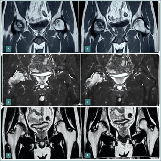
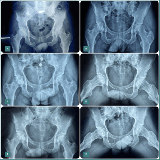
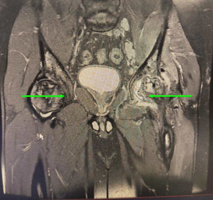
- AVN: quote PREVALENCE and INCIDENCE separately - femoral head about 10% at any one time, humeral head about 6%, but accruing at 2-4.5 per 100 patient-years, which is why older cohorts report far higher cumulative figures. Often bilateral and multi-joint
- Osteomyelitis: SALMONELLA is most common organism (unique to SCD, unlike general population)
- Perioperative: Avoid hypothermia, hypoxia, dehydration, acidosis - simple transfusion to Hb 10 g/dL for most surgery; benefit of exchange to HbS less than 30% for the highest risk is unsettled
- “Salmonella osteomyelitis
- “H-shaped vertebrae
- “Bilateral AVN common
- “Hydration critical perioperatively
Overview and Epidemiology
Sickle cell disease (SCD) is a hereditary haemoglobinopathy with significant orthopaedic manifestations. It is autosomal recessive, the result of a single substitution in the HBB gene, glutamate to valine at position 6 of the beta-globin chain (Glu6Val), which produces HbS.
Genotypes. Severity follows the genotype:
- HbSS - the most severe, with a worse prognosis than HbSC
- HbSC - milder
- HbS-beta thalassaemia
Where it is found. Over 300,000 affected children are born each year worldwide, roughly 75-80% of them in sub-Saharan Africa, with India and the Middle East also high-burden. The distribution follows the historical malaria belt because the HbS trait confers resistance to falciparum malaria, a balanced polymorphism. Migration is raising prevalence in Europe, North America and Australasia, and diaspora populations have made SCD an orthopaedic problem everywhere rather than a regional one.
Survival. Median survival is 40-60+ years in high-resource settings with hydroxyurea, vaccination and transfusion programmes, and remains markedly lower in limited-resource regions where childhood mortality is high. Hydroxyurea reduces crisis frequency and improves survival, and stem cell transplant is potentially curative in selected patients.
Pathophysiology and Mechanisms
Sickling. A single nucleotide mutation in the beta-globin gene gives HbS, whose molecules aggregate when deoxygenated. The long polymer fibres they form distort the red cell membrane into the sickle shape, and repeated sickling damages the membrane irreversibly.
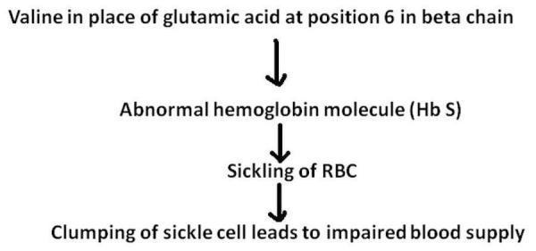
Vaso-occlusion. Sickled cells are rigid and adhere to the endothelium, obstructing the microvasculature, the capillaries and sinusoids. The tissue downstream becomes ischaemic, and reperfusion then adds injury with an inflammatory response.
What it does to bone. Four processes act on the skeleton:
- Marrow infarction leads to painful crises and bone necrosis
- End-artery occlusion falls particularly on the epiphyses (AVN) and the vertebral endplates (H-shaped vertebrae)
- Marrow hyperplasia, a compensatory expansion against chronic haemolysis, thins the cortex
- Infection risk rises because functional asplenia (autosplenectomy, a small or absent spleen) combines with micro-infarcts that create a nidus
Triggers. Sickling, and so a crisis, is precipitated by:
- Cold
- Hypoxia, including high altitude
- Acidosis
- Infection
- Dehydration
- Physical exertion
Clinical Presentation
The painful vaso-occlusive crisis is the most common acute complication requiring hospitalisation. The pain is severe and often diaphyseal: in children under 3 years it takes the form of dactylitis (described below), and in older children and adults it settles in the long bones.
Examination. The affected bones are swollen and tender, often at several sites at once, with no fluctuance or abscess. There is no fever, or only a mild low-grade temperature. An episode typically lasts 4-7 days.
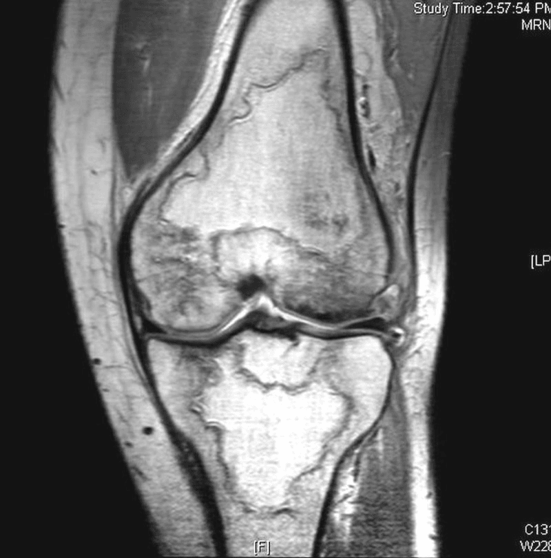
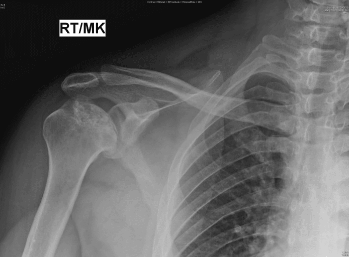
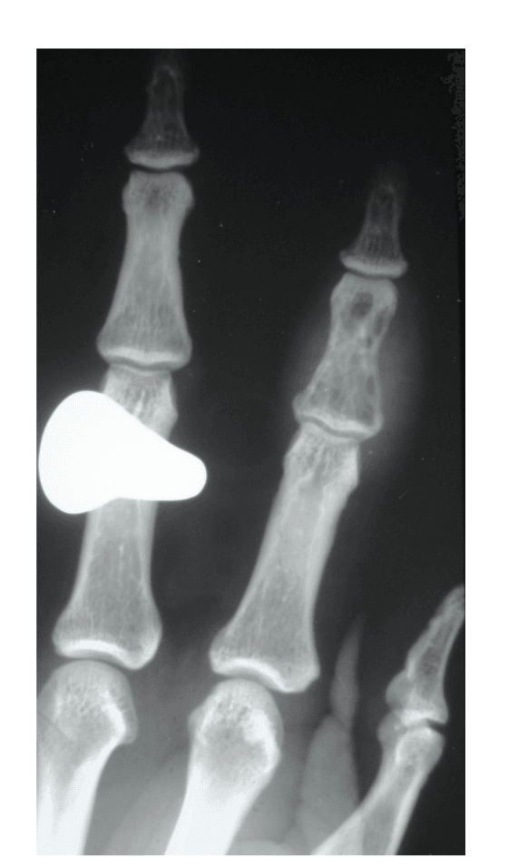
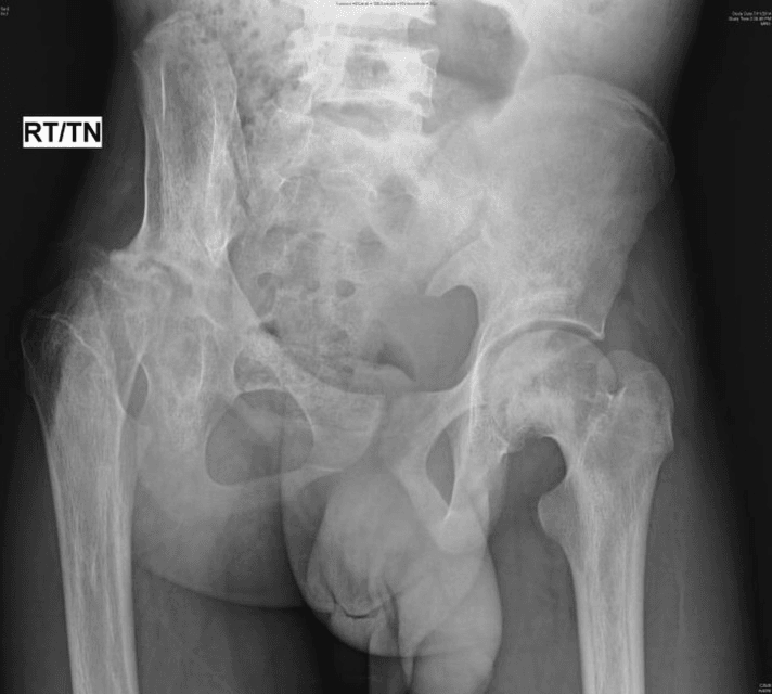
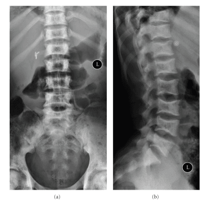
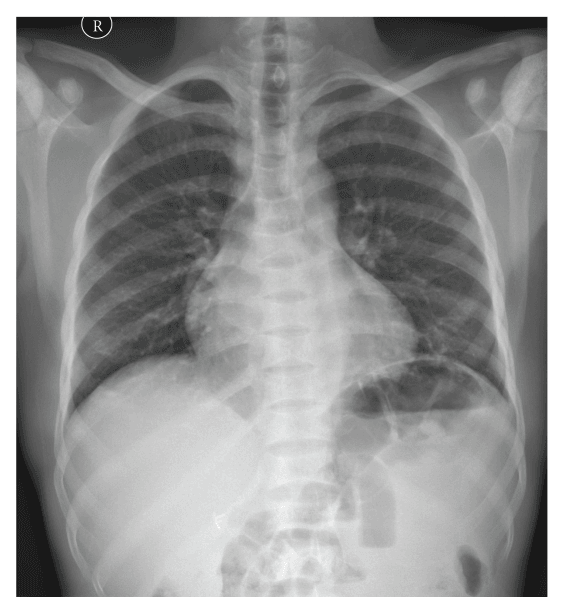
Investigations
Haematology. Four tests:
- Full blood count - chronic anaemia, Hb 6-9 g/dL typical
- Reticulocyte count - raised by chronic haemolysis
- Blood film - sickled cells, and Howell-Jolly bodies from asplenia
- Haemoglobin electrophoresis - confirms HbSS, HbSC or HbS-thal, and gives the HbS percentage used in surgical planning
Infection. Take blood cultures if the patient is febrile. CRP and ESR rise in infection; a very high WCC suggests infection, where a crisis gives only a mild rise, and procalcitonin may help separate the two.
Before surgery. Group and hold or crossmatch, coagulation studies, renal function because sickle nephropathy may be present, and liver function for iron overload from transfusions.
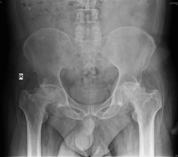
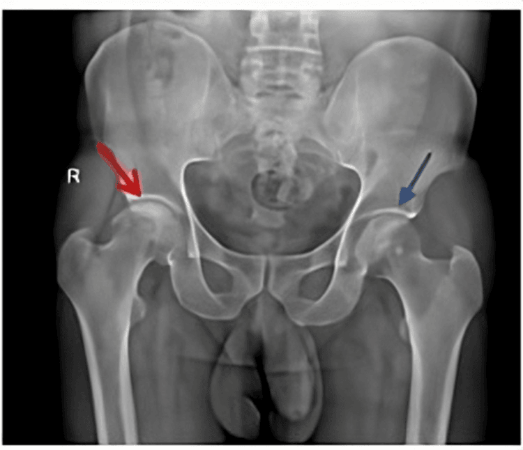
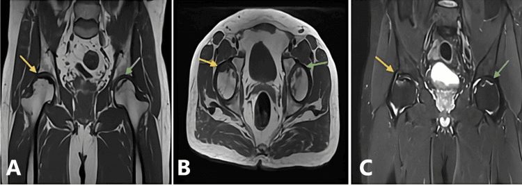
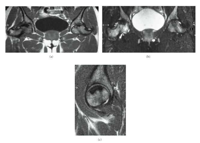
Crisis or osteomyelitis. Both present with a painful, swollen bone; these are the features that separate them:
- Vaso-occlusive Crisis
- Acute (hours)
- Osteomyelitis
- Subacute (days to weeks)
- Vaso-occlusive Crisis
- Low-grade or absent
- Osteomyelitis
- High-grade (38.5C or higher)
- Vaso-occlusive Crisis
- Normal or mildly elevated
- Osteomyelitis
- Significantly elevated
- Vaso-occlusive Crisis
- Mildly elevated (under 100)
- Osteomyelitis
- Markedly elevated (over 100)
- Vaso-occlusive Crisis
- Often multifocal
- Osteomyelitis
- Usually single focus
- Vaso-occlusive Crisis
- Marrow oedema only
- Osteomyelitis
- Periosteal reaction, soft tissue changes, abscess
- Vaso-occlusive Crisis
- 4-7 days typical
- Osteomyelitis
- Weeks if untreated
- Vaso-occlusive Crisis
- Good improvement
- Osteomyelitis
- Minimal improvement
The wider differential. The painful limb or joint in SCD has six diagnoses to consider:
- Distinguishing Features
- Multifocal, symmetrical, recurrent, low-grade or no fever, responds to hydration/analgesia
- Key Investigation
- MRI: marrow oedema, no periosteal reaction
- Distinguishing Features
- Single focus, high fever, persistent despite hydration, soft-tissue signs
- Key Investigation
- MRI plus blood/bone cultures (Salmonella greater than Staph)
- Distinguishing Features
- Hot, swollen joint, refusal to move, often hip in children with AVN
- Key Investigation
- Joint aspiration (urgent)
- Distinguishing Features
- Insidious groin/shoulder pain, mechanical, reduced internal rotation, often bilateral
- Key Investigation
- MRI (earliest), staged by Ficat/ARCO
- Distinguishing Features
- Chest/back pain, fever, hypoxia, new infiltrate - may mimic rib infarct
- Key Investigation
- CXR plus SpO2; high mortality
- Distinguishing Features
- Monoarticular, hyperuricaemia from cell turnover, crystals
- Key Investigation
- Joint aspiration for crystals
Management
Acute treatment. Treat the crisis with:
- Hydration - IV fluids at 1.5x maintenance
- Analgesia - often opioids (morphine PCA); NSAIDs with caution for renal function; antiemetics if needed
- Oxygen - if SpO2 is below 95%
- Warmth - maintain normothermia
- Transfusion - simple transfusion if severe
- Rest, and monitoring for complications
When to escalate. Escalate for any of:
- Acute chest syndrome (fever, respiratory symptoms, new infiltrate)
- Severe anaemia, Hb below 5 g/dL
- Stroke symptoms
- Priapism lasting more than 4 hours
Complications
Orthopaedic. Beyond AVN, infection and growth disturbance, repeated crises and immobilisation leave joint contractures, and recurrent or incompletely treated infection becomes chronic osteomyelitis.
Surgical. Hypoxia or hypothermia can trigger a sickling crisis around the operation, and acute chest syndrome is a potentially life-threatening postoperative complication. Delayed wound healing is common, and implants loosen earlier and need revision sooner (see Outcomes).
Systemic.
- Acute chest syndrome - the most common cause of death in SCD
- Stroke - in 10% of children with SCD
- Renal failure - sickle nephropathy
- Pulmonary hypertension - from chronic haemolysis
- Iron overload - from chronic transfusion
Acute Chest Syndrome: the Perioperative Killer
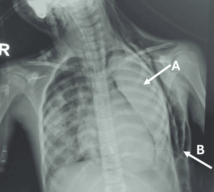
Definition. ACS is a new pulmonary infiltrate on chest imaging involving at least one complete lung segment, plus a new respiratory sign or symptom: fever, cough, chest or back pain, tachypnoea, wheeze or hypoxia. It is clinically and radiologically indistinguishable from pneumonia at onset, so the two are managed together.
Why orthopaedic care precipitates it. Surgery opens three routes:
- Fat and marrow embolism from infarcted long-bone marrow, as bone-marrow necrosis showers fat emboli to the lungs; relevant after fracture, marrow-cavity instrumentation or a severe long-bone crisis
- Hypoventilation and atelectasis from postoperative pain, opioid respiratory depression, and splinting after thoracic, upper-abdominal, rib or sternal incisions or infarcts
- Immobility, over-transfusion or fluid overload, and infection in the perioperative period
Recognition. After surgery, look for new or worsening hypoxia, tachypnoea, chest or back pain, fever and a new infiltrate on the chest radiograph. Distinguish it from pneumonia, pulmonary embolism, fluid overload and, in the trauma patient, classic fat-embolism syndrome, though in SCD these mechanisms overlap.
Treatment. Escalate early to HDU or ICU. Give supplemental oxygen titrated to saturations, incentive spirometry, bronchodilators if there is wheeze, and broad antibiotics covering atypical and encapsulated organisms.
Analgesia and blood. Provide analgesia adequate to prevent splinting yet titrated to avoid opioid-induced hypoventilation. Simple transfusion improves oxygen-carrying capacity, and exchange transfusion is indicated for severe or deteriorating hypoxia.
Prevention is the surgeon's lever. Incentive spirometry, early mobilisation, judicious rather than excessive fluids, and regional analgesia to limit systemic opioids, layered on the normothermia-oxygenation-transfusion protocol.
Post-operative hypoxia in a sickle-cell patient is acute chest syndrome until proven otherwise. It complicated roughly 1 in 10 operations in the PTSCD trial and was the only cause of death in that study, so transfusing to Hb 10 g/dL does not remove the risk.
Dactylitis (Hand-Foot Syndrome): Often the First Manifestation
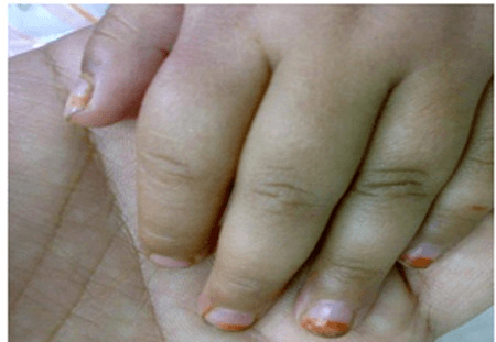
Dactylitis, the "hand-foot syndrome" of infancy, is frequently the earliest skeletal manifestation of sickle cell disease and is often the event that first brings the child to diagnosis.
Mechanism. Vaso-occlusive infarction of the marrow of the small tubular bones: the metacarpals, metatarsals and phalanges. It clusters in infants and children younger than 3 years of age, the period before the red (haematopoietic) marrow of the hands and feet converts to yellow marrow. As that conversion completes, dactylitis largely disappears and crises shift to the long bones and axial skeleton.
Clinical picture. Painful, diffuse dorsal swelling of the hands, the feet or both, with warmth and tenderness, often a low-grade fever and reluctance to use the limb. It is characteristically bilateral, symmetrical and multi-digit, a pattern that helps separate it from a single-focus infection.
Radiographs lag the pain. Films are normal or show only soft-tissue swelling in the first days. After roughly one to two weeks, periosteal reaction, patchy medullary lucency and sclerosis appear from the infarct, occasionally a bone-within-bone appearance. The changes usually resolve without deformity, but rare epiphyseal or physeal damage can leave a shortened digit (brachydactyly) or a metacarpal or metatarsal length discrepancy.
Differentials. Consider each in the swollen hand or foot of an infant:
- Osteomyelitis or septic arthritis - single focus, higher and persistent fever, unresponsive to hydration
- Non-accidental injury in the swollen infant
- Tuberculous dactylitis (spina ventosa) in tuberculosis-endemic regions
Management is that of any vaso-occlusive crisis: hydration, analgesia, warmth and reassurance, as the episode is self-limiting over one to two weeks.
Outcomes
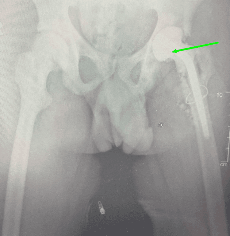
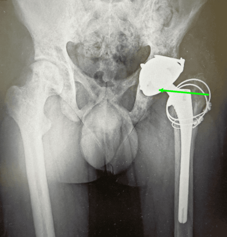
Arthroplasty. THA in SCD survives less well and fails more often than in the general population, and the revision rate is higher because of loosening and infection.
- SCD
- 80-90%
- General Population
- 95-98%
- SCD
- 10-15%
- General Population
- 1-2%
- SCD
- Increased
- General Population
- 1-3%
- SCD
- Earlier failure
- General Population
- 1-2% at 10 years
- SCD
- Higher
- General Population
- Standard
Prognostic factors. Beyond genotype, three things shape the long-term course:
- Frequent crises - associated with more complications
- Age - earlier AVN is associated with worse long-term outcomes
- Transfusion burden - iron overload complications
Guidelines, Registries & Global Practice
Global epidemiology:
- Over 300,000 affected births per year worldwide; the great majority in sub-Saharan Africa, with India and the Middle East also high-burden.
- Diaspora migration has made SCD a routine consideration in orthopaedic units across Europe, North America and Australasia.
- Femoral head osteonecrosis affects roughly 10% (higher with age and HbSS + alpha-thalassaemia); osteomyelitis and septic arthritis are over-represented compared with the general population.
Side-by-side society guidance:
- Position
- Preoperative simple transfusion to Hb approximately 10 g/dL for most surgery under GA; extended antigen matching to limit alloimmunisation
- Position
- Multidisciplinary haematology-led perioperative planning; hydroxycarbamide (hydroxyurea) for recurrent crises; transfusion to reduce HbS for high-risk surgery
- Position
- Hydroxyurea for frequent crises/acute chest syndrome; chronic transfusion for primary stroke prevention; penicillin prophylaxis and full vaccination in children
- Position
- No SCD-specific implant mandate; emphasis on thermoregulation, oxygenation, hydration, controlled transfusion and anticipating sclerotic/obliterated canals
- No dedicated SCD arthroplasty registry exists; national joint registries (NJR, AOANJRR, AJRR, Swedish/Norwegian) capture osteonecrosis as an indication but rarely sub-stratify SCD, so most outcome data come from single-centre and pooled series (e.g. the Hernigou experience).
- High-resource: newborn screening, hydroxyurea, vaccination, antigen-matched blood, MRI access, planned exchange transfusion and elective protocol-driven arthroplasty.
- Limited-resource: late presentation, restricted safe-blood and MRI availability, higher childhood mortality, and a greater burden of advanced osteonecrosis and chronic osteomyelitis presenting for salvage rather than elective reconstruction.
- Universal principles regardless of setting: haematology co-management, avoidance of the sickling triggers, infection coverage for Salmonella plus Staph, and informed counselling about elevated surgical risk.
Controversies and Areas of Uncertainty
Tourniquet use. Traditionally avoided for fear of regional hypoxia/acidosis precipitating sickling, but several series report safe use with limited time and good oxygenation. There is no high-level evidence either way - practice varies; if one is needed, use it with caution.
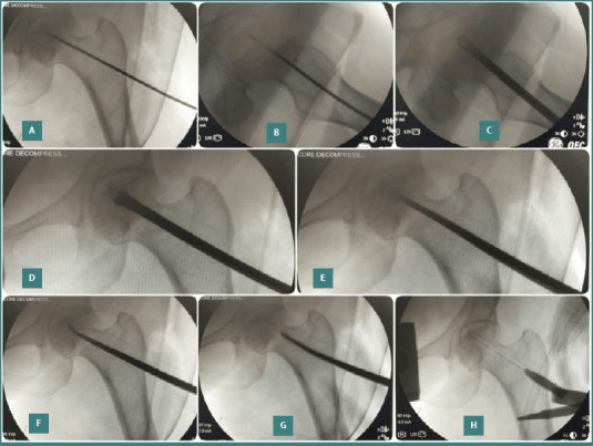
Core decompression and biologics. Outcomes of core decompression (with or without concentrated bone marrow / cell therapy) are less predictable than in non-SCD osteonecrosis; patient selection (pre-collapse, small lesions) is key and the evidence base is largely observational.
Cemented vs cementless fixation. Abnormal sclerotic bone and obliterated medullary canals complicate both techniques. No consensus exists; choice is guided by bone quality, canal anatomy and surgeon experience rather than robust comparative data.
Distinguishing infarct from osteomyelitis. Even MRI plus inflammatory markers can be equivocal; some advocate early aspiration/biopsy, others a trial of conservative management with close monitoring. Over-diagnosis leads to unnecessary surgery; under-diagnosis risks sepsis.
Curative therapies. Allogeneic stem cell transplant and emerging gene therapies may alter the future orthopaedic disease burden, but their long-term effect on established bone disease is unknown.
MCQ Practice Points
Q: What is the most common cause of osteomyelitis in patients with sickle cell disease?
A: Salmonella species is the most common organism causing osteomyelitis in sickle cell disease (approximately 50%), unlike the general population where S. aureus predominates. Reason: Splenic dysfunction (autosplenectomy) impairs clearance of encapsulated organisms and Salmonella. S. aureus is still the second most common. Clinical challenge: Differentiating bone infarction (vaso-occlusive crisis) from osteomyelitis - both present with fever, pain, and elevated inflammatory markers. MRI helps differentiate (osteomyelitis shows soft tissue abscess, cortical destruction).
Q: What are the orthopaedic manifestations of sickle cell disease?
A: Avascular necrosis: Femoral head (most common - prevalence about 10%, rising cumulatively with age), humeral head (about 6%, often silent), vertebral bodies; due to vaso-occlusive crisis affecting end-arterial blood supply. Osteomyelitis: Increased risk, Salmonella most common pathogen. Bone infarcts: Long bone diaphyses; may mimic osteomyelitis. Dactylitis ("hand-foot syndrome"): Painful swelling of hands/feet in infants - first manifestation of SCD. Growth disturbance: Vertebral end-plate collapse ("H-shaped" or "Lincoln log" vertebrae). Pathological fractures from weakened bone.
Q: How do you differentiate bone infarction from osteomyelitis in sickle cell disease?
A: Clinical overlap: Both cause pain, fever, swelling, elevated WBC/CRP/ESR. Favoring osteomyelitis: Localized warmth, erythema, soft tissue abscess, single bone involvement, persistent fever despite hydration/analgesia. Favoring infarction: Multiple bone involvement, symmetric, responds to hydration/pain management. Imaging: MRI - osteomyelitis shows soft tissue collection, cortical destruction, enhancing abscess; infarction shows serpentine enhancement pattern. Aspiration/biopsy: Definitive - culture positive in osteomyelitis. When in doubt, treat empirically for osteomyelitis covering Salmonella and S. aureus.
Q: What perioperative considerations are important in patients with sickle cell disease undergoing orthopaedic surgery?
A: Preoperative: Hematology consultation; consider preoperative transfusion to achieve HbS less than 30% and Hb 10g/dL (exchange transfusion if needed); optimize hydration. Intraoperative: Avoid hypoxia, acidosis, hypothermia, dehydration (all precipitate sickling); use supplemental oxygen; warm IV fluids; careful tourniquet use (controversial - limit time, ensure adequate oxygenation). Postoperative: Continue supplemental oxygen; aggressive pain management; early mobilization; incentive spirometry (prevent acute chest syndrome); maintain hydration. Higher risk of VTE, infection, and wound complications.
Q: What are the treatment options for avascular necrosis of the femoral head in sickle cell disease?
A: Treatment mirrors AVN from other causes but with specific considerations: Early stages (Ficat I-II): Protected weight-bearing, core decompression (mixed results in SCD). Advanced stages (Ficat III-IV): Total hip arthroplasty - higher complication rate in SCD (infection, wound problems, perioperative crisis) but outcomes improving with modern perioperative protocols. Specific considerations: Younger patient age often; higher revision rates than non-SCD; cement may be preferred (abnormal bone quality); aggressive perioperative transfusion reduces complications. Preoperative optimization critical.
Self-Assessment Quiz
Viva Scenarios
Practise clinical reasoning and management decisions out loud
“10-year-old with known sickle cell disease (HbSS) presents with fever 39C, right tibial pain and swelling for 1 week. Mum says it's different from his usual pain crises.”
“Same patient now 30 years old requires total hip arthroplasty for Ficat Stage IV AVN of right hip. How do you prepare him for surgery?”
“22-year-old woman with HbSS sickle cell disease presents with 3-month history of bilateral hip pain and limp. X-rays show early sclerosis of both femoral heads without collapse.”
“You are shown a lateral spine X-ray of a 15-year-old with sickle cell disease showing characteristic central endplate depression of multiple vertebral bodies. Describe the findings and explain the pathophysiology.”
PATHOPHYSIOLOGY
- HbS: Glu6Val mutation
- Sickling under hypoxia/acidosis
- Vaso-occlusion causes infarcts
- Autosomal recessive
ORTHOPAEDIC ISSUES
- AVN: hip ~10%, shoulder ~6% prevalence; 2-4.5 per 100 patient-years
- Osteomyelitis: SALMONELLA before Staph
- Bone crises: Painful vaso-occlusion
- H-shaped vertebrae: Endplate infarcts
PERIOPERATIVE (CHANT)
- Cold avoidance
- Hydration critical
- Acidosis prevention
- Normal oxygen (SpO2 95%+)
- Transfuse: HbS less than 30%, Hb 10
THA OUTCOMES
- Infection: 10-15%
- Survivorship: 80-90% at 10yr
- Higher revision rates
- Counsel about complications
Evidence Base
Vichinsky EP et al - Preoperative Transfusion in Sickle Cell Disease Study (PTSCD)
- Multicentre RCT, 604 operations randomised to aggressive (target HbS less than 30%) vs conservative (target Hb 10 g/dL) transfusion
- Serious complications similar between groups (31% vs 35%)
- Aggressive arm had double the transfusion-related complications (14% vs 7%)
- Acute chest syndrome in 10% of both groups
Milner PF et al - Cooperative Study of Sickle Cell Disease (femoral head)
- 2590 patients followed mean 5.6 years; femoral head osteonecrosis prevalence approximately 10% at entry
- Highest incidence in HbSS with co-inherited alpha-thalassaemia (4.5 cases/100 patient-years)
- Frequent painful crises and higher haematocrit associated with osteonecrosis
- Hip arthroplasty results poor - 5 of 27 needed reoperation within 11-53 months
Milner PF et al - Cooperative Study (humeral head)
- 2524 patients; humeral head osteonecrosis prevalence 5.6% at entry
- Highest incidence in HbSS with alpha-thalassaemia and S/beta-zero-thalassaemia
- Only 20.9% had pain or restricted movement at diagnosis (often silent)
- Frequent in children and young adults
Chambers JB et al - Paediatric osteoarticular infection series
- 22-year review; 10 osteomyelitis and 4 septic arthritis cases in children with SCD
- Salmonella was the offending organism in 8 of 10 osteomyelitis cases
- Plain films and bone scans were unreliable for separating infection from infarct
- Aspiration/biopsy recommended in the ill, febrile (greater than 38.2C) child
Charache S et al - Multicenter Study of Hydroxyurea (MSH)
- Double-blind RCT, 299 adults with 3 or more crises/year
- Hydroxyurea reduced median crises from 4.5 to 2.5 per year (p less than 0.001)
- Fewer acute chest syndrome episodes (25 vs 51) and fewer transfusions (48 vs 73)
- No important short-term adverse effects
Hernigou P et al - THA for sickle cell osteonecrosis: perioperative guidelines
- Synthesis of 2126 single-surgeon SCD THAs over 40 years plus 3742 from the literature
- Provides structured perioperative recommendations to reduce the high complication burden
- Emphasises haematology co-management, controlled transfusion and meticulous thermoregulation/oxygenation
- Acknowledges higher infection, loosening and revision rates than primary OA
ASH Clinical Practice Guidelines - Transfusion support in SCD
- Preoperative simple transfusion to Hb approximately 10 g/dL advised for most patients undergoing surgery with general anaesthesia
- Extended red-cell antigen matching to limit alloimmunisation
- Aggressive exchange not routinely superior to simple transfusion for moderate-risk surgery
- Individualise in chronically transfused or highly alloimmunised patients