SPONK/SONK | Subchondral Insufficiency Fracture | Medial Femoral Condyle
- SPONK is now understood as subchondral insufficiency fracture rather than primary avascular necrosis
- Sudden onset medial knee pain in elderly female without trauma is the classic presentation
- MRI is essential for early diagnosis and lesion size measurement
- Lesions involving less than 40 percent of condylar width often heal with conservative care
- Large lesions or collapse greater than 2 mm usually require surgical intervention
- “Differentiate from secondary osteonecrosis (steroids, alcohol, sickle cell)
- “Weight-bearing AP and Rosenberg views show early flattening of medial condyle
- “Lesion size on MRI (area and percentage of condyle) determines prognosis
- “High tibial osteotomy unloads the medial compartment in younger active patients
Overview and Epidemiology
Spontaneous osteonecrosis of the knee (SPONK/SONK) is the most common form of osteonecrosis around the knee. It must be distinguished from secondary osteonecrosis, which carries systemic risk factors and often multifocal disease. The current understanding is that SONK begins as a subchondral insufficiency fracture in osteopenic bone rather than as primary vascular occlusion.
Why early diagnosis matters. Recognition on MRI before radiographic collapse allows a trial of protected weight bearing that can prevent progression in small lesions. Larger lesions or established collapse usually require unloading osteotomy or arthroplasty, which makes accurate lesion sizing and staging essential for surgical planning.
Who. Peak age is 60-70 years, with a female predominance of 2-3:1; the classic patient is a woman over 60. The incidence is underestimated, because many cases are labelled as osteoarthritis or a meniscal tear.
Where. The medial femoral condyle is involved in greater than 90 percent of cases, at its weight-bearing surface, and the disease is bilateral in less than 5 percent, unlike secondary osteonecrosis. Lateral condyle or tibial plateau involvement suggests an alternative diagnosis or secondary osteonecrosis. Lateral-condyle SONK does occur, though less commonly, so inspect both condyles rather than assuming every lesion is medial.
How fast. The sudden severe pain is often mistaken for a meniscal tear or a flare of osteoarthritis. Collapse can occur within weeks to months.
Pathophysiology
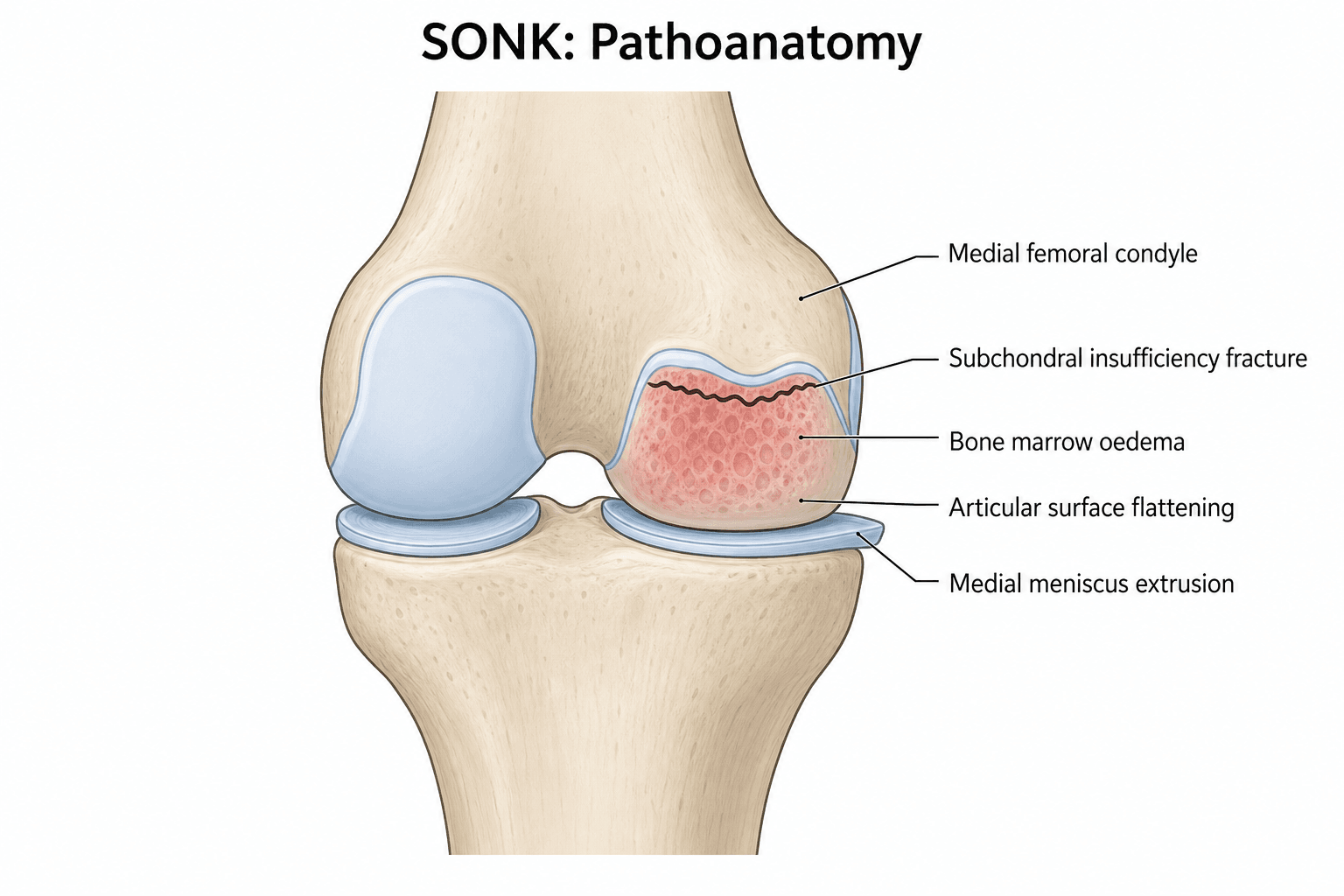
The fracture comes first. Current evidence supports that SONK is initiated by a subchondral insufficiency fracture in osteopenic bone rather than by primary avascular necrosis. Reduced trabecular thickness and connectivity in elderly patients lower the threshold for microfracture, so osteopenic subchondral bone fails under normal physiological load. Repetitive daily weight bearing then propagates the fracture line.
The necrosis follows. The fracture disrupts the local blood supply, isolating a segment of subchondral bone and producing focal osteonecrosis. This explains the typical single-site medial condyle lesion in elderly patients without systemic risk factors.
The meniscus concentrates the load. Medial meniscal extrusion increases focal load on the condyle. A posterior root tear destroys hoop stress, the meniscus extrudes, and load concentrates on exactly the medial condyle that fails, so root tears both predispose to and accelerate the overload.
From fracture to collapse. Loss of structural support lets the weight-bearing surface flatten under continued loading. Joint incongruity then accelerates cartilage wear and secondary osteoarthritis; the medial joint line narrows and varus malalignment develops, which further increases medial compartment load in a vicious cycle. Fibrocartilage or fibrovascular tissue fills the defect as a repair attempt, and if collapse persists the end stage is secondary degenerative change.
Secondary osteonecrosis is a different disease. It involves true vascular occlusion from fat emboli, coagulopathy or steroid-induced adipocyte hypertrophy, and is often multifocal.
- SPONK/SONK
- Elderly female, no risk factors
- Secondary ON
- Younger, though any age; steroid, alcohol, sickle cell or SLE history
- Key Discriminator
- History of systemic risk factors points to secondary ON
- SPONK/SONK
- Sudden severe pain, no trauma
- Secondary ON
- Insidious or after risk exposure
- Key Discriminator
- Temporal relationship to steroids
- SPONK/SONK
- Subchondral insufficiency fracture
- Secondary ON
- Vascular occlusion (fat emboli, steroids)
- Key Discriminator
- Mechanical versus vascular aetiology
- SPONK/SONK
- Osteopenic elderly bone
- Secondary ON
- Normal bone with superimposed insult
- Key Discriminator
- DEXA scan useful in SONK workup
- SPONK/SONK
- Single focus, medial femoral condyle in greater than 90 percent
- Secondary ON
- Multiple foci, often bilateral
- Key Discriminator
- Single-site medial condyle favours SONK; screen hips and shoulders if secondary suspected
- SPONK/SONK
- Focal subchondral fracture line + oedema
- Secondary ON
- Diffuse serpiginous geographic lesions, metaphyseal/diaphyseal extension
- Key Discriminator
- Serpiginous pattern indicates secondary ON; pattern recognition avoids misdiagnosis
The Third Type: Post-Arthroscopic Osteonecrosis
Examiners classically divide osteonecrosis around the knee into three types: spontaneous and secondary, contrasted above, and post-arthroscopic osteonecrosis (PAON), which is equally testable.
What it is. New osteonecrosis appears in the operated compartment, not present pre-operatively, in an older patient weeks to months after knee arthroscopy, classically after partial medial meniscectomy. The leading explanation is again a subchondral insufficiency fracture in the now mechanically altered, and often chondromalacic, compartment, rather than thermal or laser injury alone. Management mirrors SONK and is size- and collapse-based: protected weight bearing for small lesions, UKA or osteotomy for large or collapsed lesions.
The medicolegal trap. Because it appears after surgery, it can be mistaken for a surgical complication, so a pre-operative MRI documenting normal subchondral bone is invaluable.
Classification and Staging
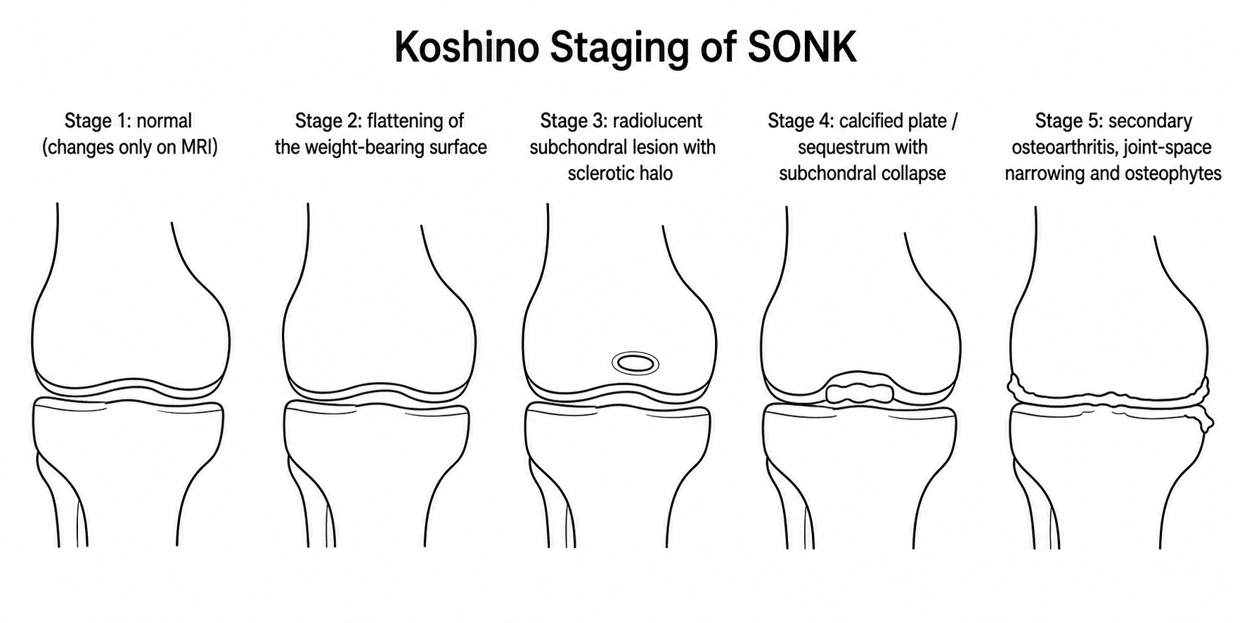
Two measures run side by side: the stage, which describes how far the structure has failed, and the size of the lesion.
- Radiographic Findings
- Normal or subtle sclerosis
- MRI Findings
- Bone marrow oedema, possible linear fracture line
- Treatment Implications
- Protected weight bearing, high healing potential
- Radiographic Findings
- Subchondral radiolucency or flattening
- MRI Findings
- Oedema + subchondral fracture line visible
- Treatment Implications
- Bisphosphonate trial, close radiographic follow-up
- Radiographic Findings
- Crescent sign, subchondral collapse
- MRI Findings
- Fragmentation, fluid-filled cleft
- Treatment Implications
- Consider high tibial osteotomy or UKA
- Radiographic Findings
- Secondary OA, joint space loss
- MRI Findings
- Full-thickness cartilage loss, osteophytes
- Treatment Implications
- UKA or TKA based on compartment involvement
Lesion size. Size drives the treatment decision, and the teaching bands combine width and area.
- Width Percentage
- Less than 40 percent
- Area (cm2)
- Less than 3.5
- Prognosis with Conservative Care
- Greater than 80 percent heal without surgery
- Width Percentage
- 40-60 percent
- Area (cm2)
- 3.5-5.0
- Prognosis with Conservative Care
- 50-60 percent heal, monitor closely
- Width Percentage
- Greater than 60 percent
- Area (cm2)
- Greater than 5.0
- Prognosis with Conservative Care
- Less than 20 percent heal, surgery recommended
Collapse depth. Depth of collapse greater than 2 mm on sagittal images is an independent predictor of failure of conservative treatment.
Know where the area figures come from, because the classic threshold is lower than the bands above. Rozing, Insall and Bohne studied 90 knees and found the prognosis depends on the size of the radiolucent lesion, with osteoarthritis likely to develop once the lesion exceeds 2.3 square centimetres, a figure still quoted as the original cut-point. The 40 percent width band is the one used in the later literature to separate lesions. The bands in the table are a practical composite rather than a single validated rule, so do not reassure a patient with a 3 square centimetre lesion on the strength of the "small" row: by Rozing's threshold that lesion is already in the range that tends toward arthritis. Treat area and width as concordant risk signals and act on the worse of the two.
The Reversible Mimic: Transient Bone Marrow Oedema Syndrome
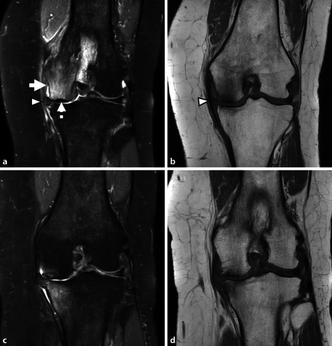
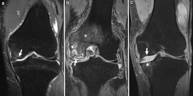
Not every painful older knee with bone marrow oedema is SONK. Transient bone marrow oedema syndrome (transient osteoporosis) of the knee is a self-limiting, reversible cause of pain and oedema that must not be over-treated as if it were a collapsing insufficiency fracture.
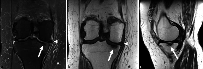
On MRI. Transient bone marrow oedema syndrome shows diffuse oedema without a discrete subchondral fracture line, without a focal subchondral low-signal collapse zone and without articular flattening. SONK has the focal subchondral fracture line and, later, collapse; a subchondral band of marked hypointensity adjacent to the endplate favours an insufficiency fracture, not transient oedema.
Natural history. Transient bone marrow oedema syndrome resolves spontaneously over weeks to months with protected weight bearing and analgesia, and does not progress to collapse. It can be regional or migratory, and the hip is the classic site.
Why it matters. Misreading transient oedema as SONK risks unnecessary surgery; misreading SONK as transient oedema risks allowing a preventable collapse. Look specifically for the subchondral fracture line.
Clinical Assessment
History. The pain is sudden and severe, medial, often after minor activity or at night, and the patient frequently denies any injury. Night pain that wakes the patient distinguishes it from mechanical meniscal symptoms. The pain may improve slightly and then worsen with collapse, and function suffers: antalgic gait, reduced walking distance, difficulty with stairs.
Risk factors. Ask specifically about steroids, alcohol, haemoglobinopathy such as sickle cell, and Gaucher disease. Their presence points to secondary osteonecrosis.
Examination.
- Gait: antalgic; a varus thrust may be present if collapse is advanced
- Swelling: mild effusion, with medial joint-line tenderness
- Range of motion: limited by pain, with a possible flexion contracture if chronic
- Stability: usually stable, unless there is secondary ligamentous laxity from collapse
- Alignment: measure the mechanical axis; varus deformity is common in advanced cases
Telling it apart. Sudden onset without trauma in an elderly patient with night pain should raise suspicion for SONK. Mechanical locking or catching suggests meniscal pathology, and a gradual increase in pain over months is more typical of primary osteoarthritis. Always obtain MRI in this demographic with unexplained medial knee pain.
- Onset
- Sudden, no trauma
- Night Pain
- Prominent
- Key Discriminating Feature
- MRI shows subchondral fracture line and oedema
- Onset
- Twisting or squatting
- Night Pain
- Rare
- Key Discriminating Feature
- MRI shows meniscal signal to articular surface
- Onset
- Gradual over weeks
- Night Pain
- Mild
- Key Discriminating Feature
- Joint space narrowing, osteophytes on X-ray
- Onset
- Sudden after minor fall
- Night Pain
- Moderate
- Key Discriminating Feature
- MRI shows linear fracture in proximal tibia
- Onset
- Variable, often insidious
- Night Pain
- Variable
- Key Discriminating Feature
- Risk factor history, multifocal lesions on MRI
Investigations
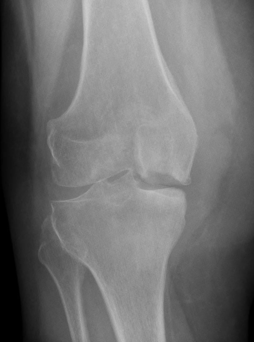
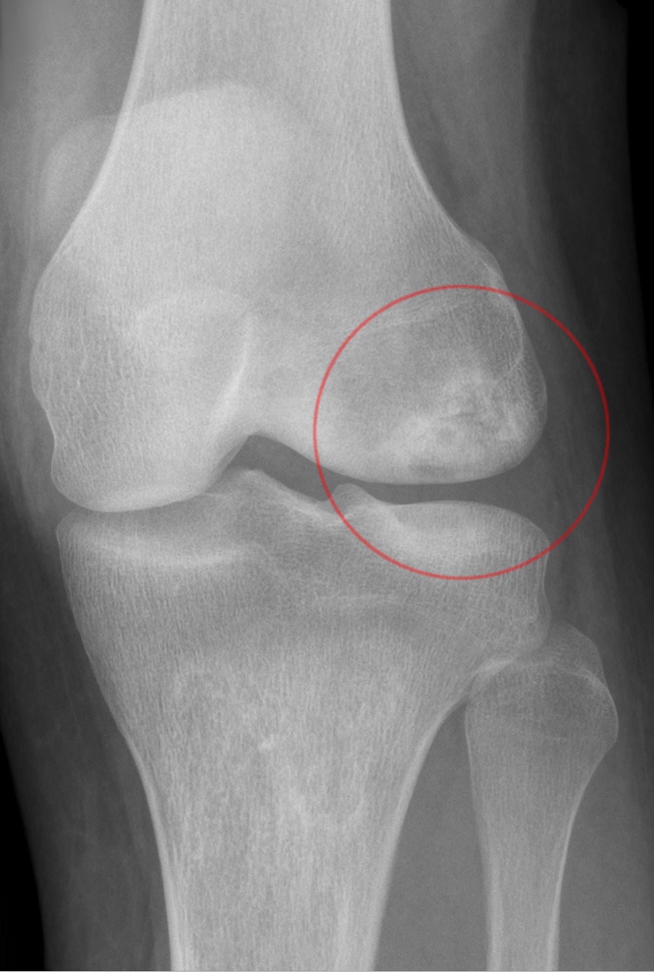
Radiographs first. They are the first-line test, but a normal radiograph does not exclude early SONK.
- Views: weight-bearing AP, Rosenberg (45-degree flexion PA), lateral and skyline
- Look for: subtle flattening of the medial femoral condyle, subchondral sclerosis, early joint-space narrowing
- Compare with the contralateral knee; varus alignment greater than 5 degrees increases suspicion
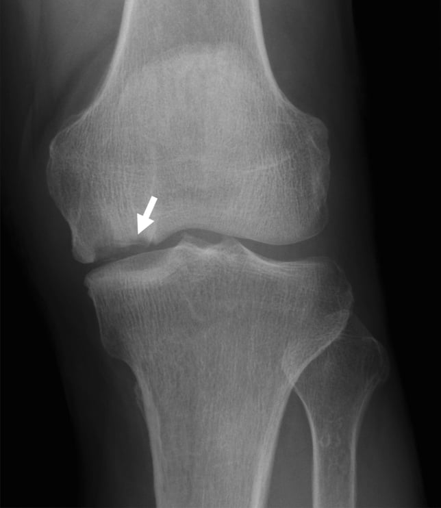
MRI is the gold standard. It makes the diagnosis before radiographic change. Obtain coronal and sagittal T1, T2, STIR and proton-density sequences; staging is performed on the coronal and sagittal T2-weighted and STIR images.
The diagnostic finding is the line, not the oedema. Look for a subchondral T2 hypointense line in the affected condyle with extensive surrounding bone marrow oedema-like signal. The oedema is the eye-catching part and the part that is not specific: avascular necrosis, osteochondritis dissecans and osteoarthritis all produce it. The subchondral line, its length and its distance from the articular surface are what make the diagnosis.
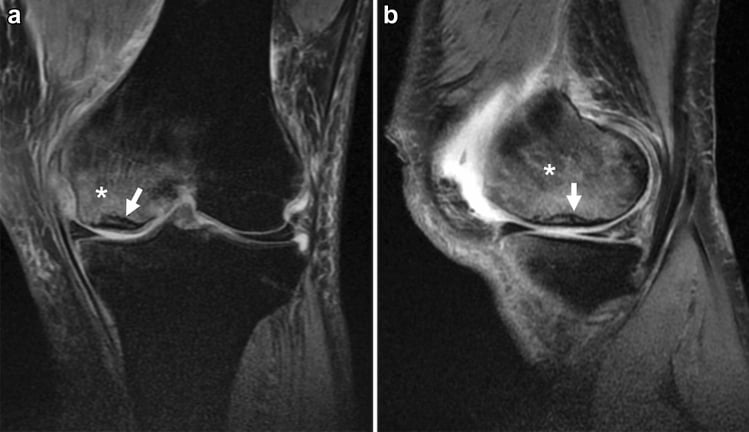
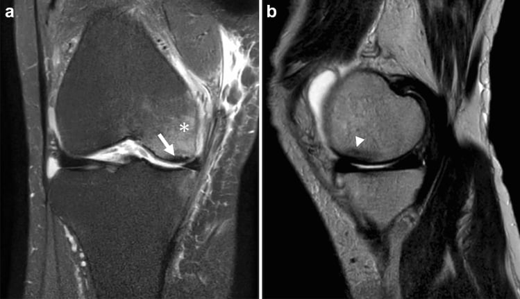
What to measure. Measure on the coronal slice of maximum width, and size the low-signal subchondral component rather than the surrounding oedema field.
- Width as a percentage of the entire medial femoral condyle width on the same slice
- Area in square centimetres
- Depth of collapse in millimetres, on sagittal images
- Extent of bone marrow oedema on STIR, which predicts pain, healing time and healing potential, although oedema extent alone does not define prognosis
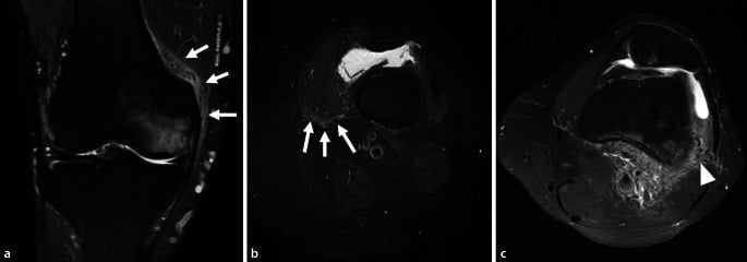
Look for a medial meniscus posterior root tear, do not merely exclude one. Root tears and chondral lesions are found in many patients with subchondral insufficiency fracture and are considered contributory. Finding one does not overturn the diagnosis, it often explains it, and in a suitable patient it is a separate problem that may need addressing in its own right. Also look for ligament injury and for the secondary osteonecrosis pattern: serpiginous, geographic lesions, often bilateral or multifocal.
Soft-tissue signs. The metaphyseal burst sign and oedema tracking beside the distal femur or vastus fascia can support the diagnosis. They do not replace identification of the subchondral lesion.
Long-leg alignment film. Obtained when planning high tibial osteotomy or UKA, to measure the mechanical axis deviation, medial proximal tibial angle and lateral distal femoral angle. It determines whether a corrective osteotomy can unload the lesion or whether arthroplasty is required.
Management Algorithm
The decision. Lesion size, collapse and the patient decide treatment, and the early aim is to unload the condyle, by protected weight bearing or osteotomy. Before choosing, assess the lateral compartment, the patellofemoral joint, and the patient's age and activity.
- Width less than 40 percent, collapse less than 2 mm and a patient compliant with protected weight bearing: conservative care. Small lesions with minimal oedema may be observed with protected weight bearing and repeat MRI at 8-12 weeks
- Width 40-60 percent: borderline; consider bisphosphonates and close monitoring
- Width greater than 50 percent or collapse greater than 2 mm: a high likelihood of progression, so counsel toward surgical options early
Conservative care. Protected weight bearing is the mainstay, with a bisphosphonate as an adjunct, and review is expectant: serial radiographs and clinical review until the pain settles or collapse occurs.
- Weeks 0-6: touch weight bearing with crutches or a walker; avoid full weight bearing, to prevent propagation of the subchondral fracture
- Weeks 6-12: partial (50 percent) weight bearing, increasing gradually if pain allows; repeat radiographs or MRI to assess healing
- Months 3-6: full weight bearing as tolerated once pain subsides and imaging shows resolution of oedema or stabilisation of the lesion
Some centres use intravenous zoledronic acid or oral alendronate in early SONK to reduce bone turnover and support healing of the subchondral fracture. Evidence is limited to small series but the rationale is biologically plausible given the insufficiency fracture mechanism. Discuss with patient as off-label use.
Surgery. Large lesions and collapse usually come to surgery, as can failed conservative care.
- Ideal Candidate
- Younger (less than 65), active, varus alignment, preserved lateral cartilage
- Key Technical Points
- Medial opening wedge, aim for 3-5 degrees valgus overcorrection
- Outcome Considerations
- Good pain relief, preserves native joint, 10-15 year survival
- Ideal Candidate
- Older patient, large lesion or collapse, low activity demand
- Key Technical Points
- Fixed-bearing medial UKA, ensure intact ACL and lateral compartment
- Outcome Considerations
- Excellent function, faster recovery than TKA, 90 percent 10-year survival
- Ideal Candidate
- Multicompartment disease, inflammatory arthritis, ligament deficiency
- Key Technical Points
- Standard posterior-stabilised or cruciate-retaining implant
- Outcome Considerations
- Most reliable long-term option when UKA contraindicated
Osteotomy or UKA. The choice depends on age, activity level, alignment and lesion size. Osteotomy is preferred in the younger, active patient with correctable varus and preserved lateral cartilage. UKA is favoured in the older patient with a larger lesion, collapse or failed conservative care, where joint preservation is less critical.
Size stops mattering once you replace the joint. In a series of 71 knees taken to arthroplasty for end-stage SONK, there was no correlation between the size of the necrotic lesion and the failure rate, and unicompartmental arthroplasty gave slightly better function than total knee arthroplasty (WOMAC 1.0 vs 1.6, p=0.04) at a mean of 6.6 years. Lesion size predicts whether conservative care will fail; it does not predict how the arthroplasty will do, so it should not be used to argue a patient out of a unicompartmental implant.
Complications
Untreated disease. Subchondral collapse of the weight-bearing condyle is the pivotal event, converting a potentially reversible lesion into an irreversible one. Secondary osteoarthritis of the medial compartment follows, often rapidly once collapse occurs, with progressive varus malalignment, chronic pain and functional decline.
Complications of treatment.
- High tibial osteotomy: delayed union or nonunion, loss of correction, over- or under-correction, intra-articular fracture, common peroneal nerve injury, hardware irritation
- Unicompartmental knee arthroplasty: aseptic loosening, polyethylene wear, bearing dislocation (mobile-bearing designs), progression of disease in the other compartments, persistent pain, periprosthetic fracture, conversion to TKA
- Bisphosphonate therapy (off-label adjunct): rare atypical femoral fracture and osteonecrosis of the jaw with prolonged use
- Prolonged protected weight bearing: deconditioning, falls and venous thromboembolism risk in the elderly
Subchondral collapse greater than 2 mm and a lesion involving more than about 50 percent of the condylar width are the key tipping points: beyond them there is a high likelihood of progression, and joint-preserving osteotomy outcomes become unpredictable. Counsel toward arthroplasty (or osteotomy in the young) before, not after, irreversible collapse and secondary osteoarthritis supervene.
Guidelines, Registries & Global Practice
- SONK incidence increasing with ageing population and greater MRI access worldwide
- Medial condyle predominance consistent across all ethnic groups and regions
- Female predominance 2-3:1 in every published series from Europe, Asia, North America
- Under-diagnosis common in low-resource settings where MRI is not readily available
- High-resource: Early MRI, lesion size quantification, bisphosphonate protocols, UKA or osteotomy in specialist centres
- Limited-resource: Diagnosis often delayed until radiographic collapse, protected weight bearing with crutches, TKA when advanced
- Universal principle: Lesion size and collapse depth remain the critical decision factors regardless of geography
- Surgery availability: UKA and osteotomy concentrated in high-volume arthroplasty centres globally
- Diagnosis Emphasis
- MRI for all unexplained medial knee pain in elderly
- Conservative Care
- Protected weight bearing 6-12 weeks, consider bisphosphonates
- Surgical Threshold
- Greater than 40 percent lesion or greater than 2 mm collapse
- Diagnosis Emphasis
- Clinical suspicion plus MRI staging
- Conservative Care
- Non-operative first line for small lesions
- Surgical Threshold
- Individualised, lesion size and patient factors
- Diagnosis Emphasis
- Differentiate SONK from secondary ON early
- Conservative Care
- Weight-bearing restriction, metabolic workup
- Surgical Threshold
- Osteotomy in younger, UKA/TKA in older
- Diagnosis Emphasis
- High incidence in elderly Asian females noted
- Conservative Care
- Similar conservative protocols
- Surgical Threshold
- UKA popular due to smaller stature and lower demand
There is no dedicated SONK registry. Data are derived from national joint registries (NJR, AJRR, AOANJRR) reporting outcomes of UKA and TKA performed for osteonecrosis, and from small prospective series on conservative care and osteotomy. The evidence base remains level 3-4. Key principle across all guidelines: measure lesion size accurately on MRI and base treatment on percentage of condyle involved and depth of collapse.
Controversies & Areas of Uncertainty
Small series suggest zoledronic acid or alendronate may accelerate healing of subchondral fractures in SONK, but no high-quality randomised trials exist. Use remains off-label and centre-dependent.
Six to twelve weeks is conventional, but exact duration and degree of unloading lack high-level evidence. Serial MRI at 8-12 weeks is increasingly used to guide progression.
Patients aged 60-70 with medium-sized lesions and mild collapse present a decision dilemma. Some surgeons favour joint preservation with osteotomy, others prefer the reliability of UKA. Shared decision-making with patient activity goals is essential.
Limited evidence supports core decompression or bone grafting for early SONK. Most centres reserve these for rare cases in very young patients or as part of research protocols.
Exam Viva Scenarios
Practise clinical reasoning and management decisions out loud
“A 68-year-old woman presents with sudden onset severe medial right knee pain for 3 weeks. No history of trauma or steroid use. Pain wakes her at night. Weight-bearing radiographs are normal. MRI shows a 2.8 square centimetre lesion of the medial femoral condyle with bone marrow oedema but no subchondral collapse. The lesion involves 32 percent of the condylar width. What is your diagnosis and initial management?”
“A 72-year-old active man presents with 4-month history of progressive medial knee pain. He recalls a minor twisting injury 5 months ago. Radiographs show 3 mm collapse of the medial femoral condyle with varus alignment of 7 degrees. MRI demonstrates a 6.2 square centimetre lesion involving 58 percent of the condylar width with a fluid-filled cleft. The lateral compartment and patellofemoral joint are preserved and he has an intact ACL. What surgical options would you discuss?”
Key Demographics and Presentation
- Elderly female (greater than 60 years), sudden medial knee pain without trauma
- Night pain is characteristic, distinguishes from meniscus tear or OA flare
- Greater than 90 percent involve the medial femoral condyle weight-bearing surface
- No systemic risk factors (steroids, alcohol, sickle) - these suggest secondary ON
MRI Staging and Sizing
- Stage 1: Normal X-ray, bone marrow oedema only - high healing potential
- Stage 2-3: Subchondral lucency or collapse - measure width percentage and depth
- Small lesion (less than 40 percent width) with no collapse - conservative trial; note the original Rozing area cut-point is 2.3 cm2, lower than the composite bands, so an area between 2.3 and 3.5 cm2 is NOT reassuring on area alone
- Large lesion (greater than 50 percent) or greater than 2 mm collapse - surgical candidate
Differential Diagnosis
- Secondary ON: multifocal, serpiginous lesions, risk factor history
- Medial meniscus tear: mechanical symptoms, twisting injury, MRI meniscal signal
- Insufficiency fracture tibia: linear fracture line in proximal tibia on MRI
- Primary OA: gradual onset, joint space narrowing and osteophytes on X-ray
Treatment Thresholds
- Conservative: touch weight bearing 6-12 weeks, consider bisphosphonate
- High tibial osteotomy: younger active patient, correctable varus, preserved lateral compartment
- UKA: older patient, large lesion or collapse, intact ACL and lateral cartilage
- TKA: multicompartment disease, ligament deficiency, inflammatory arthritis
Prognosis and Follow-up
- Small lesions without collapse have a good chance of healing conservatively - but the often-quoted 'over 80 per cent' is a composite of small series, not a figure from Rozing, whose 90 knees were only enrolled once a lesion was radiographically visible
- Large lesions with collapse: high progression risk, counsel early for surgery
- UKA survival in SONK: 90-93 percent at 10 years when selection criteria met
- Serial MRI at 8-12 weeks for conservatively managed patients to detect collapse
Evidence Base
Original description of spontaneous osteonecrosis of the knee
- Seminal paper that first described spontaneous osteonecrosis of the knee as a distinct entity affecting predominantly the medial femoral condyle in older patients
- Characterised the classic presentation of sudden-onset knee pain without preceding trauma
- Documented radiographic progression and used biopsy/radionuclide assessment in its evaluation
Subchondral insufficiency fracture as the cause of SONK
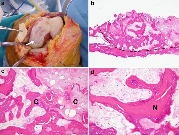
- Histopathological review of 14 patients (8 women, 6 men, aged 59 to 88) operated on for SONK, concluding that the primary event is a subchondral insufficiency fracture and that the osteonecrosis is its RESULT
- Only 9 of the 14 were interpretable: 3 stage-2 lesions were pure subchondral insufficiency fractures and 6 stage-3 lesions showed fracture with focal osteonecrosis confined between the fracture line and the articular surface, while 5 were indeterminate because the lesion had detached from the condyle
- Fracture callus and granulation tissue consistent with reparative response to mechanical failure
- Supported the insufficiency fracture hypothesis over avascular necrosis theory
Unicompartmental knee arthroplasty for osteonecrosis (spontaneous and secondary)
- Retrospective review of 30 patients (31 knees) undergoing UKA for osteonecrosis - 21 spontaneous and 10 secondary; mean age 71, mean follow-up 7 years (range 3 to 16)
- Reliable pain relief and functional improvement (Knee Society scores) in 30 of 31 knees (96 percent)
- Appropriate lower-limb mechanical axis restored in 27 knees (88 percent)
- Kaplan-Meier survivorship 96.7 percent at 12 years