Post-ACL Reconstruction Arthrofibrosis | Loss of Extension | Anterior Nodule
- Cyclops lesion = localised anterior arthrofibrotic nodule at the ACL graft tibial tunnel entrance
- Presents with loss of terminal extension after ACL reconstruction, typically 3-12 months post-operatively
- Named for its appearance: a bulbous fibrous nodule with a central dimple resembling a single eye
- Diagnosis is clinical (extension deficit) plus MRI (nodule anterior to tibial tunnel, graft impingement)
- Arthroscopic excision restores extension in the great majority of cases
- “Loss of terminal extension after ACLR is never normal and must be investigated
- “The nodule wedges between the graft and the intercondylar roof in full extension, causing a mechanical block
- “MRI sensitivity is high but clinical suspicion is the key to ordering the right scan
- “Arthroscopic findings are pathognomonic: a firm nodular mass with a pale fibrous surface at the tibial tunnel aperture
Overview and Epidemiology
Loss of terminal extension after ACL reconstruction is one of the most common reasons for re-operation and prolonged rehabilitation, and a cyclops lesion is a specific, treatable cause of it. The surgeon who recognises the entity spares the patient months of misdirected physiotherapy and can offer a definitive arthroscopic solution. The topic joins graft biology, imaging interpretation and arthroscopic decision-making, which is why it is examined.
What it is. A cyclops lesion is a localised nodular mass of fibrovascular scar tissue at the entrance of the tibial bone tunnel, anterior to the reconstructed ACL graft. In terminal extension the nodule is trapped between the graft and the roof of the femoral intercondylar notch, producing a mechanical block to full extension. The name is from its arthroscopic appearance: a pale, bulbous mass with a central dimple, resembling a single eye.
Incidence: say which question you are answering. There are three different numbers here, and the commonest error on this topic is quoting one of them for all three.
- Cyclops lesion, an imaging or arthroscopic finding that is frequently asymptomatic: seen in up to about a quarter of second-look arthroscopies; one second-look series found lesions in 15 of 55 knees (27.3%)
- Cyclops syndrome, the lesion plus pain or an extension block: roughly 3-11% across series; 3.61% after double-bundle reconstruction, 10.9% in that same second-look series, and 10.2% when counted prospectively in 588 consecutive primary reconstructions
- Re-operated: about 6% of that 588-patient cohort, so a substantial minority of symptomatic patients never come to surgery
The widely repeated 1-2% belongs to none of these cleanly; it sits below even the lowest measured symptomatic figure. An international survey found most surgeons estimate 1-5%, so the profession as a whole appears to under-estimate how often this happens. Most cyclops lesions are asymptomatic incidental findings, and what that means for the MRI report is dealt with under classification.
Risk factors. Non-anatomic tunnel placement, a tibial tunnel too far anterior, is the dominant one and now has a number: OR 4.15 (95% CI 2.17-7.96). A tunnel drilled too far anterior creates roof impingement and debris accumulation; graft impingement against the intercondylar roof and excessive soft-tissue debris left at the tibial tunnel aperture are the classic technical contributors, and inadequate notchplasty at the index operation predisposes by leaving insufficient roof clearance. Also independently associated are a higher BMI (OR 1.09 per unit, 1.02-1.16) and a narrower notch apex (OR 0.77, 0.67-0.89).
Patellar tendon autograft has higher reported rates in older series, but graft type was not an independent risk factor on multivariate analysis in the 588-patient cohort; nor were tibial slope, sex, graft diameter or a lateral extra-articular procedure.
Rehabilitation: a genuine contradiction worth knowing. The topic is traditionally taught with aggressive early rehabilitation as a risk factor, forceful early extension exercises promoting fibrosis at the healing tunnel aperture. The largest risk-factor analysis found post-operative bracing to be independently associated (OR 2.41, 1.18-4.94), which points toward too little motion rather than too much. The two claims cannot both be the main mechanism. The bracing finding is level III and single-centre, so treat it as a real challenge to the traditional teaching rather than a replacement for it, and say so if asked.
When. Peak presentation is 3-12 months after reconstruction, once scar tissue has matured into a discrete nodule. No strong sex predilection is reported.
Pathophysiology
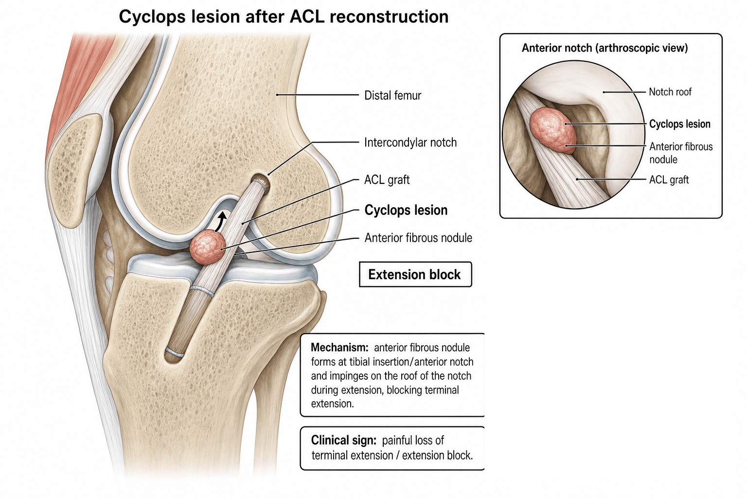
How the nodule forms. Debris at the tibial tunnel aperture (remnant ACL fibres, cancellous bone debris, synovial tissue and graft material) organises into a firm nodule. The sequence runs in four steps:
- Tunnel debris. Bone debris, remnant ACL fibres and synovial tissue accumulate at the tibial tunnel aperture at drilling or graft passage; there is more of it with larger tunnels, non-anatomic placement and aggressive graft preparation
- Fibrovascular proliferation. The debris acts as a scaffold for fibroblast and vascular ingrowth, forming a nodular scar mass over weeks to months; this is the body's healing response to intra-articular foreign material
- Nodule maturation. The scar organises into a firm, discrete nodule attached to the anterior margin of the tibial tunnel, with a characteristic pale fibrous surface; it usually reaches mature size by 3-9 months, which is when the mechanical block becomes clinically apparent
- Mechanical impingement. In terminal extension the nodule is wedged between the ACL graft, which lies posterior to it, and the femoral intercondylar roof; the result is a hard end-point block that physiotherapy cannot overcome
Where it sits. The nodule lies anterior to the tibial tunnel aperture, between the graft and the intercondylar notch. The graft is intact and functional: the problem is the nodule, not graft failure. In full extension the nodule abuts the femoral intercondylar roof, replicating the mechanism of notch impingement.
Histology. Dense collagen with fibroblasts, consistent with scar. Early lesions contain active fibrovascular tissue with vascular channels and become more fibrotic as they mature, and cancellous bone fragments from tunnel drilling may be embedded. The nodule is separate from the ACL graft, which remains viable.
Classification and Types
By location. The classical lesion is infrapatellar, at the tibial tunnel entrance; a supratibial variant and a combined form with diffuse fibrosis are described.
- Location
- Nodule at tibial tunnel entrance, anterior to ACL graft
- Frequency
- Most common (majority of cases)
- Clinical Features
- Isolated extension deficit, hard end-point
- Location
- Nodule at proximal exit of tibial tunnel
- Frequency
- Less common
- Clinical Features
- Similar presentation, higher on the tibial slope
- Location
- Nodule plus surrounding scar tissue
- Frequency
- Uncommon, may occur with delayed diagnosis
- Clinical Features
- Both extension and flexion loss; harder to treat
Cyclops versus cyclopoid: the histological distinction that predicts symptoms. Not every nodule seen on post-reconstruction MRI is a true symptomatic cyclops; the Tonin histological subdivision matters.
- True cyclops lesion: a firm nodule with a central core of bone and/or fibrocartilage within the fibrovascular tissue. It is harder, larger and mechanically symptomatic, produces the classic hard-end-point extension block, and is the lesion that needs excision
- Cyclopoid lesion: soft fibrous granulation tissue only, without the bone or cartilage core. It is softer and frequently asymptomatic, often an incidental MRI finding that does not cause a mechanical block
An MRI report of "a small nodule anterior to the graft" in a patient with full, pain-free extension may be an incidental cyclopoid lesion and does not mandate surgery. The decision to excise is driven by the clinical mechanical block, a hard-end-point extension deficit, not by the mere presence of a nodule on the scan: treat the patient, not the scan.
Clinical Assessment
History. The patient has had an ACL reconstruction within the last 3-12 months, rarely beyond 24 months, and reports progressive loss of terminal extension that did not improve despite physiotherapy. There is anterior knee pain on extension, worse with attempts at terminal extension, and a mechanical blocking or catching sensation. There is no new trauma: this is a post-surgical complication, not a new injury.
Giving way is not typical, because the graft is intact, though the mechanical block can be perceived as instability. Functionally the patient cannot lock the knee straight and has difficulty with running, cycling and descending stairs. If the lesion is missed, the nodule perpetuates inflammation and can contribute to diffuse scarring.
Examination. The extension deficit is the single most important finding, typically 10-30 degrees; measure it supine with the heel on a flat surface and compare both knees. On inspection the knee is held in slight flexion and the patient cannot achieve full lock, with an antalgic gait and quadriceps wasting. Flexion is preserved, which distinguishes the lesion from diffuse arthrofibrosis; an effusion may be present but is mild, and a firm nodule anterior to the tibial tunnel is occasionally palpable.
Lachman, anterior drawer and pivot shift should be negative because the graft is intact; positive tests suggest graft failure, not cyclops. Patella mobility is typically preserved, which helps exclude infrapatellar contracture, and hamstring tightness and effusion are checked as other causes of the deficit.
Hard versus soft end-point. The character of the end-point is the examination finding that directs the investigation pathway. A hard end-point means a mechanical block (cyclops nodule, loose body, meniscal tear, hardware impingement) and requires imaging and likely arthroscopy. A soft end-point means a soft-tissue restriction (hamstring tightness, capsular contracture, patellofemoral tightness) and may respond to physiotherapy. Cyclops lesion produces a hard end-point.
- Extension
- Lost (10-30 deg)
- Flexion
- Preserved
- End-Point
- Hard (mechanical)
- Key Discriminator
- MRI: nodule at tibial tunnel; graft intact
- Extension
- Lost
- Flexion
- Also lost
- End-Point
- Hard or soft
- Key Discriminator
- MRI: diffuse scar throughout Hoffa fat pad and suprapatellar pouch
- Extension
- Lost
- Flexion
- Lost
- End-Point
- Soft (capsular)
- Key Discriminator
- Patella baja, decreased patellar mobility, anterior knee pain
- Extension
- Lost
- Flexion
- Preserved
- End-Point
- Hard
- Key Discriminator
- MRI: graft against roof but no cyclops mass; anterior tunnel placement
- Extension
- Mildly lost
- Flexion
- Preserved
- End-Point
- Soft (elastic)
- Key Discriminator
- Improves with stretching; MRI normal; no nodule
- Extension
- Variable
- Flexion
- Variable
- End-Point
- Variable
- Key Discriminator
- Positive Lachman / pivot shift; MRI shows graft discontinuity
Global stiffness is a different problem. If the patient has lost both flexion and extension, think diffuse knee arthrofibrosis instead: a different problem with a different prognosis, and one that responds far less well to arthroscopic excision. The index procedure itself is covered in ACL injuries, and in the skeletally immature in paediatric ACL injury.
Investigations
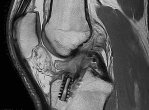
MRI is first-line. Request a post-operative protocol:
- Sagittal T1
- Sagittal PD fat-sat
- Coronal PD fat-sat
- Axial PD fat-sat
Look for a well-circumscribed nodular soft-tissue mass at the anterior opening of the tibial tunnel, anterior to the ACL graft and lying between the graft and the intercondylar notch roof. It is typically low-to-intermediate signal on T1 and low signal on T2, reflecting dense fibrous tissue, and is most conspicuous on the sagittal sequences, where its relationship to the notch roof in extension is visible. The graft posterior to it should appear intact and in continuity with the tibial and femoral tunnels, and the nodule is seen impinging between graft and roof.
MRI is highly sensitive for cyclops lesions when the appropriate protocol is used. Gadolinium is not required, though the nodule may show mild peripheral enhancement after it. The extension deficit on examination should correlate with the size of the nodule impinging on the notch. Do not confuse it with a meniscal cyst, which is in a different location, or with graft hypertrophy, which is the graft itself rather than a separate nodule.
Plain radiographs. AP, lateral and Rosenberg (45-degree PA flexion weight-bearing) views are routine. They rule out hardware complications (screw migration, tunnel widening, loose body) and show tunnel position, in particular a tibial tunnel placed too far anterior, which predisposes to cyclops lesion and graft impingement. The nodule itself is soft tissue and is not visible on plain films.
CT arthrogram. Reserved for MRI artefact from hardware such as interference screws obscuring the tibial tunnel region; the nodule shows as a filling defect at the tibial tunnel aperture. It is rarely needed if MRI is performed with metal artefact reduction sequences.
- Location
- Anterior to tibial tunnel, between graft and roof
- MRI Signal
- Low signal on T1 and T2 (fibrous)
- Key Distinguishing Feature
- Discrete nodule with central dimple; graft intact; extension block
- Location
- Graft against intercondylar roof
- MRI Signal
- Graft signal normal or mildly increased
- Key Distinguishing Feature
- No discrete nodule; may respond to notchplasty; no cyclops mass
- Location
- Throughout the joint, especially infrapatellar
- MRI Signal
- Low signal scar throughout Hoffa fat pad and suprapatellar pouch
- Key Distinguishing Feature
- Global ROM loss, not just extension; thickened scar, not a nodule
- Location
- Adjacent to meniscus body or horn
- MRI Signal
- High T2 signal (fluid)
- Key Distinguishing Feature
- Communicates with meniscal tear; different location; not at tibial tunnel
- Location
- Hoffa fat pad region
- MRI Signal
- Variable, often intermediate signal
- Key Distinguishing Feature
- Less defined than cyclops; no discrete nodule; no mechanical block pattern
Management Algorithm
The decision. The textbook position is that a cyclops lesion is a mechanical block caused by a discrete fibrous nodule, which physiotherapy cannot resolve, so arthroscopic excision is definitive once a mechanical block is established and restores extension in a high proportion of cases. Do not say non-operative treatment has no role: in an international survey of 250 surgeons across 25 countries, initial management was predominantly conservative, with most advocating an extension-focused rehabilitation trial first and reserving excision for a persistent deficit. Both positions are defensible; say which one you are describing.
Non-operative management. In the textbook framing its role is limited to:
- Very early lesions (under 3 months), where the scar tissue may still be immature and could potentially remodel
- Mild extension deficits (less than 5 degrees) that are not functionally limiting
- Patients unfit for surgery
If a trial is warranted, months 0-3 are intensive rehabilitation: aggressive quadriceps control, patellar mobilisation, hamstring stretching and posterior glide mobilisations, aiming to improve the soft-tissue component of the deficit, with serial extension measurements every 2-4 weeks. The threshold for surgery is no improvement after 6-8 weeks of focused physiotherapy.
Once the patient has a clear mechanical block with a hard end-point and MRI confirms a cyclops nodule, continued physiotherapy is futile and delays definitive treatment. The lesion is a physical obstruction that requires removal, and prolonged conservative treatment risks allowing it to mature further and promotes secondary arthrofibrosis from chronic inflammation.
Arthroscopic excision. The goal is to remove the fibrous nodule and restore terminal extension while preserving the intact ACL graft.
Surgical Protocol
Confirm the diagnosis: MRI showing a cyclops nodule plus a clinical extension deficit with a hard end-point, and graft integrity confirmed on MRI and on clinical stability tests. Consent covers graft damage during excision, recurrence, continued stiffness, infection and neurovascular injury. Equipment is a standard arthroscopy set, shaver, radiofrequency ablator and possibly a small osteotome.
- Standard anterolateral and anteromedial portals
- Diagnostic sweep first: suprapatellar pouch, medial and lateral gutters, menisci and graft integrity, before touching the nodule
- Identify the nodule anterior to the tibial tunnel aperture, between the graft and the notch roof; it is typically firm and vascular. Bring the knee into extension under vision to see it impinge against the roof
- Debulk with the shaver from the surface, then radiofrequency to clean the base at the tibial tunnel aperture. The graft lies immediately posterior to the nodule: keep the shaver oriented away from it
- Bring the knee into full extension under arthroscopic vision to confirm no residual impingement, and check the graft is intact and undamaged
- If the intercondylar roof contributes to ongoing impingement, or the graft still impinges on the roof, perform a limited notchplasty
- Debride any surrounding fibrotic tissue but avoid excessive bone removal from the tibial tunnel
The procedure is usually straightforward, but graft proximity demands care throughout.
The difference from index reconstruction rehabilitation is the emphasis on immediate and aggressive extension regain, because the mechanical block has been removed. From day 1: cryotherapy, quadriceps exercises, and passive and active-assisted extension exercises, aiming for full passive extension in weeks 1-2. Weeks 2-6 add progressive range of motion, closed-chain strengthening and cycling as tolerated; from week 6, sport-specific rehabilitation, with return to sport when full range and strength are achieved.
Complications
- Incidence
- Reported but uncommon
- Risk Factors
- Incomplete excision, ongoing impingement, predisposing tunnel position
- Management
- Revision arthroscopic excision; consider tunnel repositioning if anterior placement confirmed
- Incidence
- 5-10 percent
- Risk Factors
- Inadequate rehabilitation, concurrent capsular contracture, incomplete excision
- Management
- MUA + aggressive physiotherapy; revision arthroscopy if mechanical block persists
- Incidence
- Rare but possible
- Risk Factors
- Graft adherent to nodule, poor visualisation, over-aggressive shaver use
- Management
- Minor: protect and monitor; major: revision ACL reconstruction
- Incidence
- Low if treated promptly
- Risk Factors
- Delayed diagnosis, prolonged inflammation from impingement
- Management
- More aggressive rehabilitation; rare cases require further arthroscopic release
- Incidence
- Standard arthroscopy risk (less than 1 percent)
- Risk Factors
- Immunocompromised, prolonged procedure, wound contamination
- Management
- Urgent arthroscopic washout + antibiotics
Prevention at index surgery. Cyclops lesion is largely a preventable complication, and these steps at the time of ACL reconstruction dramatically reduce the risk:
- Place the tibial tunnel at the anatomic centre of the native ACL footprint, not too anterior, using fluoroscopy or navigation
- Perform adequate notchplasty if the intercondylar notch is narrow
- Irrigate the joint thoroughly, use a probe to clear debris, and debride remnant tissue from the tibial tunnel aperture after graft passage
- Avoid aggressive forced terminal extension in the early post-operative period; guide rehabilitation with controlled early motion
Timing: the Shelbourne anti-arthrofibrosis principle. Beyond the technical steps, the single biggest preventable driver of post-reconstruction stiffness, cyclops and diffuse arthrofibrosis alike, is operating on the wrong knee at the wrong time. Shelbourne's classic teaching is that reconstruction of a knee that is hot, effused and lacking full extension carries a markedly higher arthrofibrosis and cyclops rate. Quiet the knee first: defer reconstruction until it has regained full symmetric extension and near-full flexion, the effusion has settled and the patient has good quadriceps control ("prehabilitation"). That state matters more than a fixed number of days or weeks from injury.
Full extension is the priority before and immediately after surgery. Heel-prop and prone hangs, aimed at full terminal extension, are the most protective rehabilitation goal, because a knee that never regains extension is the one that scars. A quiet knee with good range and meticulous tunnel technique is the combination that minimises the fibrovascular response at the tunnel aperture.
Outcomes and Prognosis
After excision. Full or near-full extension is restored in over 90%: the figure is widely quoted but cannot be traced to a primary source, so treat it as conventional rather than measured. Greater than 90% return to their pre-injury level of sport, with rehabilitation usually taking 2-3 months after excision. The graft is separate from the lesion, so excision does not compromise it and it remains stable and functional. Recurrence is low with complete excision and higher if predisposing factors, an anterior tunnel above all, are not addressed. Patient satisfaction is high: the operation is minimally invasive and effective, and patients are often relieved to have a definitive mechanical explanation.
Prognostic factors. The best results follow:
- Early diagnosis, within 12 months
- A discrete nodule without diffuse fibrosis
- Complete arthroscopic excision
- Immediate post-operative extension rehabilitation
The prognosis is worse with:
- Delayed diagnosis, over 24 months
- Associated diffuse arthrofibrosis
- Incomplete excision
- Anterior tunnel malposition that is not corrected
Guidelines, Registries & Global Practice
- Cyclops lesion is reported worldwide following ACL reconstruction across all graft types and fixation methods
- Incidence is declining in centres using anatomic ACL reconstruction techniques with precise tunnel positioning
- Higher historical incidence was associated with transtibial drilling techniques that produced anterior tibial tunnels
- No strong geographic or ethnic variation; incidence relates to surgical technique rather than patient factors
- High-resource: MRI is readily available; early diagnosis; arthroscopic excision as day-case surgery with structured rehabilitation
- Limited-resource: Diagnosis may rely more on clinical examination and plain radiographs; CT arthrogram if MRI unavailable; excision still possible with basic arthroscopy equipment
- Universal principle: outcome depends on early recognition of the extension deficit and prompt arthroscopic intervention
- Prevention: Meticulous tunnel placement and joint debridement at index surgery are achievable in all settings
- Prevention emphasis
- Anatomic single-bundle or anatomic double-bundle ACLR; adequate notchplasty; debridement of tunnel debris
- Diagnosis
- MRI for persistent extension deficit after ACLR; clinical hard end-point is key finding
- Treatment
- Arthroscopic excision; structured post-operative rehabilitation with immediate extension focus
- Prevention emphasis
- Individualised anatomic tunnel placement using fluoroscopic or navigation guidance; graft harvesting and preparation to minimise debris
- Diagnosis
- MRI with fat-suppressed sequences; correlate with clinical examination for hard end-point block
- Treatment
- Arthroscopic excision recommended; rehabilitation guided by sport-specific timelines
- Prevention emphasis
- Anatomic ACLR techniques emphasised in BOAST guidelines; notchplasty as needed
- Diagnosis
- Clinical suspicion drives imaging request; MRI first-line for any unexplained post-ACLR stiffness
- Treatment
- Arthroscopic excision as day-case procedure; physiotherapy-led rehabilitation
- Prevention emphasis
- Tunnel positioning at native ACL footprint; soft tissue debridement; careful graft passage technique
- Diagnosis
- MRI protocol for post-operative knee; assess tunnel position on radiographs
- Treatment
- Arthroscopic debridement of the nodule; consider tunnel revision if malpositioned
There is no dedicated registry for cyclops lesions. However, national ACL reconstruction registries (Danish ACL Registry, Swedish National Knee Ligament Register, Norwegian Knee Ligament Registry, NZJR) capture re-operation rates after ACLR, which include arthroscopic procedures for cyclops lesions. Registry data suggest that re-operation for stiffness and arthrofibrosis after ACLR accounts for a small but important proportion of all re-operations. The trend toward anatomic ACL reconstruction is reflected in declining re-operation rates for extension loss in registry data.
Record in every ACL reconstruction follow-up:
- Passive and active extension range measured against the contralateral knee
- Whether the end-point is hard (mechanical) or soft (capsular/muscular)
- If a hard block is present, document suspicion for cyclops lesion and order MRI
- At index surgery, document tunnel positions, notchplasty performed, and adequacy of joint lavage
Failure to diagnose a cyclops lesion after months of physiotherapy for a mechanical block is a recurring theme in clinical negligence claims. Always investigate a persistent extension deficit after ACLR with imaging rather than attributing it to rehabilitation failure.
Controversies & Areas of Uncertainty
There is no consensus on the minimum interval after index ACLR before performing cyclops excision. Most surgeons wait at least 3 months to allow the nodule to mature into a discrete, excisable mass, but earlier intervention may prevent secondary arthrofibrosis. The balance is between nodule maturity (easier excision when well-formed) and preventing chronic impingement.
There is limited evidence for pharmacological prevention of cyclops lesion. Some centres use NSAIDs or steroid injections post-operatively to modulate scar formation, but this is not evidence-based for cyclops prevention specifically and carries its own risks.
Whether routine notchplasty should be performed at all ACL reconstructions to prevent cyclops and graft impingement, or only when the notch is objectively narrow, remains debated. Routine notchplasty may increase bone debris and potentially paradoxically contribute to cyclops formation if debris is not adequately cleared.
Some early series reported higher cyclops rates with patellar tendon bone-tendon-bone autograft, but contemporary anatomic techniques using hamstring autograft also report cyclops lesions. The relative risk by graft type is not definitively established, and surgical technique (tunnel position, debridement) likely matters more than graft choice.
MCQ Practice Points
Q: What is a cyclops lesion? A: A cyclops lesion is a localised nodular mass of fibrovascular scar tissue that forms at the entrance of the tibial bone tunnel, anterior to the reconstructed ACL graft, after ACL reconstruction. It causes a mechanical block to terminal extension by impinging between the graft and the intercondylar notch roof. Named for its arthroscopic appearance resembling a single eye.
Q: How do you diagnose a cyclops lesion? A: Clinical: Isolated loss of terminal extension (10-30 degrees) with a hard end-point after ACLR, preserved flexion, intact graft (negative Lachman). MRI: Well-circumscribed low-signal nodule on T1 and T2 at the anterior tibial tunnel entrance, anterior to an intact ACL graft, impinging on the intercondylar roof. The diagnosis is clinical plus imaging; arthroscopy is both diagnostic and therapeutic.
Q: What is the definitive treatment for cyclops lesion? A: Arthroscopic excision using shaver and radiofrequency ablation. The nodule is debulked from its base at the tibial tunnel aperture while protecting the ACL graft posterior to it. Post-operative rehabilitation emphasises immediate extension regain, and the great majority of patients regain full extension (over 90 percent is the conventional figure, though it is not traceable to a primary source). I would be careful not to say that non-operative treatment has no role: international practice is to trial extension-focused rehabilitation first and reserve excision for a persistent deficit, so I would offer surgery for a block that has failed to respond rather than as the automatic first answer.
Q: How do you distinguish cyclops lesion from diffuse arthrofibrosis after ACLR? A: Cyclops lesion: Isolated loss of extension with preserved flexion, hard end-point, discrete nodule on MRI, excellent response to excision. Diffuse arthrofibrosis: Global loss of both flexion and extension, diffuse scar throughout the Hoffa fat pad and suprapatellar pouch on MRI, guarded prognosis, requires more extensive arthroscopic release.
Q: What are the key preventable risk factors for cyclops lesion at ACLR? A: Anterior tibial tunnel placement (the single most important factor), inadequate notchplasty leaving insufficient roof clearance, debris left at the tibial tunnel aperture after drilling and graft passage, and aggressive forced terminal extension in early post-operative rehabilitation. Prevention is achieved by anatomic tunnel positioning, adequate notchplasty, thorough joint irrigation and debridement, and guided rehabilitation.
Exam Viva Scenarios
Practise clinical reasoning and management decisions out loud
“A 24-year-old netball player had an ACL reconstruction 6 months ago using hamstring autograft. She has made good progress with flexion but cannot fully extend her left knee. Examination reveals a 15-degree extension deficit with a hard end-point. Her Lachman test is negative. She is frustrated because her physiotherapist says she is not working hard enough. What is your assessment and management?”
“A 30-year-old footballer had an ACL reconstruction 4 years ago. Two years after the reconstruction, he underwent arthroscopic excision of a cyclops lesion with good initial restoration of extension. He now presents again with a 10-degree extension deficit and similar symptoms. MRI suggests a recurrent cyclops-like nodule. How would you manage this recurrence?”
Definition and Pathogenesis
- Cyclops lesion = localised anterior arthrofibrotic nodule at the tibial tunnel entrance after ACLR
- Named for arthroscopic appearance: bulbous nodule with central dimple (single eye)
- Forms from organised fibrovascular scar on a scaffold of tunnel debris, remnant ACL, and bone dust
- Nodule wedges between the ACL graft and the intercondylar roof in full extension, causing mechanical block
Clinical Presentation
- Isolated loss of terminal extension (10-30 degrees) with preserved flexion, typically 3-12 months post-ACLR
- Hard end-point block (not soft/elastic), anterior knee pain on extension
- ACL graft is intact: negative Lachman, negative pivot shift (this distinguishes from graft failure)
- Cyclops syndrome roughly 3-11 percent across series; the widely quoted 1-2 percent sits below every measured symptomatic figure
Risk Factors
- Anterior tibial tunnel placement (the single most important modifiable factor)
- Inadequate notchplasty leaving insufficient roof clearance
- Debris left at the tibial tunnel aperture after drilling and graft passage
- Aggressive forced terminal extension in early post-operative rehabilitation
Investigations
- MRI (first-line): low-signal nodule on T1 and T2 at tibial tunnel entrance, anterior to graft
- Plain radiographs: rule out hardware issues and assess tunnel position
- CT arthrogram: reserved for cases where MRI artefact from screws obscures the region
Treatment
- Non-operative: very limited role; mechanical block requires physical removal
- Arthroscopic excision: definitive treatment, restores extension in over 90 percent
- Technique: shaver and radiofrequency excision from base at tibial tunnel, protect graft, check extension under vision
- Post-op: immediate extension rehabilitation, aggressive quadriceps work, return to sport 2-3 months after excision
Complications
- Recurrence (uncommon if complete excision; higher if tunnel malposition persists)
- Persistent extension deficit (incomplete excision or concurrent capsular contracture)
- Graft damage during excision (rare; graft lies immediately posterior to nodule)
- Progression to diffuse arthrofibrosis if diagnosis delayed
Evidence Base and Key Trials
Cyclops syndrome: loss of extension following intra-articular anterior cruciate ligament reconstruction
- Original description of the cyclops syndrome as a cause of loss of extension after intra-articular ACL reconstruction
- Reported nodular scar tissue at the tibial tunnel aperture producing a mechanical block to full extension
- Named the lesion for its arthroscopic appearance: a bulbous nodule resembling a single eye (cyclops)
- Demonstrated that arthroscopic excision restored extension in affected patients
MR imaging evaluation of the postoperative knee
- Comprehensive review of MRI findings after ACL reconstruction including cyclops lesion appearance
- Described the cyclops lesion as a low-signal nodular mass at the tibial tunnel entrance on sagittal sequences
- Emphasised that the lesion is separate from the ACL graft and the graft itself remains intact
- Discussed MRI protocol optimisation to minimise artefact from interference screws
Localized anterior arthrofibrosis (cyclops lesion) after reconstruction of the anterior cruciate ligament: MR imaging findings
- First dedicated study of MRI findings in cyclops lesions after ACL reconstruction
- Described the cyclops lesion as a focal nodular soft-tissue mass anterior to the ACL graft at the tibial tunnel aperture
- Demonstrated low signal on T1-weighted and intermediate-to-low signal on T2-weighted images consistent with fibrous tissue
- Established MRI criteria for distinguishing cyclops lesion from graft impingement and other post-operative masses