SC Joint Injury | Anterior vs Posterior | Mediastinal Emergency | Reconstruction Risk
- Sternoclavicular joint is a saddle-type synovial joint with the least bony stability of any major joint
- Posterior dislocation is a surgical emergency - mediastinal compromise of vessels, trachea, oesophagus
- CT scan (with vascular contrast) is the investigation of choice - plain films miss up to 50 percent
- Anterior dislocations: most managed non-operatively with sling and analgesia
- Reconstruction risks: instability, recurrent dislocation, implant-related complications, medial clavicle resection concerns
- “Posterior SC dislocation = trauma call, not clinic referral
- “CT with IV contrast to define both bony anatomy and mediastinal structure involvement
- “Anterior SC dislocations are common and stable - do not reduce operatively unless chronic instability
- “Atraumatic SC subluxation in young women is usually multidirectional laxity, not trauma
Overview and Epidemiology
The sternoclavicular joint is the only true articulation between the upper limb and the axial skeleton, and the least stable major joint in the body. Dislocation is rare, under 1% of all joint dislocations.
Direction. Anterior dislocation is about nine times commoner than posterior, and posterior dislocation accounts for under 1% of shoulder girdle injuries. The anterior injury is a cosmetic prominence with mild pain and is often well tolerated. The posterior injury is rare but life-threatening: the medial clavicle is driven towards the mediastinum, and dyspnoea, dysphagia or vascular compromise make it an emergency.
Mechanism. Indirect high-energy trauma, a force applied to the lateral shoulder, is the most common cause.
- Fall onto the shoulder - indirect force through the clavicle
- Road traffic collision - restrained driver or passenger, lateral compression; a high-energy restraint injury
- Contact and collision sport - a direct blow to the medial clavicle or shoulder
- Fall onto the outstretched hand - force transmitted medially through the clavicle, levering its medial end
- Atraumatic subluxation - multidirectional laxity in young patients
Who it matters in. Chronic instability limits overhead athletes. In adolescents, and in anyone under 25 years, a medial clavicular physeal separation often mimics dislocation.
Anatomy and Pathophysiology
The joint. A saddle-type (sellar), ginglymoarthrodial synovial joint with a complete intra-articular disc of fibrocartilage that divides it in two. The medial clavicle is larger than the manubrial fossa: more than 50 percent of its articular surface sits outside the socket, and only a small portion articulates. There is no bony constraint, so stability is entirely ligamentous. The range is small: 40 degrees of elevation and 30 degrees of rotation.
The restraints. The extra-articular ligaments (interclavicular, costoclavicular and capsular) and the disc hold the joint reduced. The posterior capsule is the strongest single restraint, and the anterior capsule is the weakest; anterior dislocations are the commoner. The interclavicular ligament crosses the suprasternal notch between the two clavicles, and the costoclavicular (rhomboid) ligament runs from the clavicle to the first rib.
The costoclavicular ligament. It is the primary restraint to superior and medial-lateral displacement, but sectioning studies show it contributes little to the anterior-posterior translation that produces a dislocation. That is why the capsular ligaments, not the costoclavicular, are what modern reconstruction recreates.
- Origin-Insertion
- Anterior manubrium to anterior clavicle
- Primary Function
- Resists anterior translation
- When Injured
- First torn in anterior dislocation
- Origin-Insertion
- Posterior manubrium to posterior clavicle
- Primary Function
- Strongest restraint to anterior displacement
- When Injured
- Failed in posterior dislocation
- Origin-Insertion
- First rib to clavicle (medial)
- Primary Function
- Resists medial-lateral and superior-inferior; contributes little to anterior-posterior translation
- When Injured
- Intact in subluxation, usually torn in dislocation
- Origin-Insertion
- Clavicle to clavicle across suprasternal notch
- Primary Function
- Resists superior displacement
- When Injured
- Torn in complete dislocations
- Origin-Insertion
- First rib to clavicle through joint
- Primary Function
- Shock absorber, force transmission
- When Injured
- Often torn in dislocation, may block reduction
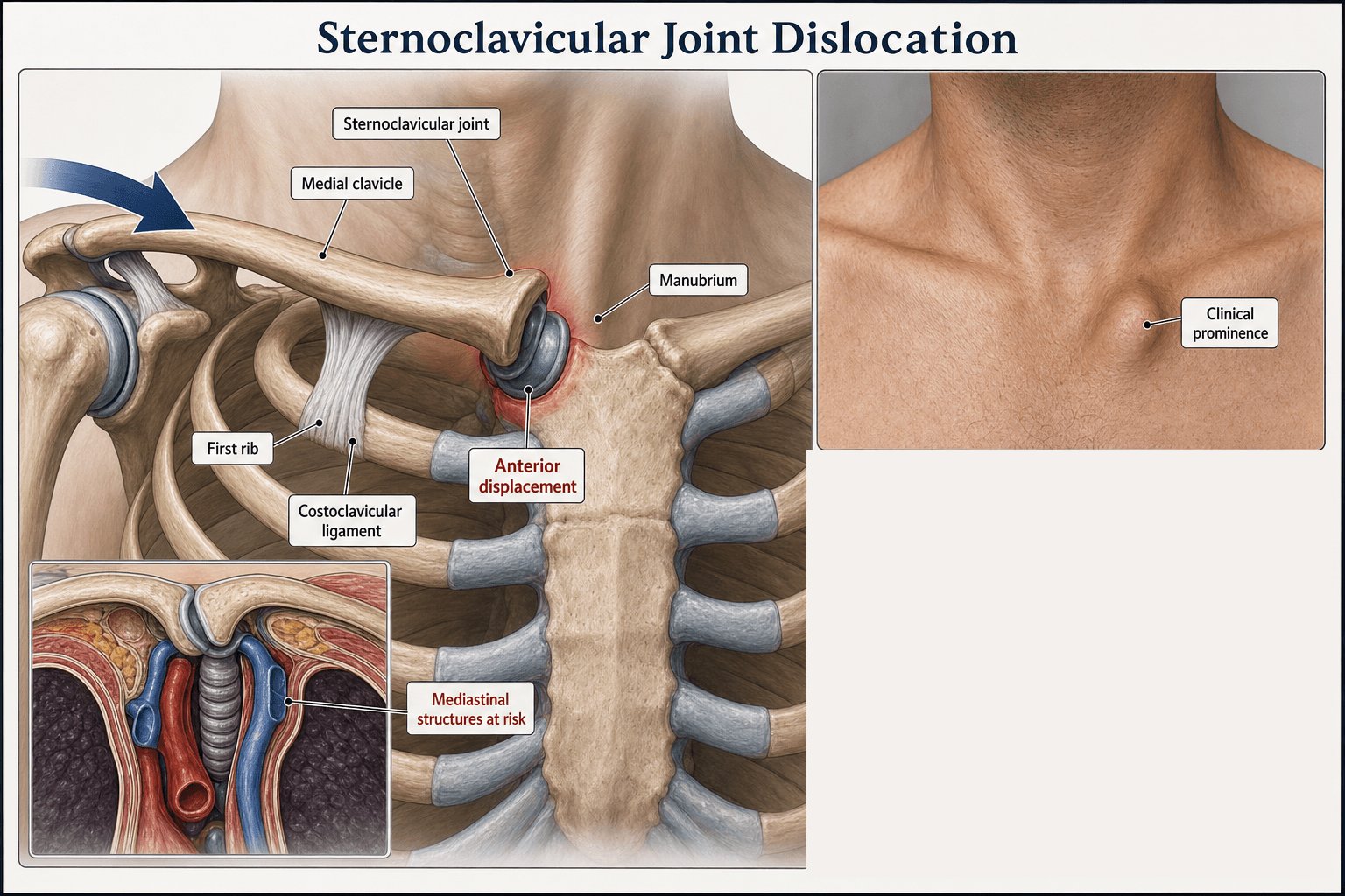
How it dislocates. An indirect force to the lateral shoulder drives the medial clavicle, which pivots at the costoclavicular ligament.
- Anterior force vector (most common) - the clavicle is levered forwards out of the manubrium, tearing the anterior capsule, costoclavicular and interclavicular ligaments, with the posterior capsule intact
- Posterior force vector (less common, high-energy) - posterior dislocation, with the posterior capsule torn
- Direct blow to the medial clavicle - can drive it posteriorly
What lies behind the joint. A posteriorly displaced clavicle can compress any of these, and the internal jugular vein as well:
- Immediately posterior to the medial clavicle - brachiocephalic vein, common carotid, subclavian artery and vein
- Deeper plane - trachea, oesophagus, vagus nerve, phrenic nerve, and on the left the thoracic duct
The physis. The medial clavicular physis is the last to fuse, at 23-25 years, and in adolescents it is the weakest link. A physeal injury in an adolescent often mimics a posterior dislocation on imaging.
Classification and Types
The injury is described by the direction of displacement, the time since injury, and the severity of the ligament disruption.
- Frequency
- Most common
- Mechanism
- Indirect force, anterior vector
- Urgency
- Usually non-operative
- Frequency
- Less common
- Mechanism
- Indirect high-energy or direct posterior blow
- Urgency
- Emergency - mediastinal risk
- Frequency
- Common in young hypermobile patients
- Mechanism
- Spontaneous or with minor movement
- Urgency
- Reassurance, physiotherapy
- Time from Injury
- Under 3 weeks
- Findings
- Swelling, deformity, pain
- Treatment
- Closed reduction (posterior) or non-operative (anterior)
- Time from Injury
- Over 3 weeks, or repeated episodes
- Findings
- Established instability, functional deficit
- Treatment
- Reconstruction if symptomatic and failed physio
- Time from Injury
- Under 23-25 years
- Findings
- Medial clavicle physis is weakest link
- Treatment
- Reduction if symptomatic, usually conservative
Severity grade. Direction and timing are two axes; the severity grade of the ligamentous injury is the third, and it dictates whether the joint is stable.
- Grade I, sprain - the capsular ligaments are stretched but intact; the joint is stable and minimally displaced, with tenderness and mild swelling only. Sling and analgesia.
- Grade II, subluxation - partial capsular and ligament disruption with the costoclavicular ligament still intact; the joint subluxes but is not frankly dislocated, and is partially stable. Sling, analgesia, and avoidance of provocative positions.
- Grade III, dislocation - complete disruption of the capsular ligaments (and usually the costoclavicular ligament); the joint is frankly dislocated anteriorly or posteriorly and unstable. This is where the anterior-versus-posterior distinction and the mediastinal emergency apply.
A Grade I-II injury does not need reduction, only symptomatic care. The dramatic management decisions (urgent CT, theatre reduction, reconstruction) belong to the Grade III dislocation, so do not over-treat a sprain or subluxation.
Clinical Assessment
History. Establish the mechanism: high-energy trauma, contact sport, a road traffic collision or a fall. Then ask for the posterior red flags - dyspnoea, stridor, dysphagia, voice change, arm paraesthesia or ischaemia. Previous episodes of recurrent subluxation suggest multidirectional laxity, and in a patient under 25 years consider a physeal injury rather than a true dislocation.
Examination. Look for asymmetry, swelling, ecchymosis and skin tethering, and palpate the position of the medial clavicle relative to the suprasternal notch and the other side. Elevating the arm in extension stresses the joint. In a suspected posterior injury, look for neck vein engorgement and tracheal deviation, and check the pulses and the neurology.
- Anterior SC Dislocation
- Prominent medial clavicle, often visible lump
- Posterior SC Dislocation
- Depression or sulcus at SC joint
- Anterior SC Dislocation
- Rare
- Posterior SC Dislocation
- Common - dyspnoea, stridor, dysphagia, venous congestion
- Anterior SC Dislocation
- Erythema, swelling, possible skin tenting
- Posterior SC Dislocation
- May have skin dimple, cyanosis, surgical emphysema
- Anterior SC Dislocation
- Painful but usually full
- Posterior SC Dislocation
- Markedly limited by pain and mediastinal symptoms
Assess and document in every SC joint injury:
- Respiratory distress, stridor, voice change, cyanosis
- Neck vein distension (SVC or brachiocephalic vein compression)
- Subclavian and axillary pulses, capillary refill - vascular injury may be occult
- Brachial plexus sensation and motor (C5-T1)
- Swallowing difficulty (oesophageal impingement)
- Phrenic nerve (diaphragm screening with ultrasound or fluoroscopy if intubated)
- Vagus (recurrent laryngeal - voice change)
A posterior SC dislocation with any of these signs mandates urgent reduction with cardiothoracic or vascular surgery on standby.
Differential diagnosis. Not every painful or deformed medial clavicle is a dislocation.
- Direction of Deformity
- Anterior prominence
- Discriminating Finding
- Lateral compression reproduces pain, palpable step
- Key Test / Imaging
- CT confirms anterior position
- Direction of Deformity
- Loss of medial clavicle contour
- Discriminating Finding
- Mediastinal signs, asymmetric pulses
- Key Test / Imaging
- CT with IV contrast to define mediastinal relationship
- Direction of Deformity
- Anterior or posterior
- Discriminating Finding
- Adolescent (under 25), mechanism matches trauma
- Key Test / Imaging
- CT shows physeal line; MRI can confirm physis
- Direction of Deformity
- Variable, often bilateral
- Discriminating Finding
- Multidirectional laxity, no high-energy event
- Key Test / Imaging
- Clinical; imaging normal or minor asymmetry
- Direction of Deformity
- No acute deformity
- Discriminating Finding
- Insidious pain, crepitus, older patient
- Key Test / Imaging
- X-ray and CT show joint space narrowing, osteophytes
- Direction of Deformity
- No acute deformity
- Discriminating Finding
- Middle-aged women, pain without laxity
- Key Test / Imaging
- X-ray: sclerosis of medial clavicle
- Direction of Deformity
- No acute deformity
- Discriminating Finding
- Insidious medial clavicle pain
- Key Test / Imaging
- MRI shows AVN changes
- Direction of Deformity
- Swelling, warmth, fluctuance
- Discriminating Finding
- Fever, IVDU, immunocompromise, recent infection
- Key Test / Imaging
- Bloods, joint aspiration, MRI
Septic arthritis. A swollen, warm, tender SC joint is not always traumatic. Septic arthritis of the SC joint is a distinct, examinable entity that is easily missed, and because the joint sits over the mediastinum, untreated infection can spread to a chest-wall abscess, sternoclavicular osteomyelitis and mediastinitis, a potentially life-threatening complication.
- Host risk factors - intravenous drug use (classic), diabetes, immunosuppression, chronic kidney disease, an indwelling central line or catheter, and a distant source of bacteraemia; it can also occur with no obvious risk factor
- Organisms - Staphylococcus aureus is commonest overall; Pseudomonas aeruginosa is classically associated with intravenous drug use
- Diagnosis - raised inflammatory markers, blood cultures, CT or MRI to define abscess, osteomyelitis or retrosternal extension, and joint aspiration for Gram stain and culture
- Management - targeted IV antibiotics for early disease; established septic arthritis with abscess or osteomyelitis usually needs surgical debridement or en bloc resection of the SC joint (medial clavicle and manubrial portion), which is generally well tolerated functionally
In an IV drug user or immunocompromised patient with a hot, painful medial clavicle and fever, think SC septic arthritis with possible mediastinal extension: image and aspirate, and do not treat it as a sprain.
Atraumatic subluxation. Common in young, hypermobile women, and not a dislocation. The medial clavicle subluxes spontaneously or with minor movements and reduces again. It needs no reduction, immobilisation or surgery; education, reassurance and avoidance of provocative positions are the management, and operating on it has high failure and complication rates.
Investigations
Plain films cannot reliably diagnose or exclude SC dislocation. Any patient with clinical suspicion (mechanism, deformity, mediastinal symptoms) requires CT with IV contrast. Do not delay CT to obtain multiple plain views.
Plain radiographs. A limited role: the AP chest film is the initial screen and excludes associated rib or lung injury. Plain films are notoriously hard to interpret here and miss up to 50 percent of SC dislocations; they cannot define the direction accurately or show the relationship to the mediastinum. The serendipity view (40-degree cephalic tilt) is now largely superseded, a historical curiosity.
CT with IV contrast is the investigation of choice, indicated in all suspected SC dislocations. Acquire thin slices (1 mm) from chin to diaphragm with IV contrast, and add 3D and multiplanar reformats for surgical planning. It gives the definitive diagnosis and maps the vessels:
- Direction of dislocation and distance displaced
- Relationship to the mediastinal vessels, trachea and oesophagus, including tracheal or vessel deviation
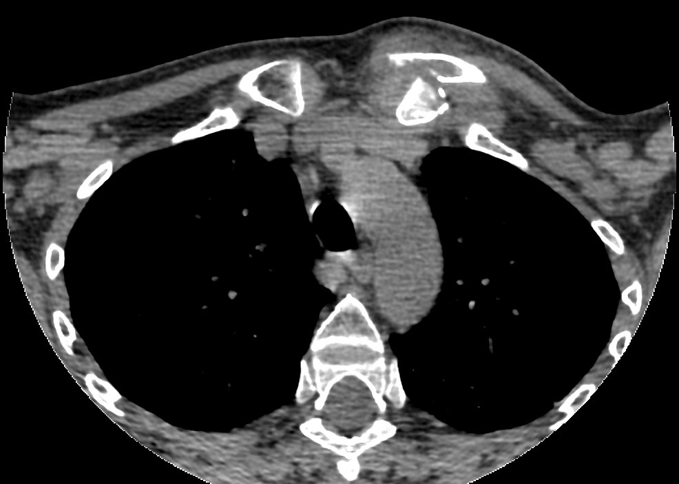
CT angiography is added for a posterior dislocation with a pulse deficit, an expanding haematoma or asymmetry. It shows vessel compression, intimal injury, pseudoaneurysm or occlusion, and the surgical plan is made jointly with vascular surgery, with image sharing and on-table angiography as needed.
MRI distinguishes a physeal injury from a true dislocation in adolescents. Physeal oedema with intact ligaments and no true joint disruption justifies conservative treatment and avoids an operation on the physis.
Management
The goal is symptom relief and return to function. Anterior dislocations are generally stable after closed reduction, even if the joint re-subluxes cosmetically, and are well tolerated even when the medial clavicle remains prominent. Non-operative management is the default.
Days 0-7. Analgesia with oral paracetamol, an NSAID and a short opioid course if needed; a broad-arm sling for 1-3 weeks for comfort; ice and rest for the first 48-72 hours. Explain that the lump may persist cosmetically, and that this is acceptable.
Weeks 1-3. Progressive range of motion: pendulum, then active-assisted, then active. Rotator cuff and scapular stabiliser strengthening starts at 3-4 weeks, and heavy lifting and contact sport are avoided for 6-12 weeks.
Weeks 6-12. Sport-specific rehabilitation for overhead athletes, throwers and contact sports. Most return to full function with acceptable cosmesis; surgery is reserved for persistent instability, functional limitation or failed rehabilitation.
Do not operate on a first-time anterior SC dislocation. Operative reduction and stabilisation has high failure and complication rates (recurrent instability, hardware failure, neurovascular injury). Even if the medial clavicle remains prominent, function is generally excellent with conservative care. Surgery is reserved for chronic, symptomatic instability that has failed structured rehabilitation.
Complications
- Incidence
- Up to 30 percent of posterior dislocations
- Risk Factors
- High-energy mechanism, sharp displaced fragment
- Management
- Vascular surgery, repair or bypass
- Incidence
- Reported in case series
- Risk Factors
- Posterior displacement, delayed reduction
- Management
- Urgent reduction, possible tracheostomy
- Incidence
- Rare
- Risk Factors
- Penetrating component, sharp posterior fragment
- Management
- Contrast study, surgical repair
- Incidence
- 20-30 percent after reconstruction
- Risk Factors
- Ligamentous laxity, hypermobility, inadequate rehab
- Management
- Revision reconstruction or resection
- Incidence
- Catastrophic when it occurs
- Risk Factors
- Use of K-wires across SC joint
- Management
- Avoid K-wires; use plates or tendon graft
- Incidence
- Up to 50 percent with plates
- Risk Factors
- Thin soft tissue cover, repetitive motion
- Management
- Removal of hardware once ligaments healed
- Incidence
- Rare
- Risk Factors
- Posterior compression, intimal injury
- Management
- Anticoagulation, vascular intervention
K-wires and Kirschner wires across the sternoclavicular joint have migrated into the heart, aorta, pulmonary artery, lung, oesophagus, trachea, and spinal cord, with multiple reported deaths. Use plate fixation, suture anchors, or tendon graft reconstruction. If a wire is used (rare salvage scenarios), it must have a secure bend at the lateral end, and the patient must be followed with serial imaging until removal.
Outcomes and Prognosis
- Indication
- First-time anterior dislocation
- Expected Outcome
- 90 percent good function, acceptable cosmesis
- Long-term Function
- Excellent return to sport and work
- Indication
- Acute posterior dislocation, stable after reduction
- Expected Outcome
- Good function, low recurrence if costoclavicular intact
- Long-term Function
- Best reduced early, within an age-dependent window (see Management). Succeeds in only 38% of adult posterior dislocations
- Indication
- Failed closed, mediastinal compromise
- Expected Outcome
- Good but with surgical morbidity
- Long-term Function
- Good if vascular or visceral injury addressed
- Indication
- Failed non-operative, symptomatic instability
- Expected Outcome
- 70-80 percent good-to-excellent
- Long-term Function
- Variable; re-operation common
Prognostic factors. The best prognosis belongs to an acute anterior dislocation treated conservatively, an acute posterior dislocation reduced within 24 hours with an intact costoclavicular ligament, and a low-demand patient. The poor prognosis belongs to a delayed posterior dislocation, failed closed reduction, multiple reconstruction attempts, hypermobility syndromes, and contact athletes returning to high-impact sport.
Guidelines, Registries & Global Practice
- SC dislocations are rare, representing under 1 percent of all joint dislocations and 3 percent of shoulder-girdle injuries
- Anterior dislocations are 3 to 1 more common than posterior
- Posterior dislocations are predominantly high-energy - road traffic collisions, contact sport, falls from height
- Mediastinal compromise is reported in a quarter to a third of posterior dislocations in published series
- High-resource: 24-hour access to CT, vascular and cardiothoracic surgery, and image intensifier for closed reduction
- Limited-resource: Closed reduction under sedation in the emergency department may be necessary if theatre and vascular backup are unavailable - transfer to a tertiary centre is preferred if any mediastinal signs
- Universal principle: Posterior SC dislocation requires urgent reduction; do not delay transfer for a CT that is not available locally
- Reconstruction: Concentrated in specialist shoulder units; soft tissue techniques preferred over hardware
- Diagnosis emphasis
- CT with contrast in any suspected SC dislocation
- Acute treatment
- Posterior: closed reduction in theatre with thoracic backup; anterior: conservative
- Surgery / reconstruction
- Soft tissue reconstruction (figure-of-8 tendon graft) for failed non-operative
- Diagnosis emphasis
- CT for direction and mediastinal assessment
- Acute treatment
- Posterior: urgent closed or open reduction; anterior: sling and rehab
- Surgery / reconstruction
- Tendon graft preferred; plates reserved for failed soft tissue; avoid K-wires
- Diagnosis emphasis
- CT or MRI in suspected dislocation, MRI for physeal assessment
- Acute treatment
- Posterior: closed reduction under GA, thoracic surgery standby
- Surgery / reconstruction
- Tendon reconstruction (sternocleidomastoid, palmaris longus, hamstring)
- Diagnosis emphasis
- CT with contrast; vascular surgery consult for posterior
- Acute treatment
- Posterior: closed reduction in theatre; anterior: non-operative
- Surgery / reconstruction
- Soft tissue reconstruction with suture anchors preferred
There is no dedicated registry for sternoclavicular joint dislocation. Evidence is dominated by small retrospective case series and technique papers. The major arthroplasty registries (NJR, AJRR, AOANJRR) do not capture SC joint reconstruction, as it is uncommon. Practice is principle-based: CT with contrast for diagnosis, urgent closed reduction of posterior dislocations with cardiothoracic backup, and soft tissue reconstruction for chronic instability using tendon graft rather than hardware.
Record in every SC joint injury:
- Direction of dislocation (anterior or posterior) on CT
- Mediastinal structure assessment (vessels, trachea, oesophagus)
- Pulses, capillary refill, and neurological exam in the affected arm
- Voice, breathing, and swallowing symptoms at presentation
- Plan for closed versus open reduction and surgical team involved
- Counselling about recurrence, hardware, and re-operation risks if reconstruction is performed
Posterior SC dislocation with missed mediastinal injury is a recurring source of severe morbidity and litigation worldwide. Document the CT findings, the surgical team involved, and the reduction technique in every case.
Controversies & Areas of Uncertainty
Soft tissue reconstruction (figure-of-8 tendon graft, suture anchors) is biomechanically and biologically appealing but has higher reported re-dislocation rates in some series. Plate fixation provides initial stability at the cost of frequent hardware-related re-operation. There are no head-to-head trials.
Some surgeons advocate operative stabilisation of first-time anterior dislocations in young, high-demand athletes, but complication rates are high and re-dislocation is common. Most guidance reserves surgery for chronic, symptomatic instability that has failed structured non-operative care.
Sternocleidomastoid (in situ), hamstring (semitendinosus, gracilis), and palmaris longus have all been described. There is no clear winner. Sternocleidomastoid is convenient but limited in length; hamstring is robust but adds donor morbidity; palmaris longus is thin and may be absent.
Excessive resection destabilises the medial clavicle and worsens instability. The safe upper limit is generally cited as 1 to 1.5 cm in adults, with preservation of the costoclavicular ligament. Beyond this, reconstruction or arthrodesis may be required.
MCQ Practice Points
Q: What is the primary ligamentous restraint to anterior and posterior translation at the sternoclavicular joint? A: The posterior capsule is the strongest restraint to translation in BOTH directions — Spencer's sectioning study found dividing it increased posterior translation by 107% and anterior translation by 42%, against 26% (anterior only) for the anterior capsule, while dividing the costoclavicular and interclavicular ligaments had little effect. The costoclavicular ligament is the primary restraint to superior and medial-lateral displacement. Chronic instability is therefore reconstructed with a figure-of-eight capsular graft rather than by recreating the costoclavicular ligament.
Q: What is the investigation of choice for suspected sternoclavicular joint dislocation? A: CT scan with IV contrast. Plain radiographs miss up to 50 percent of dislocations, and the serendipity view is largely obsolete. CT demonstrates direction, displacement, and relationship to mediastinal structures. CT angiography is added if vascular injury is suspected.
Q: A patient has a posterior SC dislocation with dyspnoea, dysphagia, and diminished radial pulse. What is your immediate management? A: This is a mediastinal emergency. Follow ATLS, establish IV access, send bloods, obtain urgent CT with IV contrast, and contact cardiothoracic and vascular surgery. The dislocation requires reduction under general anaesthesia in theatre with surgical backup, ideally within 24 hours.
Q: What is the first-line management of an acute anterior SC dislocation? A: Non-operative management with sling, analgesia, and physiotherapy. Operative reduction and stabilisation has high failure and complication rates and is reserved for chronic symptomatic instability. Even if the medial clavicle remains prominent, function is generally excellent.
Q: Why are K-wires contraindicated in sternoclavicular joint reconstruction? A: K-wires have migrated into the heart, aorta, pulmonary artery, lung, oesophagus, trachea, and spinal cord, with multiple reported deaths. Use plate fixation, suture anchors, or autologous tendon graft reconstruction (figure-of-8 sternocleidomastoid or hamstring graft).
Q: A 17-year-old presents after a fall with apparent anterior SC dislocation. What is the most likely true diagnosis? A: Medial clavicle physeal separation. The medial clavicular physis is the last in the body to fuse (around 23-25 years) and is the weakest link in the SC region in adolescents. True joint dislocation requires disruption of the strong posterior capsule and costoclavicular ligament, which is uncommon before skeletal maturity. The injury is usually stable after closed reduction and remodels over time.
Exam Viva Scenarios
Practise clinical reasoning and management decisions out loud
“A 22-year-old rugby player presents to the emergency department after a direct tackle to his left shoulder. He has pain over the medial clavicle, difficulty breathing, and a change in his voice. On examination, there is a palpable depression at the left SC joint, the left arm is cooler than the right, and the left radial pulse is diminished. What is your immediate assessment and management?”
“A 19-year-old competitive cyclist falls over his handlebars during a race and presents to the emergency department with a visible lump over the medial end of his right clavicle. He has no dyspnoea, dysphagia, voice change, or vascular symptoms. CT confirms anterior SC dislocation with the medial clavicle displaced anterior to the manubrium and no mediastinal compromise. He is keen to return to competitive cycling. How do you manage this?”
Key Anatomy
- Saddle-type synovial joint with intra-articular disc
- Medial clavicle larger than manubrial fossa - stability is entirely ligamentous
- Posterior capsule = strongest restraint to anterior displacement
- Costoclavicular ligament = primary restraint to medial-lateral displacement
- Medial clavicle physis is the last to fuse (23-25 years) - weakest link in adolescents
Diagnosis
- Posterior = mediastinal emergency: dyspnoea, dysphagia, voice change, vascular signs
- Anterior = cosmetic lump, often stable, rarely an emergency
- CT with IV contrast is the investigation of choice - plain films miss up to 50 percent
- Always assess pulses, voice, breathing, and swallowing in any SC injury
- Serendipity view is largely obsolete - do not delay CT to obtain multiple plain views
Treatment Algorithm
- Anterior (first time) = non-operative: sling, analgesia, physiotherapy, return to sport 8-12 weeks
- Posterior (acute) = urgent closed reduction under GA with thoracic/vascular backup
- Posterior (failed closed) = open reduction via supraclavicular approach with vascular control
- Chronic instability = figure-of-8 tendon graft reconstruction; K-wires contraindicated
Reconstruction Risks
- K-wire migration into heart, aorta, lung, oesophagus, spinal cord - never use
- Plate fixation has high rate of hardware-related re-operation
- Tendon graft reconstruction - 20-30 percent re-dislocation rate
- Medial clavicle resection greater than 1.5 cm destabilises further
- Reserve surgery for chronic, symptomatic instability after failed non-operative care
Complications and Pitfalls
- Missed posterior SC dislocation with mediastinal injury is catastrophic
- Attempting closed reduction without imaging or surgical backup risks vascular injury
- Operating on atraumatic SC subluxation in hypermobile patients fails
- Anterior SC dislocation in adolescent = medial clavicle physeal separation, not true dislocation
- Document direction, mediastinal assessment, and reduction technique in every case
Evidence Base and Key Trials
Acute traumatic retrosternal dislocation of the clavicle
- The paper's contribution is a REDUCTION TECHNIQUE, not a timing rule: the authors first note that the literature shows closed reduction of retrosternal dislocation often fails
- In 6 of their 7 patients, reduction was achieved by retracting the shoulders with CAUDAL TRACTION ON THE ADDUCTED ARM while the patient lay supported on an interscapular bolster
- Note that this is not the abduction-extension manoeuvre usually taught - the arm is adducted and pulled caudally, and the authors analysed why this and other methods work
Sternoclavicular joint dislocation: a systematic review and meta-analysis
- Systematic review and meta-analysis of sternoclavicular joint dislocations - synthesises treatment outcomes across the published series
- Posterior dislocations are uncommon but carry substantially higher morbidity than anterior dislocations, with mediastinal compromise the key concern
- Operative reconstruction is reserved for chronic symptomatic instability; most anterior dislocations are managed non-operatively with good functional outcomes
Treatment of traumatic posterior sternoclavicular dislocations
- 21 patients with traumatic posterior SC injury; every patient had a trial of closed reduction
- Closed reduction was definitive in only 8 of 21 (38%) - the remaining 13 required open reduction and SC joint reconstruction. Do not counsel a patient that closed reduction usually works for a posterior dislocation
- The timing threshold in this adult series was NOT 24-48 hours: closed reduction was significantly more likely to succeed when performed within 10 DAYS of injury (P < .05)
- 18 of 21 were good or excellent on the UCLA scale, and patients definitively treated by closed or by open reduction compared favourably for pain, strength and motion
- Where closed reduction failed, open treatment aimed at reconstructing the costoclavicular ligaments gave good results
Sternoclavicular epiphyseal separation with adjacent clavicular fracture
- Classic first description of medial clavicular epiphyseal (physeal) separation - adolescents presenting with apparent SC dislocation more often have a physeal injury than a true joint dislocation
- True SC dislocation requires disruption of the strong posterior capsule and costoclavicular ligament, which is uncommon in skeletally immature patients
- Physeal injury is more often stable after closed reduction and rarely requires operative fixation
Posterior sternoclavicular joint injuries in the adolescent population: a meta-analysis
- Patient-level data on 140 adolescents aged 12 to 18 extracted from 79 published studies; mean age 15.24 years
- OPERATIVE TREATMENT WAS NOT THE EXCEPTION: only 49 (35.0 per cent) had closed treatment alone, 42 (30.0 per cent) had open treatment alone and 47 (33.6 per cent) had closed treatment FOLLOWED by open treatment - so a third of those given a closed attempt went on to surgery anyway
- TIMING IN ADOLESCENTS IS TIGHTER THAN IN ADULTS: 55.8 per cent of closed reductions attempted within 48 hours succeeded, against 30.8 per cent after 48 hours
- Final function was excellent either way - 92.31 per cent regained full function without recurrence after closed treatment and 95.83 per cent after operative treatment - but the authors note that follow-up reporting varies considerably