Patient Selection | Bone Quality | Age | Activity Level | AOANJRR Outcomes
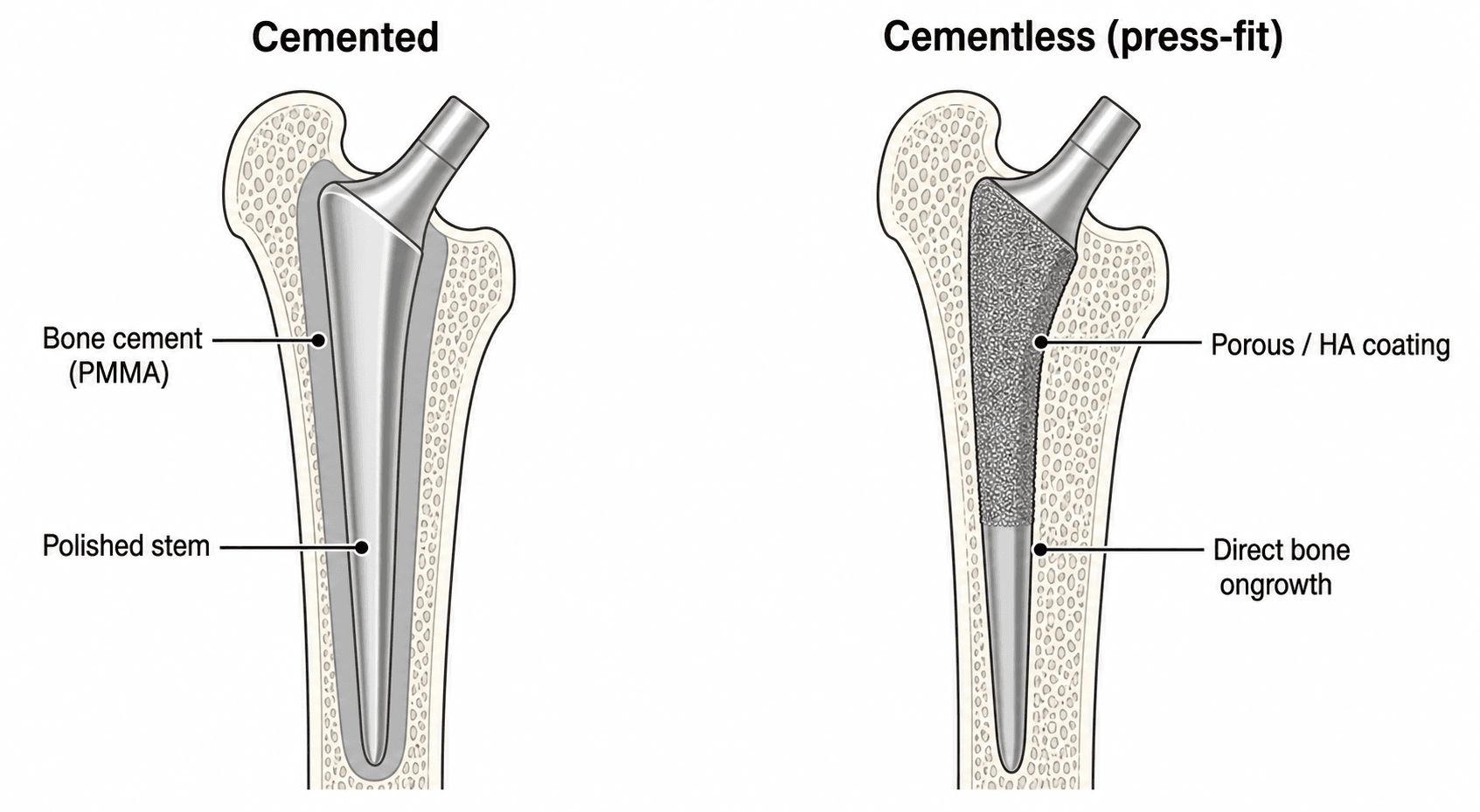
- Cemented fixation uses PMMA bone cement (polymethylmethacrylate) for immediate stability
- Cementless fixation relies on biological ingrowth into porous coating (takes 6-12 weeks)
- Patient age and bone quality are primary selection criteria
- AOANJRR data shows excellent outcomes with both methods when appropriately selected
- Hybrid THA (cementless cup + cemented stem) is widely used for intermediate-age patients
- “Cemented stems have longer track record (Charnley since 1960s)
- “Cementless relies on press-fit initial stability and osseointegration
- “Cement disease is a misnomer: aseptic loosening from particulate debris (cement, polyethylene, metal)
- “Young patients (under 65) benefit from cementless (easier revision)
Overview and Epidemiology
Fixation is the method of attaching the prosthetic components to host bone. There are two primary methods, cemented, using polymethylmethacrylate (PMMA) bone cement, and cementless, relying on biological ingrowth, and each has distinct indications, advantages and limitations. A hybrid mixes them, and the common hybrid is a cementless cup with a cemented stem.
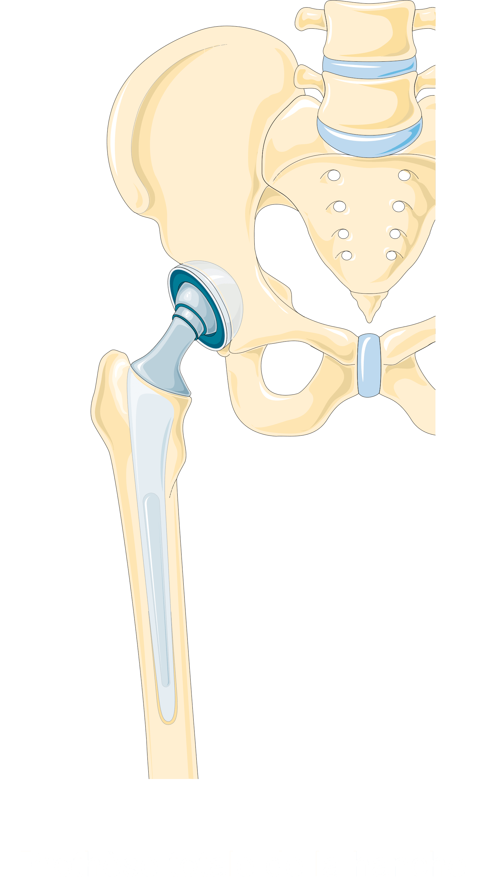
Cemented fixation. PMMA provides an immediate mechanical interlock, and it has been the gold standard since Charnley in the 1960s. It is ideal for elderly, osteoporotic bone, provided the surgeon uses proper third-generation technique.
Cementless fixation. Press-fit initial stability is required, after which bone grows into the porous coating over 6-12 weeks. It suits young, active patients with good bone, and it makes any future revision easier.
How the balance shifted. Sir John Charnley developed the low-friction arthroplasty using PMMA cement in the 1960s, and excellent long-term results in the 1970s established cemented THA as the gold standard. Concerns about "cement disease" in the 1980s led to the development of cementless implants. The 1990s brought recognition that both methods work well when appropriately indicated, and registry data in the 2000s confirmed excellent outcomes with modern techniques for both.
"Cement disease" is a misnomer - it's actually aseptic loosening caused by particulate debris (cement, polyethylene, metal) triggering osteolysis. The problem isn't cement itself but particle generation from micromotion and wear. Modern cement technique and improved polyethylene have dramatically reduced this complication.
Clinical Presentation and Indications
The "clinical presentation" for a fixation decision is the patient evaluation that determines which method to use. It happens pre-operatively and is completed intra-operatively.
Age, bone quality and activity drive the choice. Young, active patients get cementless fixation; elderly osteoporotic patients benefit from the immediate stability of cement. This is evidence-based, not surgeon preference.
- Age
- Under 50 years
- Bone Quality
- Good bone stock
- Recommended Fixation
- Cementless both components (easier revision)
- Age
- 50-65 years
- Bone Quality
- Good to moderate bone
- Recommended Fixation
- Cementless (or hybrid with cemented stem)
- Age
- 65-75 years
- Bone Quality
- Moderate bone quality
- Recommended Fixation
- Hybrid (cementless cup + cemented stem)
- Age
- Over 75 years
- Bone Quality
- Osteoporotic bone
- Recommended Fixation
- Cemented both components (immediate stability)
- Age
- Any age
- Bone Quality
- Poor bone quality
- Recommended Fixation
- Cemented both components (bone quality issue)
Age. Under 50, a future revision is likely, which is why cementless is preferred. Between 50 and 65 either method is acceptable, decided on bone quality. Over 75, osteoporosis is common and the implant is a lifetime one, which is why cement is preferred.
Activity. High-demand athletic patients suit cementless, biological fixation. In a low-demand, sedentary patient cement is acceptable at any age, and a manual labourer can have either method if the bone quality is adequate.
Comorbidities. Several change the choice and the surgical plan:
- Osteoporosis - cemented, because press-fit cannot be achieved
- Rheumatoid arthritis - cemented, for poor bone quality and protrusio
- Previous DVT/PE - consider avoiding cement (embolism risk)
- Cardiac disease - cemented fixation requires careful monitoring (hypotension risk)
Bone quality is the cornerstone. The femur is graded by Dorr type (see Classification), and bone density adds a second axis when osteoporosis is suspected:
- Normal (T-score above -1.0) - cementless suitable
- Osteopenia (T-score -1.0 to -2.5) - either method, depending on other factors
- Osteoporosis (T-score below -2.5) - cemented is the safer choice; this T-score strongly favours cemented fixation
Acetabular bone stock. Good coverage with no defects is excellent for a cementless cup, and mild dysplasia can still take a cementless cup with screws. Severe dysplasia or defects may need a structural graft, and cement should be considered.
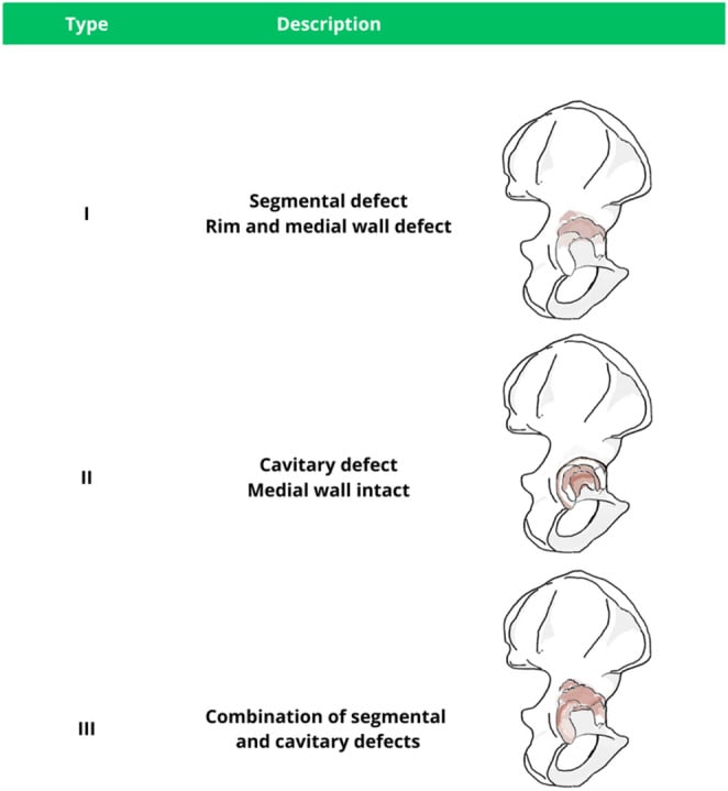
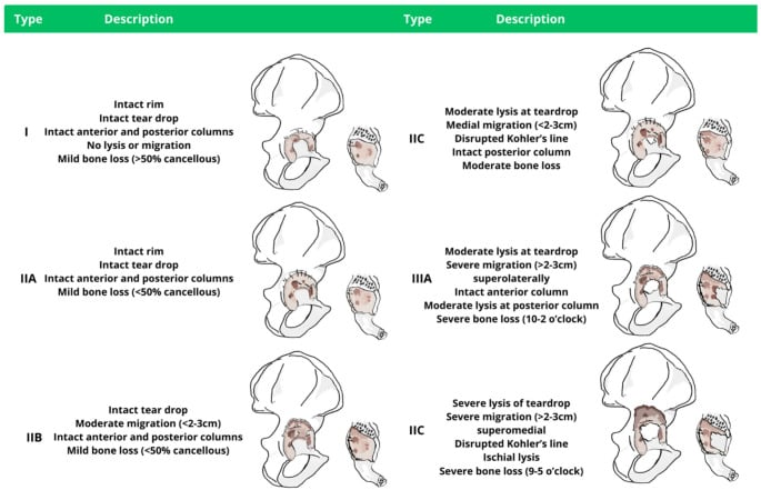
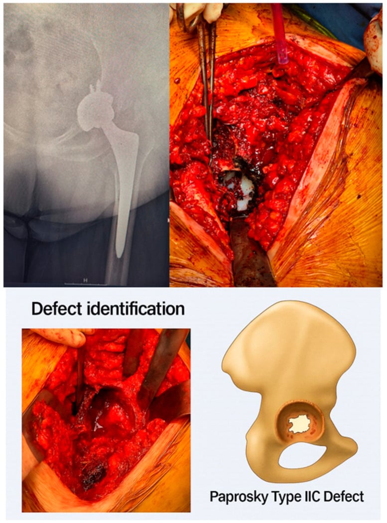
Disease-specific considerations. Each diagnosis brings its own problem:
- Primary osteoarthritis - usually good bone quality; standard age-based selection applies, and either cemented or cementless is successful
- Avascular necrosis - typically young patients, so cementless is preferred; femoral head collapse may affect the acetabular bone
- Developmental dysplasia - a high hip centre with a deficient acetabulum; cementless if there is adequate host bone contact, otherwise a structural graft, or cemented fixation with bone graft, may be needed
- Post-traumatic arthritis - bone quality varies with the fracture history, and there may be residual deformity or hardware; assess bone stock individually
- Inflammatory arthropathy - cemented preferred because of osteopenia; protrusio acetabuli is common in rheumatoid disease and may need bone grafting
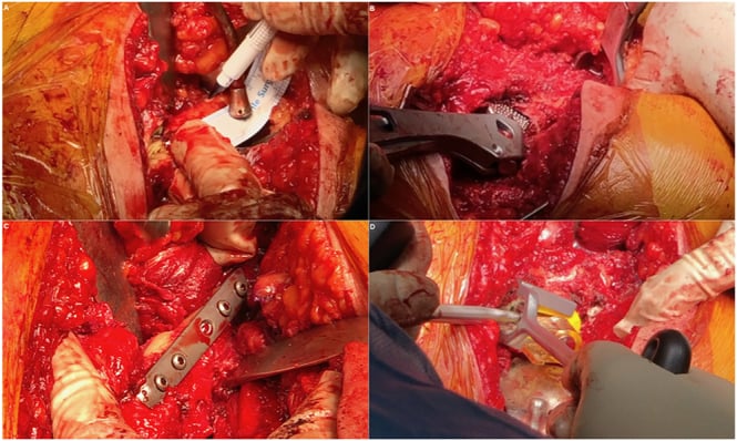
The plan can change in theatre. Even with pre-operative planning, the final fixation choice may change because of:
- The bone quality actually encountered during reaming
- Unexpected bone defects or sclerosis
- Inability to achieve adequate press-fit
- An intraoperative fracture, which may favour cement for immediate stability
If unable to achieve adequate press-fit stability for cementless component, be prepared to convert to cemented fixation. Always have cement available when performing cementless THA.
Pathophysiology of Fixation and Loosening
Cement is a grout. Cemented fixation works by mechanical interlock, not chemical bonding. PMMA penetrates into the cancellous bone, creating a mechanical interdigitation that distributes load from the implant to the bone, and the mantle fills irregularities to create a stable interface.
How cemented fixation fails. Five mechanisms are described:
- Particle disease - cement, polyethylene or metal particles activate macrophages
- Osteolysis - activated macrophages recruit osteoclasts, and periprosthetic bone is resorbed
- Mantle fracture - a thin mantle, less than 2mm, is prone to fracture
- Interface failure - the cement-bone interface weakens over time from micromotion, showing as progressive radiolucent lines, and eventually fails mechanically
- Subsidence - component migration indicates loss of fixation
Cementless fixation is biological. It relies on osseointegration, which needs initial press-fit stability, conventionally quoted as keeping micromotion below about 150 microns, so that bone can grow into the porous coating over 6-12 weeks. At maturity the biological interface is stronger than cement.
The 150-micron figure is a rule of thumb. Treat it as that rather than a measured biological cutoff. The cadaveric work cited on this page found relative displacements of up to about 35 microns occurring without any true sliding at the interface, and concluded that what governs ingrowth is whether the interface slides, not the displacement number on its own. The clinical instruction is unchanged - get a stable press-fit - but do not defend 150 as though it were a threshold anyone has measured in living bone.
How cementless fixation fails. Five mechanisms are described here too:
- Inadequate press-fit - micromotion beyond that conventional limit prevents bone ingrowth
- Fibrous interface - without osseointegration, an unstable fibrous membrane forms
- Stress shielding - stiff implants shield the proximal bone, which atrophies
- Particle-induced osteolysis - bearing surface particles still cause bone loss
- Subsidence - poor initial stability leads to progressive migration
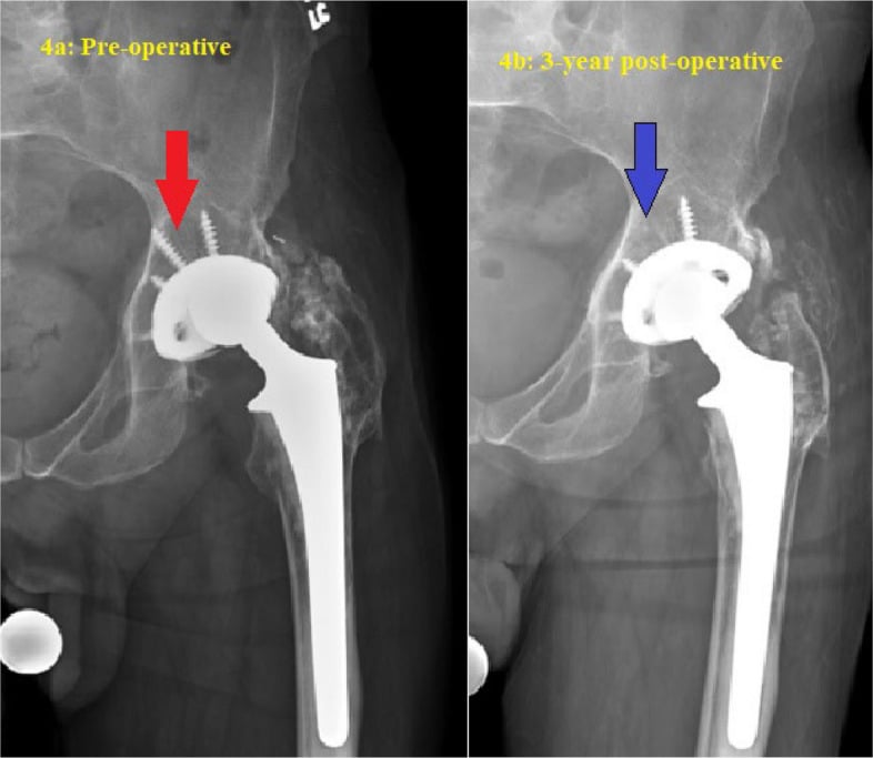
The common end point. Both methods ultimately fail from particle-induced osteolysis when the bearing surfaces generate debris. Modern highly crosslinked polyethylene has dramatically reduced this problem.
Classification
By method. Four constructs are named by which components are cemented, and the reverse hybrid is the rare one.
- Components
- Cemented cup + cemented stem
- Mechanism
- PMMA mechanical interlock
- Optimal Patient
- Elderly (over 75), osteoporosis
- Components
- Cementless cup + cementless stem
- Mechanism
- Biological osseointegration
- Optimal Patient
- Young (under 65), good bone stock
- Components
- Cementless cup + cemented stem
- Mechanism
- Combined approach
- Optimal Patient
- Age 65-75, Dorr C femur
- Components
- Cemented cup + cementless stem
- Mechanism
- Combined approach
- Optimal Patient
- Poor acetabular bone, good femur (rare)
Dorr classification of the femur. Femoral morphology, assessed on the AP pelvis radiograph, guides the choice of stem fixation:
- Type A - narrow canal, thick cortices: ideal for cementless
- Type B - intermediate: either method suitable
- Type C - wide canal, thin cortices: cemented preferred
Gruen zones. Seven zones around the femoral stem, used to locate radiolucent lines on follow-up films.
- Location
- Proximal-lateral
- Clinical Significance
- Stress shielding common here
- Location
- Mid-lateral
- Clinical Significance
- Mid-stem fixation zone
- Location
- Distal-lateral
- Clinical Significance
- Distal fixation zone
- Location
- Stem tip
- Clinical Significance
- Risk of thigh pain if modulus mismatch
- Location
- Distal-medial
- Clinical Significance
- Distal fixation zone
- Location
- Mid-medial
- Clinical Significance
- Mid-stem fixation zone
- Location
- Proximal-medial (calcar)
- Clinical Significance
- Critical load transfer zone
DeLee-Charnley zones. The acetabular equivalent divides the cup-bone interface into zone I (superior), zone II (superomedial) and zone III (inferomedial). Progressive radiolucent lines in all zones indicate loosening.
Investigations and Pre-operative Planning
Radiographs. Two views, both templated:
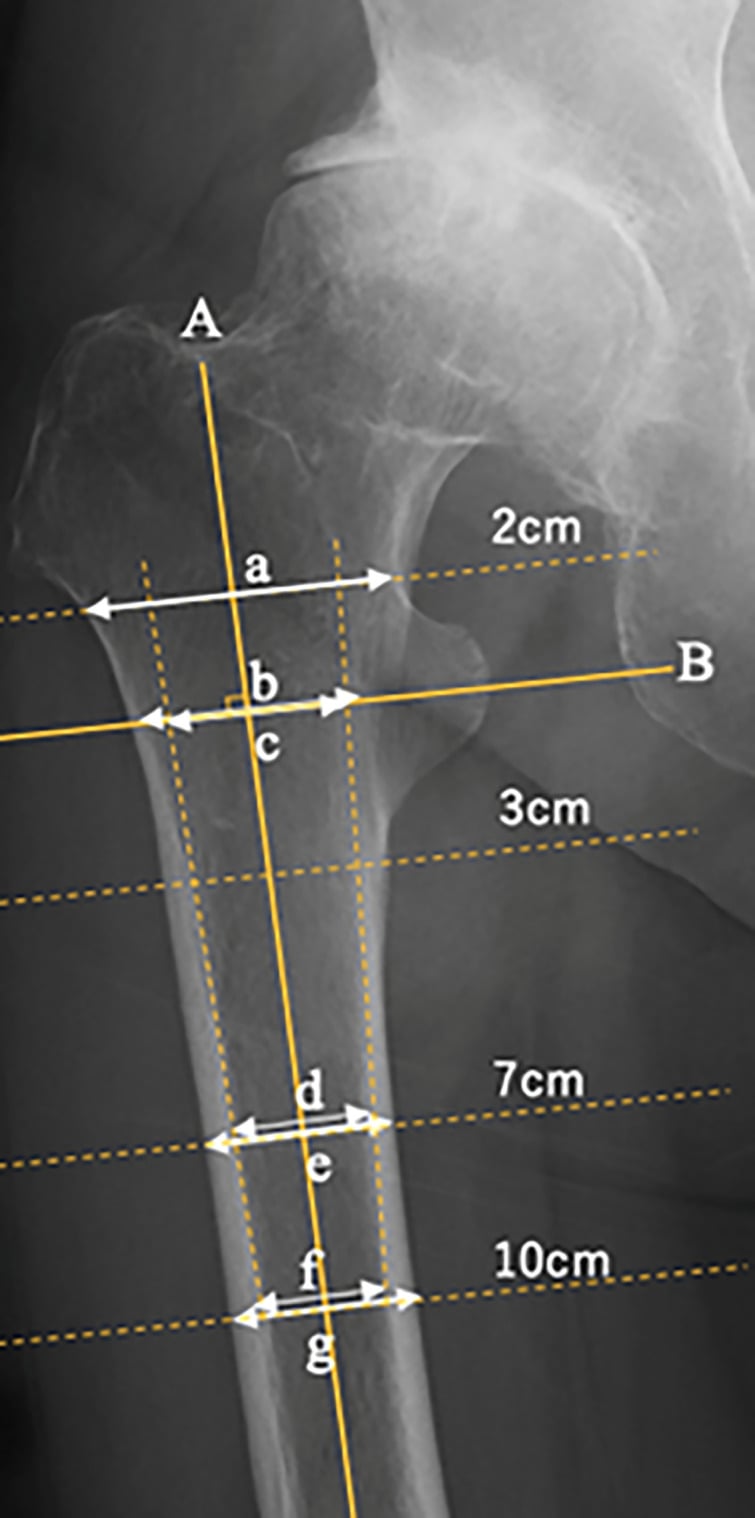
- AP pelvis (mandatory) - Dorr type, canal diameter at the isthmus, cortical thickness, dysplasia, protrusio or bone defects, and leg length discrepancy
- Lateral hip - femoral bow, anterior and posterior cortical thickness, and the stem size and design
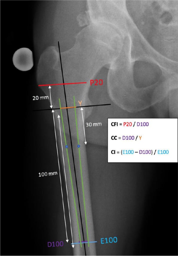
Templating. Accurate templating is essential for component selection and surgical planning. Digital software on both views determines component sizes for both the cemented and the cementless options and plans restoration of offset and leg length. It also shows whether the anatomy suits cement or press-fit:
- Narrow canal (canal-to-cortex ratio less than 0.5) - Dorr A, cementless ideal
- Wide canal (canal-to-cortex ratio over 0.75) - Dorr C, cemented preferred
- Intermediate canal - either method appropriate
Advanced imaging. Used selectively, on clinical indication:
- CT (selected cases) - severe dysplasia needing reconstruction planning, post-traumatic arthritis with deformity, bone stock assessment when radiographs are inadequate, and 3D reconstruction for complex anatomy; not routine for a standard primary THA
- MRI (rarely indicated) - AVN extent when resurfacing is being weighed against THA, or to rule out infection if inflammatory markers are raised; not for routine fixation planning
- DEXA - if osteoporosis is suspected (elderly, thin, corticosteroid use); it can measure regional BMD of the proximal femur
Blood tests. Routine pre-operative tests are an FBC (baseline haemoglobin for blood loss), UEC (renal function, for cement toxicity risk), coagulation studies if the patient is anticoagulated, and a group and hold. In special situations, check vitamin D (optimise if deficient), PTH if hyperparathyroidism is suspected, and rheumatoid factor or anti-CCP in inflammatory arthritis.
If infection is suspected. ESR and CRP should both be normal for a primary THA; if they are elevated, consider aspiration to rule out infection. Do not perform THA with active infection.
Optimisation before fixation is chosen. Correct anaemia (Hb greater than 120 for elective THA), treat osteoporosis with vitamin D and bisphosphonates if time permits, optimise cardiac and respiratory comorbidities, and cease anticoagulation per protocol.
Management - Surgical Techniques
Cemented Fixation
The material. PMMA is mixed with a liquid monomer and polymerises exothermically, and can reach 80-110°C. Working time is 5-8 minutes and setting time 10-12 minutes. It gives immediate mechanical stability.
Third-generation technique. Poor technique causes early failure, and modern third-generation technique achieves excellent outcomes. The steps, in order:
- Pulsatile lavage - clean all blood and debris from the cancellous bone
- Bone drying - hydrogen peroxide-soaked packs, dry sponges
- Distal cement restrictor - creates a closed pressurisation system
- Vacuum mixing - reduces cement porosity and improves strength; wait for the dough stage
- Cement gun delivery - retrograde filling from distal to proximal
- Pressurisation - achieve 3-5mm cement penetration into cancellous bone
- Stem insertion - a single, deliberate insertion without repositioning, then hold steady
- Maintain pressure - hold until full polymerisation, then check the mantle on fluoroscopy
A uniform 2-3mm cement mantle in all Gruen zones is essential. Mantle less than 2mm risks fracture. Direct bone-implant contact (lack of mantle) creates stress concentration and early loosening. The cement restrictor prevents distal cement escape.
Grading the mantle: the Barrack classification. Gruen zones say where to look; Barrack says how good it is, and it is the language used on postoperative radiographs and in every audit of cementing technique.
- Grade A - "white-out": the cement-bone interface cannot be distinguished at all, with complete filling of the canal
- Grade B - a slight radiolucency at the cement-bone interface, less than 50% of it
- Grade C - radiolucency over 50% or more of the interface, or a mantle defect
- Grade D - gross defect, a void, or the stem tip touching cortex with no cement between
Why the grade is worth recording. A and B are acceptable; C and D predict loosening. The grade is a property of the technique, not of the patient, so a run of C mantles is feedback about lavage, pressurisation and restrictor use.
Two stem philosophies. "Cemented stem" is not one thing, and a viva will expect you to know both designs. A composite-beam stem ("shape-closed", bonded) is designed to bond rigidly to the cement and behave as a single beam with the mantle. It has a roughened or matte (or pre-coated) surface, often with a collar, and it must not debond: if it does, micromotion against the cement generates debris and accelerates loosening. The original Charnley flat-back and matte-finish stems are examples.
Taper-slip. A taper-slip stem ("force-closed", polished tapered) is highly polished, double- or triple-tapered and collarless, and is designed to subside a small, controlled amount within the cement mantle. As the taper engages, axial load is converted into radial compressive hoop stress on the cement and bone, and fixation is maintained through controlled cement creep. The polished surface is essential: it allows that controlled slip without generating wear debris. The Exeter and the C-stem are examples, both with excellent long-term registry survivorship.
Mismatching surface finish to design is catastrophic. A polished stem in a composite-beam (bonded) design produces debris and early loosening; a roughened stem used as a taper-slip cannot subside and abrades the cement. Polished taper-slip designs (Exeter) have outstanding long-term survivorship.
The trade-off. Cement gives immediate stability, works in poor bone and has a proven track record. Against that, revision is more difficult because the cement has to be removed, and there is a risk of embolism and of cement hypotension.
Cementless Fixation
The principle. Initial stability comes from a press-fit, with the femoral stem 1-2mm larger than the reamed canal. Bone then grows into the porous coating. Press-fit stability is mandatory: it requires good bone quality, precise reaming and an appropriately sized implant, and cementless fixation is not suitable for poor bone stock.
Porous coatings. Pore size and depth of ingrowth vary with the surface:
- Titanium plasma spray (50-200 microns) - surface ongrowth
- Sintered beads (100-400 microns) - shallow ingrowth
- Trabecular metal (400-600 microns) - deep ingrowth, highest porosity
- Hydroxyapatite - bioactive, enhances early fixation
Optimal pore size for bone ingrowth is 50-150 microns. Smaller pores allow fibrous tissue only. Larger pores (trabecular metal 400-600 microns) allow deeper ingrowth and better fixation. However, initial stability is still the most critical factor - without it, no amount of porous coating will achieve osseointegration.
The cup. A hemispherical cup is press-fitted into the reamed acetabulum:
- Ream sequentially to bleeding subchondral bone
- Underream by 1-2mm relative to the cup diameter, or ream line-to-line - the scratch-fit technique
- Impact the cup to achieve a line-to-line fit at the peripheral rim, at the equator
- Add 2-3 supplemental screws if stability is questionable
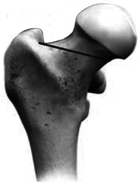
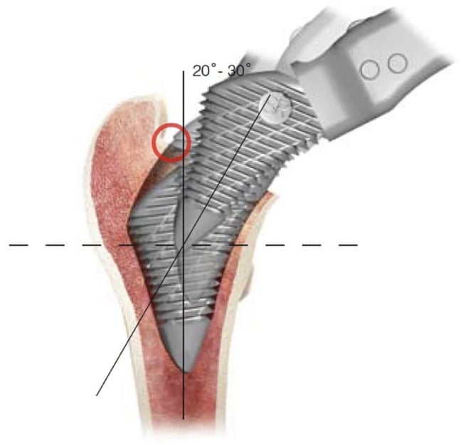
The stem. The neck cut is made perpendicular to the medullary axis, from the subcapital junction laterally into the trochanteric flare, which preserves the calcar collar needed for proximal load transfer. Fixation is either proximal, with tapered stems, or diaphyseal, with cylindrical stems. Broach sequentially to size; the last broach should feel stable in the canal, and a trial reduction assesses stability and leg length. The final implant should resist rotational stress. Before accepting the press-fit, check:
- The broach is stable to manual stress
- There is no pistoning with axial load
- There is cortical contact in the metaphysis (proximally coated stems)
- There is canal fill in the diaphysis (cylindrical stems)
The trade-off. Besides easier revision, cementless fixation leaves no cement debris and gives biological fixation. Against that, it requires good bone and weeks for ingrowth, carries a risk of thigh pain from the modulus mismatch with the femoral stem, and is more expensive. It achieves excellent outcomes when initial press-fit stability is achieved.
Hybrid Fixation
The construct. A hybrid THA is a cementless acetabular component with a cemented femoral component. It combines the advantages of both methods: the cemented stem gives immediate stability in an osteoporotic femur, and the cementless cup offers easier acetabular revision and excellent long-term fixation. Each component is optimised independently on its local bone quality.
Who gets one. It is a frequent choice internationally for patients aged 65-75, and particularly good in patients with good acetabular bone but an osteoporotic femur. A Dorr type C femur (wide canal, thin cortices) benefits from the cemented stem. The cup is prepared with standard cementless technique and supplemental screws; the stem uses third-generation cementing, which is particularly important in a Dorr C femur.
Hybrid THA is very common for this age group, particularly for patients aged 65-75. The AOANJRR data shows this combination performs excellently. Be prepared to discuss hybrid as a deliberate strategy, not a compromise. It's the most common fixation method in many centres.
Reverse hybrid. A cemented acetabular component with a cementless femoral component, used when the acetabulum has poor bone stock but the femur is good. It is less common, and less popular because the acetabulum is difficult to cement.
Complications by Fixation Method
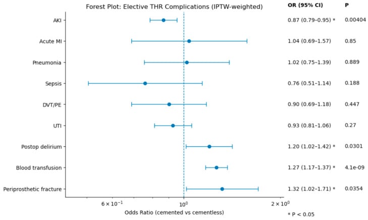
The profile differs by method. Cement brings the embolic and haemodynamic risks and a harder revision; cementless fixation brings thigh pain and fracture.
- Fixation Type
- Cemented
- Incidence
- 1-2% at 10 years
- Prevention/Management
- Modern cement technique, avoid thin mantle
- Fixation Type
- Cementless
- Incidence
- 1-2% at 10 years
- Prevention/Management
- Adequate press-fit, good bone quality
- Fixation Type
- Cementless stem
- Incidence
- 5-20% (usually mild)
- Prevention/Management
- Modulus mismatch, choose appropriate stem design
- Fixation Type
- Cemented
- Incidence
- Rare but serious
- Prevention/Management
- Pulsatile lavage, venting hole, monitor vitals
- Fixation Type
- Cemented
- Incidence
- Under 1%
- Prevention/Management
- Adequate fluid resuscitation, avoid hypovolaemia
- Fixation Type
- Cementless
- Incidence
- 1-5%
- Prevention/Management
- Careful reaming, broaching, avoid over-sizing
- Fixation Type
- Cementless
- Incidence
- 2-3% at 10 years
- Prevention/Management
- Avoid stress shielding, ensure proximal fit
- Fixation Type
- Cemented
- Incidence
- Variable
- Prevention/Management
- Cement removal tools, risk of perforation
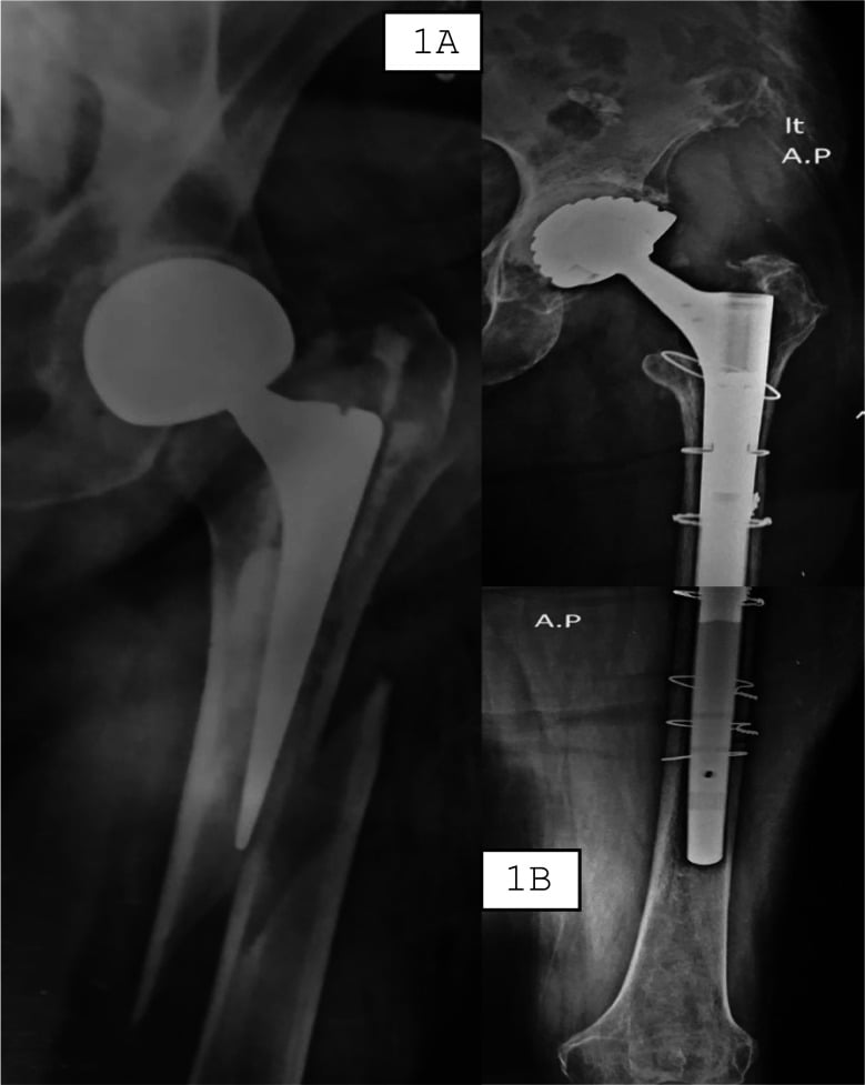
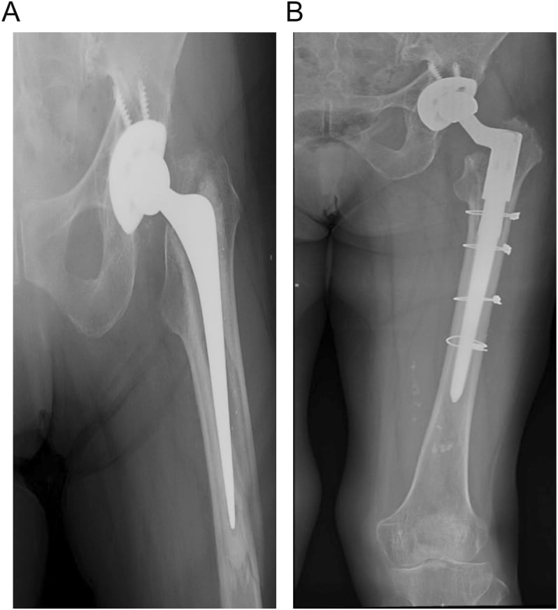
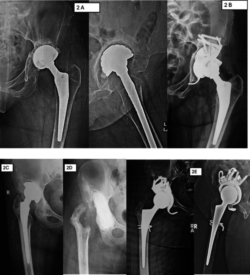
The Painful THA
Exclude infection first. A loose or failing fixation must be distinguished from the other causes of a painful hip replacement, because the management differs completely. Aseptic loosening is a diagnosis of exclusion, and periprosthetic joint infection must always be ruled out first.
- Typical clue
- Start-up pain, progressive over years, thigh/groin pain
- Key discriminating test
- Progressive radiolucent lines, subsidence; normal CRP/ESR
- Typical clue
- Rest pain, early or persistent pain, warmth, sinus
- Key discriminating test
- Raised CRP/ESR, aspiration with synovial WCC/alpha-defensin, cultures
- Typical clue
- Acute pain after a fall or low-energy trauma
- Key discriminating test
- Radiographs (Vancouver classification); assess stem stability
- Typical clue
- Pain with specific positions, sensation of giving way
- Key discriminating test
- History plus radiographs of component position (version, inclination)
- Typical clue
- Activity-related thigh pain with a well-fixed stem
- Key discriminating test
- Stable implant on serial films, proximal femoral atrophy
- Typical clue
- Pain, effusion, especially with metal-on-metal or modular junctions
- Key discriminating test
- Metal ions (Co/Cr), MARS MRI for pseudotumour
- Typical clue
- Pain not matching implant, radicular or claudicant features
- Key discriminating test
- Spine and vascular assessment; THA is a diagnosis of exclusion
Bone Cement Implantation Syndrome
Name the syndrome. The complications table lists "cement hypotension" and "fat embolism", but a viva will expect the defined entity, bone cement implantation syndrome (BCIS). It can be fatal, especially when cementing the elderly fracture patient this topic recommends, and it is commonest in the elderly cemented hip-fracture patient with a long stem.
Mechanism. Pressurised cementing and stem insertion force medullary contents - marrow fat, cement monomer, air and thrombogenic debris - into the venous circulation. The result is pulmonary embolisation, with a sharp rise in pulmonary vascular resistance and right heart strain, plus a possible monomer-mediated vasodilatory or anaphylactoid effect.
The picture. At the moment of cementation, prosthesis insertion or reduction, there is sudden hypoxia, hypotension, arrhythmia, loss of consciousness and cardiac arrest. Severity is graded by Donaldson:
- Grade 1 - moderate hypoxia (SpO2 under 94%) or hypotension (SBP fall over 20%)
- Grade 2 - severe hypoxia (SpO2 under 88%), severe hypotension (SBP fall over 40%) or unexpected loss of consciousness
- Grade 3 - cardiovascular collapse requiring CPR
Risk factors. The recognised risk factors are elderly age, ASA III/IV, pre-existing cardiopulmonary disease, osteoporosis, hip fracture (versus elective OA), pulmonary hypertension and long-stem cemented femoral components.
Prevention. Six measures are described:
- Explicit surgeon-anaesthetist communication before cementing
- Euvolaemia - avoid hypovolaemia
- Thorough pulsatile medullary lavage to clear debris
- A suction catheter or femoral venting hole to relieve intramedullary pressure
- Retrograde cementing with controlled, not excessive, pressurisation in high-risk patients
- Considering cementless fixation in the very highest-risk patient
Postoperative Care and Rehabilitation
After a cemented THA. Cement provides instant stability, so the patient is fully weight-bearing immediately, without concerns about osseointegration. Mobilise the same or next day with physiotherapy, with hip precautions based on the approach (posterior versus lateral) and DVT prophylaxis per protocol. Over weeks 1-6 the patient progresses to independent mobilisation and weans from walking aids as tolerated, with full weight-bearing encouraged and no restrictions based on the fixation method; an X-ray at 6 weeks confirms position.
After a cementless THA. Practice varies: weight-bearing as tolerated, or touch weight-bearing for 6 weeks by surgeon preference, although many surgeons now allow immediate full weight-bearing. Modern teaching favours early weight-bearing even with cementless fixation, as the press-fit is strong enough to tolerate the loads. Hip precautions depend on the approach.
The cementless timeline. Bone ingrowth is occurring through the first six weeks, so weight-bearing progresses if it was initially restricted, aiming for full weight-bearing by 6 weeks, with an X-ray at 6 weeks to assess component position. Between 6 and 12 weeks full weight-bearing is achieved and the biological fixation matures, with progressive strengthening and a return to normal activities.
Long-term surveillance. After a cemented THA, follow up annually with an X-ray for the first 5 years, then biennially if stable, watching for late loosening. After a cementless THA, X-ray annually for the first 2-3 years, watching for stress shielding and osteolysis, then follow up biennially if stable.
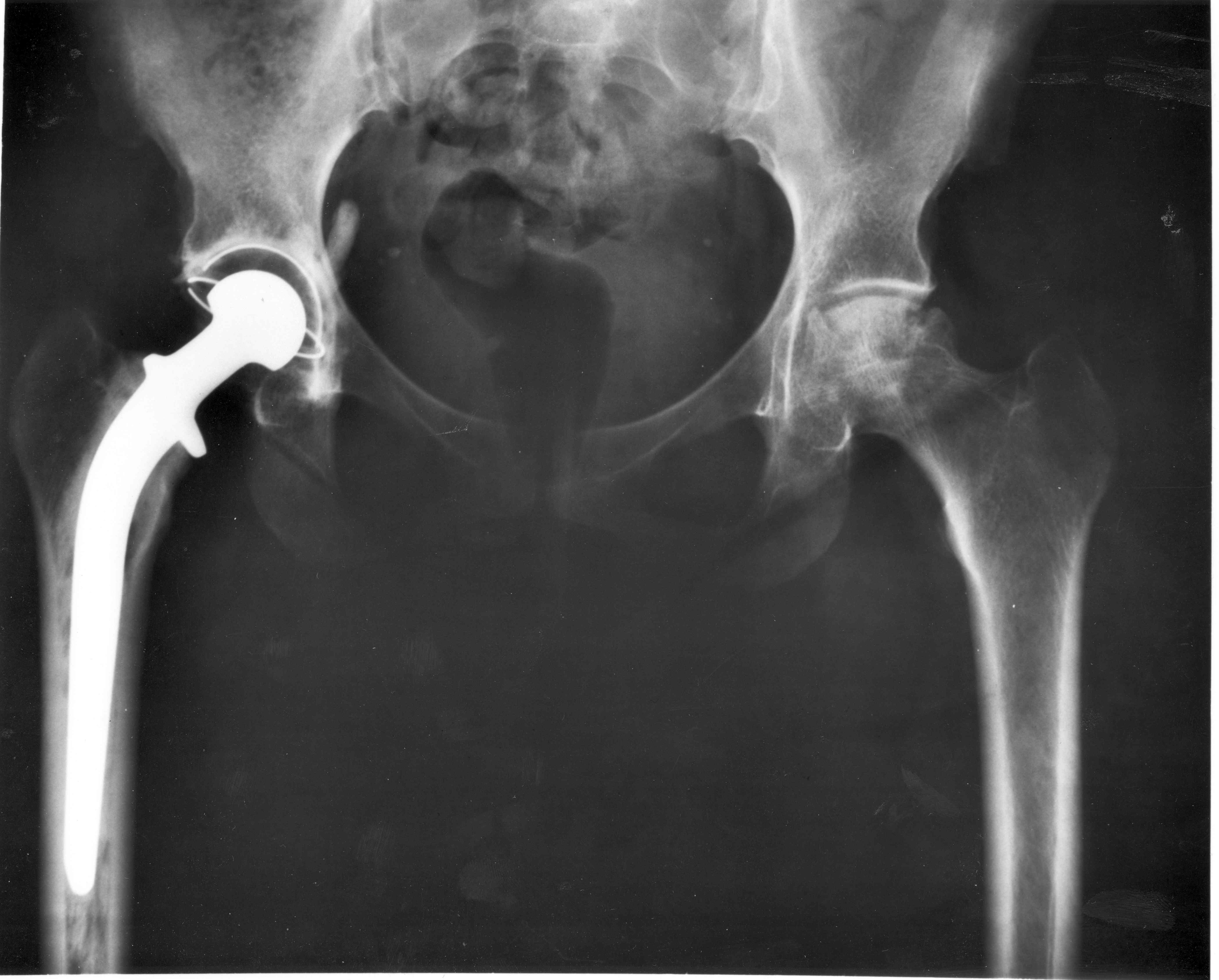
Radiographic follow-up. Take an AP pelvis (both hips for comparison) and a lateral of the operated hip, and compare them with the immediate post-operative films. Assess:
- Component position (inclination, version, offset)
- Radiolucent lines (Gruen zones for the stem, DeLee-Charnley zones for the cup)
- Osteolysis (focal bone loss)
- Subsidence (component migration)
- Heterotopic ossification (Brooker classification)
- Stress shielding (proximal femoral atrophy)
Signs of loosening. Early detection of loosening allows revision before major bone loss occurs. Look for:
- Progressive radiolucent lines wider than 2mm
- Component migration (subsidence, change in position)
- Cement mantle fracture
- Pedestal formation distal to a cemented stem
- Bone resorption around the components
AOANJRR Registry Data and Outcomes
The registry. The Australian Orthopaedic Association National Joint Replacement Registry (AOANJRR) is the world's largest national joint registry and tracks over 500,000 hip replacements. Its annual report is essential reading for exam preparation and provides evidence-based fixation recommendations; know its survival curves for cemented versus cementless fixation in different age groups.
Overall survival. Across all fixation types:
- 1 year - 98.5%
- 5 years - 97.2%
- 10 years - 95.5%
- 15 years - 94-96% (varies by fixation)
Survival by fixation. At approximately 15 years, cemented, cementless and hybrid constructs all show high survivorship, broadly in the mid-90% range, and the hybrid (cementless cup, cemented stem) is among the best-performing constructs in registry reports. It is also very common in many high-income settings. The shift towards cementless fixation has been driven by operative time and perceived simplicity rather than by superior registry survivorship in older patients. The reverse hybrid is less common and used in selected cases. Exact percentages differ between registries and between annual reports, so quote the current annual report of the relevant registry rather than a fixed number.
For exam preparation, know the AOANJRR data. Be able to state that modern techniques achieve 94-96% survival at 15 years for all fixation methods when appropriately selected. The registry supports age-based and bone quality-based selection, not dogmatic fixation choice.
Outcomes by age. The registry pattern supports stratified selection:
- Under 55 - cementless superior (easier future revision)
- 55-65 - all methods have similar outcomes
- 65-75 - hybrid gives excellent results
- Over 75 - cemented gives excellent results
Why hips are revised. In order of frequency, with the fixation-specific pattern where the registry shows one:
- Aseptic loosening - the most common reason overall
- Dislocation or instability - higher in cementless (head size effect)
- Infection - similar across all fixation types
- Periprosthetic fracture - higher in cementless (press-fit technique)
- Bearing surface wear
Cemented outcomes. The long-term track record is excellent, with Charnley data beyond 30 years, and the dislocation rate is lower (smaller head sizes historically). Cement disease is rare with modern technique and highly crosslinked polyethylene (XLPE).
Factors affecting survival. The registry identifies more than fixation:
- Surgical approach (posterior versus lateral)
- Surgeon volume (high volume, better outcomes)
- Head size (36mm optimal for dislocation versus wear)
- Bearing surface (highly crosslinked polyethylene)
- Component positioning (cup inclination, version)
Guidelines, Registries & Global Practice
Global epidemiology. Total hip arthroplasty is one of the most effective and most frequently performed elective operations worldwide, with primary osteoarthritis the leading indication across all high-income registries. Lifetime revision risk is strongly age-dependent: in a competing-risk analysis of 214,638 primary THAs from the Dutch Arthroplasty Register, the 10-year cumulative revision risk ranged from roughly 1.6% to 13%, with the highest risk in the youngest patients regardless of fixation type (Gademan et al., Acta Orthop 2021, DOI).
Registry evidence on fixation. The major national joint registries are the strongest real-world evidence base for implant survival:
- Region
- Australia / NZ
- What it shows on fixation
- All fixation types achieve high 10-15 year survivorship; cumulative revision risk higher in younger patients
- Region
- England, Wales, NI
- What it shows on fixation
- Cemented and hybrid constructs have low revision rates in older patients; very large dataset on stem and bearing comparisons
- Region
- USA
- What it shows on fixation
- Cementless fixation predominates in US practice; tracks bearing surface and head-size trends
- Region
- Sweden
- What it shows on fixation
- Historic stronghold of cemented fixation with excellent long-term survivorship data
- Region
- Global
- What it shows on fixation
- Cemented fixation carries lower all-cause revision risk than uncemented in patients over 75 across almost all registers (the 'uncemented paradox')
The multi-registry synthesis by Bunyoz et al. found that, across Australia, Denmark, England-Wales, Finland, the Netherlands, New Zealand, Norway, Sweden and Switzerland, cemented fixation was associated with a lower all-cause revision risk than uncemented fixation in patients older than 75 years, despite continued high uncemented use in many countries (Clin Orthop Relat Res 2020, DOI).
Guidance side by side. Major bodies broadly agree that fixation should be matched to age and bone quality rather than chosen dogmatically:
- Position
- Use implants with an Orthopaedic Data Evaluation Panel (ODEP) survivorship benchmark; cement strongly favoured for fragility hip-fracture arthroplasty
- Practical implication
- Evidence-driven implant selection; cement for elderly fracture patients
- Position
- Supports both cemented and cementless fixation; cementless predominates in US practice, with patient-specific selection
- Practical implication
- Either method acceptable with appropriate patient selection
- Position
- Cemented fixation preferred for arthroplasty after displaced intracapsular fracture in the elderly
- Practical implication
- Cement in fragility-fracture arthroplasty
- Position
- Cemented or hybrid femoral fixation in patients over 75; cementless reasonable in younger patients with good bone
- Practical implication
- Age- and bone-quality-stratified choice
The recommendation to cement in elderly fracture patients is supported by Level I evidence: an RCT of 400 patients (mean age 85) found lower 3-year mortality and better early mobility with cemented hemiarthroplasty and no increase in complications (Parker et al., Bone Joint J 2023, DOI).
Global practice variation. Fixation choice varies markedly by region rather than by evidence alone. Cementless fixation predominates in the USA and in several Scandinavian countries (e.g. Denmark, Norway), whereas the UK and Sweden retain higher cemented use; hybrid constructs are common in the UK and Australia for intermediate-age patients. In limited-resource settings, cost and cement availability also influence the choice. The persistence of high cementless use in older patients despite registry data favouring cement is the most clinically important practice-evidence gap in this topic - and it is worth carrying the size of it, because the numbers are more striking than the sentence. Across the registers surveyed to 2017, uncemented use in primary THA ranged from 24% in Sweden to 71% in Denmark. In patients over 75, cemented fixation carried the lower revision risk in every register examined bar one subgroup - the oldest Finnish men, where the two were equal. Yet uncemented use in that same over-75 group was stable in most countries and still rising in Denmark and Australia; Finland was the only register moving the other way, from 43% down to 24%. A practice-evidence gap that widens while the evidence accumulates is not inertia, and the authors' reading is that femoral stem fixation is probably the dominant revision risk factor in older patients - which is also why the hybrid construct, cementing only the stem, is the pragmatic answer.
Whatever board you sit, defend fixation choice with registry evidence and the age/bone-quality principle, not country-specific habit. Key points: lifetime revision risk is highest in the young (favouring revisable cementless constructs); cemented or hybrid femoral fixation lowers revision and mortality risk in the elderly; and modern highly cross-linked polyethylene reduces the wear-driven osteolysis that limits all fixation types.
MCQ Practice Points
Q: What is the most appropriate fixation method for an 82-year-old woman with osteoporosis (T-score -2.8)? A: Cemented both components. Poor bone quality cannot achieve press-fit. Registries show high survivorship (mid-90% range) and lower revision risk than uncemented over age 75.
Q: What is the most critical factor for cementless fixation success? A: Initial press-fit stability (micromotion less than 150 microns). Without it, fibrous interface forms instead of osseointegration.
Q: Which fixation method has the highest 15-year survival per AOANJRR? A: Hybrid constructs are typically among the best performers, with cementless and cemented also achieving high survivorship (all broadly mid-90% range at 15 years). All are excellent when appropriately selected; quote the current annual report for exact figures.
Q: What is the ideal cement mantle thickness for femoral stem fixation? A: 2-3mm uniform mantle. Less than 2mm risks cement fracture and early loosening. Direct bone-implant contact without cement creates stress concentration.
Q: What is the optimal pore size for bone ingrowth in cementless components? A: 50-150 microns for traditional porous coating. Smaller pores allow only fibrous tissue. Trabecular metal (400-600 microns) allows deeper ingrowth.
Q: Why is hybrid THA (cementless cup, cemented stem) a frequent choice for intermediate age? A: Combines excellent cementless acetabular fixation with reliable cemented femoral fixation in older patients with poor bone quality. AOANJRR shows excellent outcomes.
Clinical Decision Scenarios
Practise clinical reasoning and management decisions out loud
“An 82-year-old woman with severe osteoarthritis of the right hip requires total hip arthroplasty. She has osteoporosis with T-score of -2.8. She is independently mobile with a stick. What fixation method would you use and why?”
“A 45-year-old man who is a recreational runner requires total hip arthroplasty for post-traumatic arthritis following acetabular fracture 10 years ago. He has good bone stock. What fixation would you recommend and why?”
“A 68-year-old woman requires total hip arthroplasty. She has moderate activity level. Intraoperatively, you find the acetabulum has good bone stock, but the femur is Dorr Type C (wide canal, thin cortices, osteoporotic). What fixation would you use and why?”
CEMENTED FIXATION
- PMMA bone cement - immediate mechanical stability
- Indications: Age over 75, osteoporosis, rheumatoid, poor bone quality
- Third-generation technique: lavage, restrictor, vacuum mix, gun, pressurize
- Goal: 2-3mm uniform cement mantle in all Gruen zones
- Advantages: immediate stability, proven track record, works in poor bone
- Disadvantages: cement removal difficult in revision, embolism risk
- Registries: high survivorship (mid-90% range); lower revision than uncemented over 75
CEMENTLESS FIXATION
- Biological ingrowth - press-fit initial stability, osseointegration 6-12 weeks
- Indications: Age under 65, good bone quality, active patients
- Porous coating: 50-150 micron optimal for ingrowth
- Technique: underream 1-2mm acetabulum, line-to-line femur fit
- Advantages: easier revision, no cement debris, biological fixation
- Disadvantages: requires good bone, 6-12 weeks for fixation, thigh pain
- Registries: high survivorship (mid-90% range); lifetime revision risk highest in young
HYBRID FIXATION
- Cementless cup + cemented stem (most common)
- Indications: Age 65-75, mixed bone quality, Dorr Type C femur
- Combines advantages: biological cup + immediate stem stability
- A frequent choice for intermediate-age patients
- Registries: among the best-performing constructs at 15 years
- Not a compromise - deliberate strategy for specific scenarios
PATIENT SELECTION
- Under 50: cementless both components
- 50-65: cementless or hybrid
- 65-75: hybrid a frequent choice
- Over 75: cemented both components
- Dorr Type A (thick cortices): cementless ideal
- Dorr Type C (thin cortices): cemented preferred
- Bone quality trumps age in decision-making
AOANJRR KEY DATA
- All methods achieve 94-96% survival at 15 years
- Hybrid fixation: among the best-performing constructs
- Age-specific outcomes support stratified selection
- Registry shows hybrid very common for this age group
- Know annual report key findings for exam
EXAM TRAPS
- Don't choose cementless in osteoporotic bone
- Don't choose cemented in young active patients
- Hybrid is not a compromise - it's often optimal
- Know Dorr classification and implications
- Quote AOANJRR data to support decisions
- Explain specific technique for chosen fixation
Evidence Base
Caton & Prudhon - Over 25 years survival after Charnley THA
- Two long-term series of original cemented Charnley low-friction arthroplasty (22.2mm head, cemented all-polyethylene socket) confirmed excellent function beyond 25 years, with reported survivorship around 85% at 25 years. The authors emphasise that polyethylene is the weak point: mean wear was approximately 0.1mm per year and drove the late failures, not the cement itself.
Sochart & Porter - Charnley LFA in young patients
- 226 cemented Charnley arthroplasties in 161 patients of mean age 31.7 years (range 17 to 39), followed for a mean of 19.7 years. For the whole series, 25-year survival was 81 per cent for the femoral component and 68 per cent for the acetabular - the socket is what fails.
- THE DIAGNOSIS-SPECIFIC SPLIT IS THE MOST INSTRUCTIVE PART AND SHOWS A DISSOCIATION: congenital dislocation had the BEST femoral survival at 89 per cent but the WORST acetabular survival at 58 per cent, with 37 per cent aseptic cup loosening and 37 per cent cup revision. Osteoarthrosis had the worst femoral survival (74 per cent) and the highest femoral loosening and endosteal lysis. Rheumatoid arthritis had the best cup survival (79 per cent) but 43 per cent mortality.
- The wear-to-revision chain is measured, not assumed: mean acetabular wear was 0.11 mm/year overall, but 0.19 mm/year in components that were revised against 0.09 mm/year in those that survived, and a higher annual wear rate was significantly associated with cup migration and revision (p less than 0.01 for both).
Bunyoz et al. - The Uncemented Paradox Revisited (multi-registry)
- Synthesis of national hip registry reports (Australia, Denmark, England-Wales, Finland, Netherlands, New Zealand, Norway, Sweden, Switzerland) from 2010-2017. Across almost every register, cemented fixation carried a lower all-cause revision risk than uncemented fixation in patients older than 75 years, yet uncemented use remained high or rising - the 'uncemented paradox'. Femoral stem fixation appeared to be the dominant revision risk factor in older patients.
Gademan et al. - Dutch Arthroplasty Register 10-year revision risk
- Competing-risk analysis of 214,638 primary THAs from the Dutch Arthroplasty Register (LROI). The 10-year cumulative revision risk varied with age and fixation, ranging from approximately 1.6% to 13%, with consistently higher revision risk in younger patients regardless of fixation type.