Virchow's Triad | Risk Stratification | Prophylaxis Protocols | Prevention & Treatment
- Virchow's triad is the pathophysiological foundation: Stasis + Hypercoagulability + Endothelial injury
- Risk stratification is mandatory: Use Caprini score or NICE guidelines to determine prophylaxis intensity
- Get the two ACCP durations and their grades the right way round: a minimum of 10-14 days is the strong recommendation (Grade 1B); extending to up to 35 days is only a weak suggestion (Grade 2B)
- Aspirin is an ACCP Grade 1B option, the same grade as LMWH and the DOACs - the preference for LMWH over the alternatives is only Grade 2C/2B, and in EPCAT II aspirin was non-inferior to rivaroxaban. What aspirin has not been shown to do is replace anticoagulant prophylaxis from day 0
- DVT diagnosis: Compression ultrasound (sensitivity over 95% for proximal DVT)
- PE diagnosis: CTPA is gold standard; V/Q scan if contrast contraindicated
- “Virchow's triad explains why ALL orthopaedic surgery patients are at VTE risk
- “Caprini score 5 or higher = high risk requiring chemical prophylaxis
- “LMWH started 12h preop (European) or 12-24h postop (North American) - know both protocols
- “DOACs (rivaroxaban, apixaban) are non-inferior to LMWH with better compliance
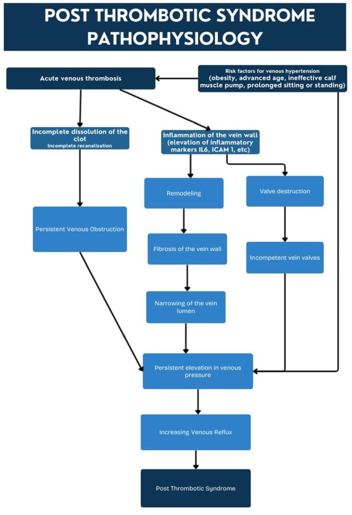
- “Post-thrombotic syndrome affects 20-50% of patients after proximal DVT - in the SOX trial compression stockings did not prevent it
Overview and Epidemiology
Venous thromboembolism (VTE), which encompasses deep vein thrombosis (DVT) and pulmonary embolism (PE), is one of the most significant preventable complications in orthopaedic surgery.
Definitions. A DVT is thrombus in the deep venous system, most commonly in the lower extremity: the iliac, femoral, popliteal and calf veins. A PE is embolisation of that thrombus to the pulmonary arterial circulation, causing ventilation-perfusion mismatch. VTE is the umbrella term for the whole spectrum, from an asymptomatic calf DVT to a fatal massive PE.
The risk without prophylaxis. Untreated, the rates across orthopaedic surgery are:
- DVT Rate
- 40-60%
- Proximal DVT
- 15-25%
- Fatal PE
- 0.5-2%
- DVT Rate
- 40-84%
- Proximal DVT
- 10-20%
- Fatal PE
- 0.5-1.5%
- DVT Rate
- 40-60%
- Proximal DVT
- 15-30%
- Fatal PE
- 2-7%
- DVT Rate
- 5-15%
- Proximal DVT
- 1-2%
- Fatal PE
- Less than 0.1%
- DVT Rate
- 15-40%
- Proximal DVT
- 5-10%
- Fatal PE
- 0.3-0.5%
- DVT Rate
- 40-80%
- Proximal DVT
- 10-20%
- Fatal PE
- 1-2%
What prophylaxis achieves. DVT rates fall to 10-20% with mechanical prophylaxis alone and to 1-5% with mechanical plus chemical prophylaxis. Fatal PE falls to less than 0.5% with adequate prophylaxis, and the number needed to treat is approximately 7 to prevent one symptomatic VTE.
A patient-safety measure. VTE remains a leading cause of preventable hospital death internationally. Many health systems mandate VTE risk assessment for all hospital admissions as a patient-safety indicator, and compliance with prophylaxis protocols is a key hospital performance metric.
Pathophysiology - Virchow's Triad
Rudolf Virchow described his triad in 1856: three factors that predispose to venous thrombosis. All three are present in major orthopaedic surgery, which explains the extremely high VTE risk in this population. Examiners expect you to explain how each component contributes to that risk, and how your prophylaxis strategy addresses each one.
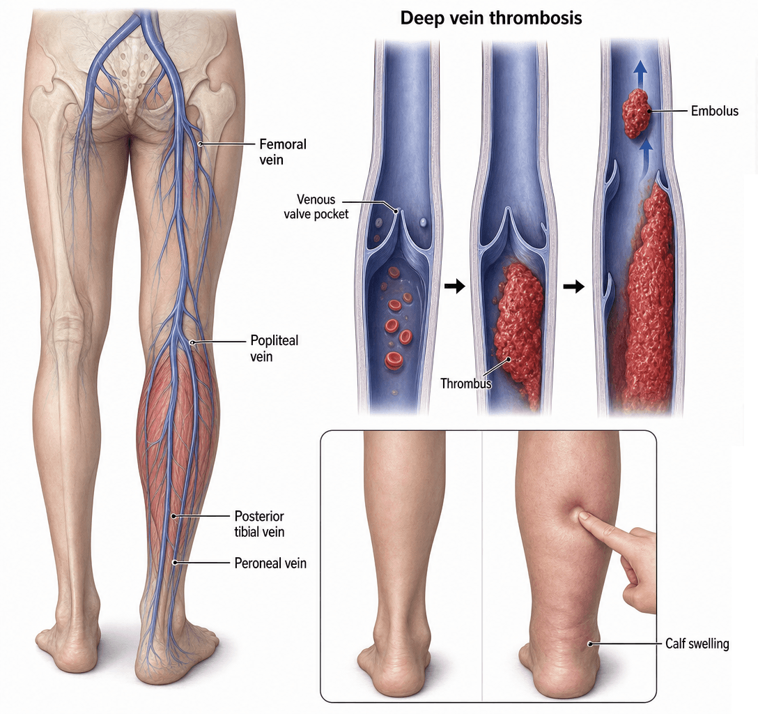
Stasis. Normal venous return depends on the calf muscle pump and competent venous valves. Immobility eliminates the pump, so blood pools and activated clotting factors accumulate locally; their reduced clearance overwhelms the natural anticoagulant systems. In orthopaedic practice stasis comes from:
- Preoperative immobility (hip fracture, multiple trauma)
- Intraoperative positioning (hip flexion, rotation)
- Postoperative bed rest and reduced mobility
- Cast immobilisation
- Lower limb paralysis (stroke, spinal cord injury)
- Venous compression
- Tourniquet application (distal stasis during inflation)
Prevention targets this limb directly: early mobilisation, mechanical prophylaxis (IPC devices, stockings), ankle exercises and calf-pump activation, avoiding prolonged hip flexion, and physiotherapy protocols.
The calf muscle pump generates pressures of 200-300 mmHg during contraction, propelling blood centrally through competent valves. Loss of this pump (immobility) reduces venous velocity by 50% and allows thrombus formation in valve pockets.
Hypercoagulability. Tissue trauma activates the extrinsic coagulation pathway through the release of tissue factor. The surgical stress response increases fibrinogen, factor VIII and platelet reactivity, the systemic inflammatory response creates a pro-thrombotic milieu, and the natural anticoagulants (protein C, protein S, antithrombin III) may be consumed. The orthopaedic triggers:
- Major tissue trauma and fractures
- Surgical trauma and manipulation
- Tourniquet use (reperfusion activates coagulation)
- Cement exothermic reaction and fat embolism
- Blood loss and transfusion (stored blood is procoagulant)
The patient's own coagulation. Inherited and acquired states add to the surgical load.
- Acquired
- Malignancy (especially pelvic, lung, pancreatic)
- Acquired
- Oral contraceptive pill and HRT
- Acquired
- Pregnancy and the postpartum state
- Acquired
- Antiphospholipid syndrome
- Acquired
- Myeloproliferative disorders
- Acquired
- Nephrotic syndrome
Do NOT routinely test for inherited thrombophilia preoperatively. Testing is indicated only if: (1) personal history of unprovoked VTE, (2) strong family history of VTE at young age, (3) VTE in unusual site. Testing does not change acute VTE management but may affect duration of anticoagulation.
Endothelial injury. Intact endothelium is anti-thrombotic: it produces prostacyclin, nitric oxide and thrombomodulin. Damage exposes subendothelial collagen and tissue factor, and platelet adhesion and activation then initiate thrombosis; surgical trauma damages the venous endothelium directly. The causes in orthopaedic practice:
- Direct vessel manipulation during surgery
- Retractor placement against vessels
- Tourniquet compression and ischaemia-reperfusion
- Fracture fragments lacerating vessels
- DVT catheter placement
The risks are specific to the procedure:
- THA - the femoral vein is stretched and compressed during dislocation and leg positioning
- TKA - the popliteal vein is at risk during posterior capsule release
- Hip fracture - injury at the time of fracture, and manipulation during surgery
- Acetabular fracture - the iliac vessels are at risk during the approach and fixation
Bone cement (PMMA) causes endothelial injury through: (1) exothermic reaction (temperatures of 60-80 degrees Celsius), (2) monomer toxicity, (3) fat and marrow embolization. This is why cemented procedures have higher VTE risk than uncemented, and why some advocate for a dose of LMWH before cementing.
The three together. In a major orthopaedic procedure all three limbs act at once, a "perfect storm": preoperative immobility supplies stasis, surgical trauma and tissue factor release supply hypercoagulability, and direct vessel injury and manipulation supply endothelial injury. This is why THA and TKA have the highest VTE risk of any elective surgical procedure, and why prophylaxis is mandatory.
Clinical Presentation
Silent in up to half. Up to 50% of DVTs are asymptomatic, detected only on screening ultrasound, and clinical diagnosis is unreliable, with a sensitivity of 50-60% and a specificity of 50%.
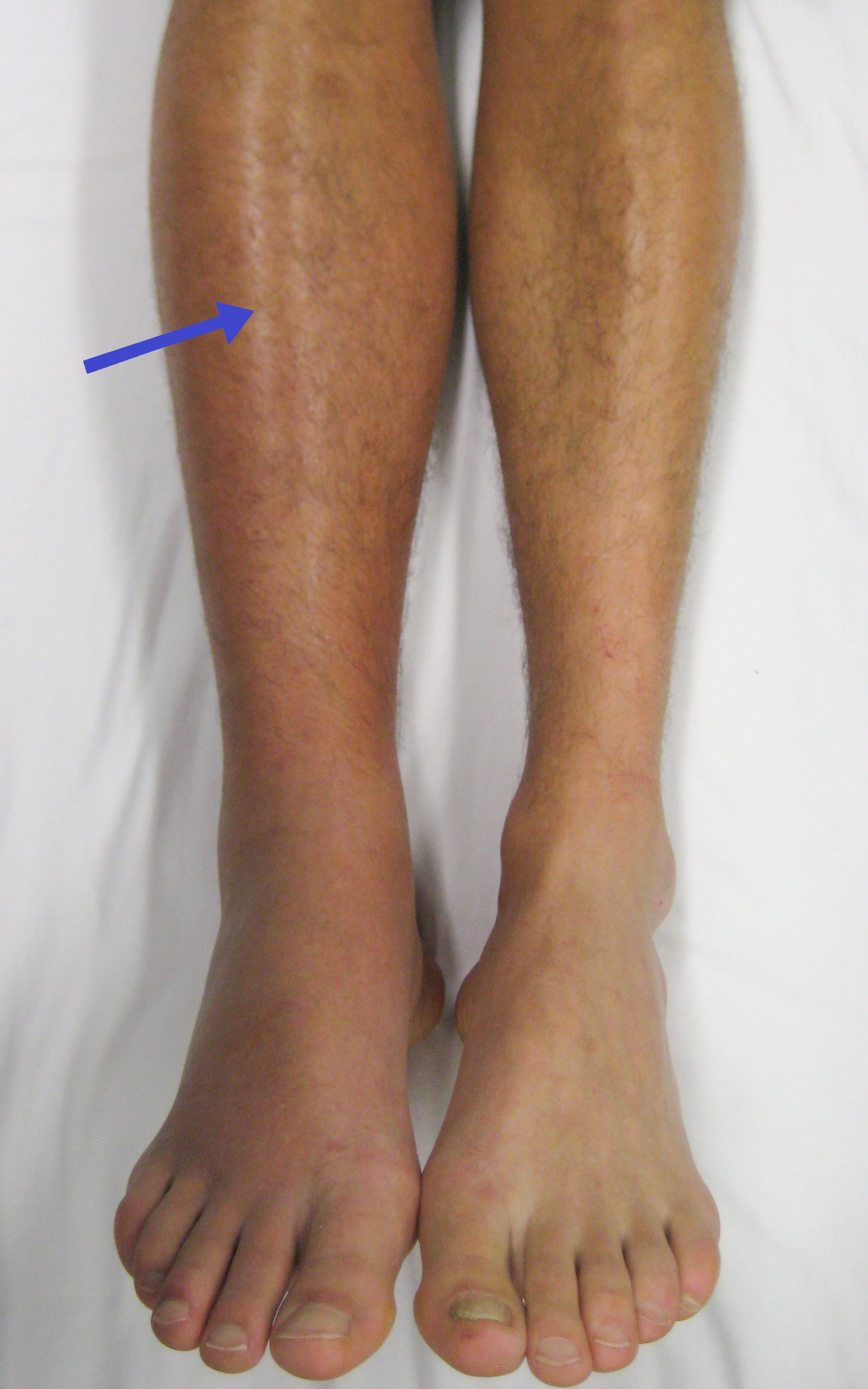
Symptoms. Calf pain is the most common, felt in the posterior calf and often described as cramping or tightness. The leg swells, acutely or insidiously, and feels heavy or aching, and the pain is worse with standing or walking.
Signs on examination:
- Unilateral leg oedema - a difference of more than 3cm between the calves is significant
- Pitting oedema
- Calf tenderness on palpation
- Increased warmth, and erythema that may be subtle
- Superficial venous distension, with visible collateral veins
- Homans' sign (calf pain on forced dorsiflexion) - unreliable, only 50% sensitive
- Points
- +1
- Points
- +1
- Points
- +1
- Points
- +1
- Points
- +1
- Points
- +1
- Points
- +1
- Points
- +1
- Points
- +1
- Points
- -2
Reading the Wells score. The total sets the pretest probability:
- 0 or less - low probability (5% DVT prevalence)
- 1-2 - moderate probability (17% DVT prevalence)
- 3 or more - high probability (53% DVT prevalence)
In postoperative orthopaedic patients, the Wells score has limited utility because multiple risk factors are already present (recent surgery, immobility). Have a LOW threshold for imaging. If clinical suspicion exists, proceed directly to compression ultrasound regardless of Wells score.
The differential of the painful, swollen postoperative leg:
- Distinguishing Features
- Unilateral calf swelling more than 3cm, calf tenderness, warmth; often asymptomatic postop
- Key Investigation
- Compression ultrasound (non-compressible vein)
- Distinguishing Features
- Erythema with well-defined advancing margin, fever, raised inflammatory markers, possible portal of entry
- Key Investigation
- Clinical; ultrasound to exclude coexisting DVT
- Distinguishing Features
- Sudden calf pain, crescent bruise around malleolus, history of knee effusion or arthritis
- Key Investigation
- Ultrasound (fluid tracking from popliteal cyst)
- Distinguishing Features
- Tender, palpable cord along a superficial vein, localised erythema
- Key Investigation
- Ultrasound (superficial vein thrombus, deep system patent)
- Distinguishing Features
- Localised swelling and bruising near wound, recent surgery, possible falling haemoglobin
- Key Investigation
- Ultrasound or CT; assess coagulation
- Distinguishing Features
- Pain out of proportion, pain on passive stretch, tense compartment, paraesthesia
- Key Investigation
- Clinical diagnosis; compartment pressures
- Distinguishing Features
- Bilateral or chronic, non-pitting (lymphoedema) or pitting with skin changes, no acute onset
- Key Investigation
- Clinical; ultrasound if acute change
- Distinguishing Features
- Sudden pain during activity, localised tenderness at musculotendinous junction
- Key Investigation
- Clinical; ultrasound if uncertain
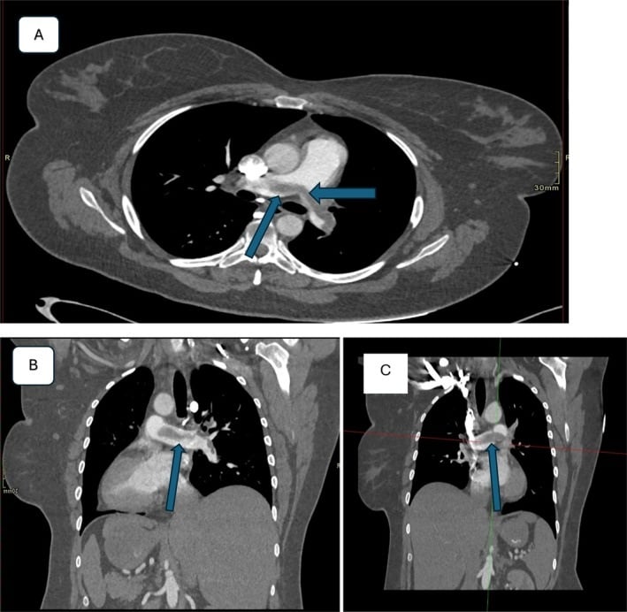
The classic exam trap in the breathless post-trauma or post-arthroplasty patient is to reach straight for PE and forget fat embolism syndrome (FES) - a clinically distinct entity that needs a completely different response (supportive care, not anticoagulation).
When to think FES: classically after long-bone (femur, tibia) or pelvic fractures, intramedullary reaming/nailing, or arthroplasty, with a characteristic latent interval of about 24 to 72 hours after the injury (PE can occur at any time but FES has this delay).
The classic triad (Gurd and Wilson criteria):
- Respiratory - hypoxia and an ARDS-like picture (the most common and earliest feature).
- Neurological - confusion, drowsiness or agitation, often fluctuating and usually reversible.
- Petechial rash - over the axillae, conjunctivae, chest and neck; appears late (in around 60 percent) and is effectively pathognomonic.
- Supportive minor features: tachycardia, pyrexia, retinal changes, fat in urine/sputum, thrombocytopenia, anaemia and a falling haematocrit.
Distinguishing it from PE: FES has the latent interval, petechiae, prominent neurological features and thrombocytopenia, and the CTPA does not show a filling defect (it may show ARDS-pattern change). PE improves with anticoagulation; FES does not - management is supportive (oxygen and ventilatory support, fluid resuscitation, organ support), and the key preventive measure is early fracture stabilisation.
Exam point: postoperative/post-trauma hypoxia is PE until proven otherwise, but consider fat embolism syndrome - suspect it with the 24 to 72 hour latency after long-bone/pelvic fracture or reaming, the respiratory-cerebral-petechial triad and thrombocytopenia; it is a clinical diagnosis managed supportively, not with anticoagulation.
Investigations
D-dimer. Highly sensitive (95%) but poorly specific (50%): it is raised by surgery, trauma, pregnancy, cancer, infection and inflammation. A negative D-dimer reliably excludes DVT only in patients with a low pretest probability.
D-dimer has NO role in ruling out DVT in postoperative orthopaedic patients. It is almost always elevated after surgery and does not add diagnostic value. Proceed directly to imaging if DVT is suspected.
Compression ultrasound is the gold standard. Inability to fully compress the vein with probe pressure is diagnostic, and the scan also shows thrombus echogenicity and venous flow patterns. Sensitivity is 95% for proximal DVT and 70-80% for calf DVT, specificity 98%, and the test is non-invasive, radiation-free and available at the bedside.
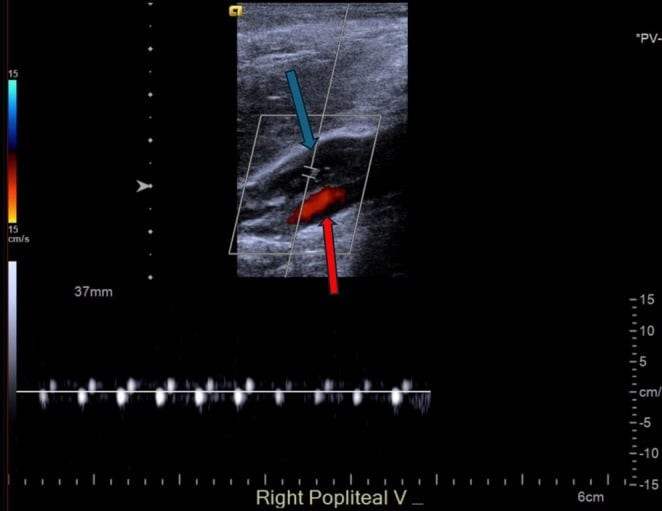
When to image. The threshold is low:
- Any clinical suspicion of DVT postoperatively
- Unexplained leg swelling, pain or warmth
- A diagnosed PE - to look for the DVT source
Other imaging options:
- CT venography - pelvic and IVC extension, or when ultrasound is non-diagnostic
- MR venography - an alternative for the pelvic veins, without radiation
- Contrast venography - the historical gold standard, rarely used now because it is invasive
Putting it together. Pretest probability decides whether D-dimer has any place:
- D-dimer
- If negative: DVT excluded
- Ultrasound
- If D-dimer positive: proceed to US
- Interpretation
- Serial US if negative but high suspicion
- D-dimer
- Not reliable
- Ultrasound
- Proceed directly
- Interpretation
- Consider repeat US in 5-7 days if negative
- D-dimer
- Do not use
- Ultrasound
- Proceed directly
- Interpretation
- Consider venography if negative and high suspicion
- D-dimer
- Do not use
- Ultrasound
- Proceed directly
- Interpretation
- Low threshold for imaging
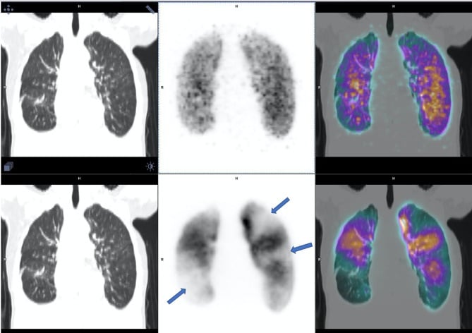
Management
The Caprini score is the most widely used tool for VTE risk stratification in surgical patients.
- 2 Points Each
- Age 61-74
- 3 Points Each
- Age 75 or more
- 5 Points Each
- Hip, pelvis, leg fracture
- 2 Points Each
- Arthroscopic surgery
- 3 Points Each
- Prior VTE
- 5 Points Each
- Stroke (less than 1 month)
- 2 Points Each
- Major surgery over 45 min
- 3 Points Each
- Family history VTE
- 5 Points Each
- Spinal cord injury
- 2 Points Each
- Malignancy
- 3 Points Each
- Positive thrombophilia
- 5 Points Each
- Elective arthroplasty
- 2 Points Each
- Bed rest over 72h
- 3 Points Each
- 5 Points Each
Reading the Caprini score. The total sets the intensity of prophylaxis:
- 0 - very low risk (0.5% VTE): early ambulation only
- 1-2 - low risk (1.5% VTE): mechanical prophylaxis
- 3-4 - moderate risk (3% VTE): mechanical plus chemical prophylaxis
- 5 or more - high risk (6% VTE): mechanical plus chemical plus extended prophylaxis (up to 35 days)
NICE. The UK National Institute for Health and Care Excellence provides the alternative risk assessment. Every orthopaedic inpatient needs a VTE risk assessment, and every patient undergoing THA, TKA or hip fracture surgery is automatically high risk. Bleeding risk must be assessed alongside it, and prophylaxis individualised.
Prophylaxis must balance VTE risk against bleeding risk. Consider bleeding risk factors: active bleeding, severe thrombocytopenia (platelets less than 50), coagulopathy, high-bleeding-risk surgery (spine, intracranial), recent major bleeding. If bleeding risk is high, use mechanical prophylaxis only until bleeding risk diminishes.
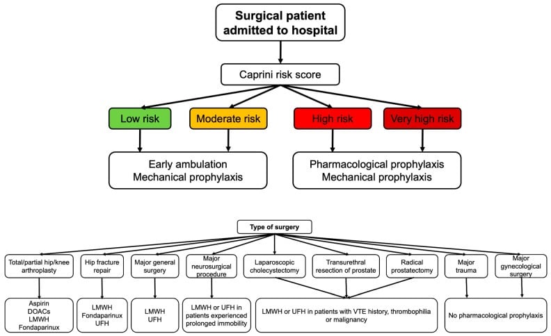
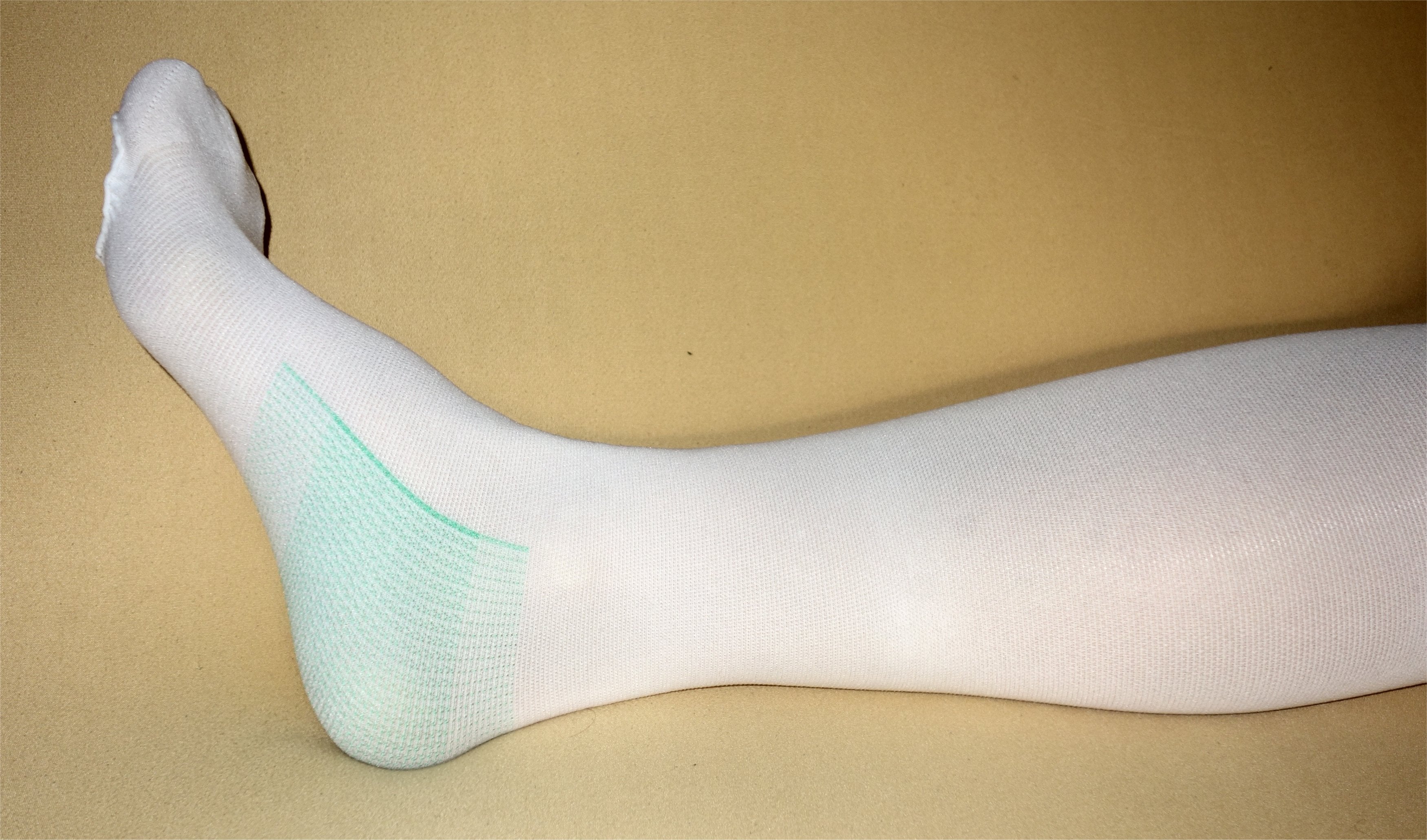
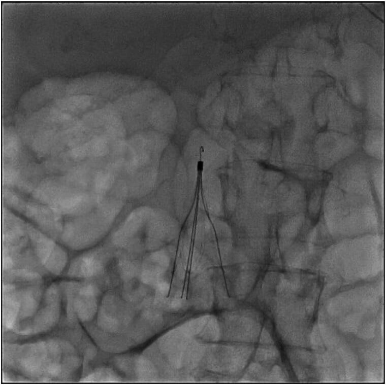
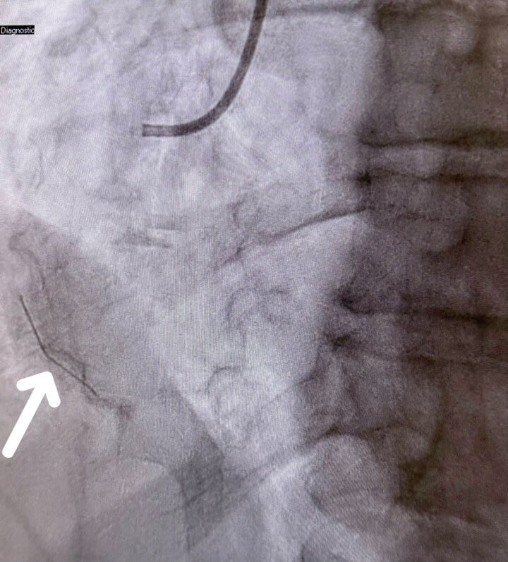
Surgical Management - IVC Filters
Absolute indications for a filter:
- Acute proximal DVT or PE with an absolute contraindication to anticoagulation
- Recurrent PE despite adequate anticoagulation
- Complications of anticoagulation requiring it to stop (major bleeding)
Relative indications, all controversial:
- Prophylactic filter in very high-risk trauma (controversial, not recommended routinely)
- Free-floating iliofemoral thrombus (some advocate, others anticoagulate)
- Massive PE with residual DVT, where further embolisation would be fatal
- Poor cardiopulmonary reserve, where any PE would be catastrophic
Filters are not indicated for:
- Routine prophylaxis in trauma or orthopaedic surgery
- As an alternative to anticoagulation when anticoagulation is feasible
- Calf DVT only
Do NOT place IVC filters for VTE prophylaxis in trauma or orthopaedic patients who can receive anticoagulation. The PREPIC-2 trial showed no benefit of prophylactic filters in addition to anticoagulation. Filters are for patients who CANNOT be anticoagulated.
Complications
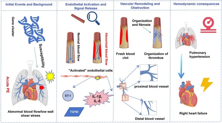
Post-thrombotic syndrome is covered under Clinical Presentation. The other complications:
- Incidence
- 5-10% at 1 year on anticoagulation
- Presentation
- New DVT or PE symptoms
- Management
- Ensure therapeutic anticoagulation, consider extended/indefinite therapy
- Incidence
- 2-4% after PE
- Presentation
- Persistent dyspnea 6+ months post-PE
- Management
- Referral to pulmonary hypertension centre, pulmonary endarterectomy
- Incidence
- Rare
- Presentation
- Massive iliofemoral DVT, cyanotic limb, compartment syndrome risk
- Management
- Urgent surgical thrombectomy or catheter-directed thrombolysis, fasciotomy if compartment syndrome
- Incidence
- 3-5% major bleeding on therapeutic anticoagulation
- Presentation
- GI bleed, intracranial hemorrhage, surgical site bleeding
- Management
- Hold anticoagulation, reversal agents, transfusion, surgical hemostasis if needed
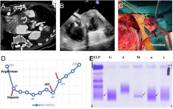
Heparin-induced thrombocytopenia. The incidence is 0.5% with LMWH and 2-3% with UFH. The platelet count drops by more than 50% at 5-14 days, with paradoxical thrombosis.
Heparin-Induced Thrombocytopenia is a prothrombotic emergency despite low platelets. 4Ts score (Thrombocytopenia, Timing, Thrombosis, oTher causes) helps assess probability. If HIT suspected: (1) Stop ALL heparin immediately, (2) Start alternative anticoagulant (argatroban, fondaparinux - NOT warfarin alone), (3) Send HIT antibody testing. HIT can cause limb loss, stroke, PE, and death.
Guidelines, Registries & Global Practice
Global Epidemiology:
VTE is a major global cause of preventable death and disability. A systematic review for the International Society on Thrombosis and Haemostasis (Raskob et al, Arterioscler Thromb Vasc Biol 2014, PMID 25304324) reported an annual VTE incidence of 0.75-2.69 per 1000 population across Western Europe, North America, Australia and Southern Latin America, rising to 2-7 per 1000 in those aged 70 years or more. Incidence is lower in Chinese and Korean populations, but the disease burden remains high because of population ageing. Hospital-associated VTE - including surgery and major lower-limb trauma - is the dominant contributor and the leading cause of disability-adjusted life-years lost in low- and middle-income countries. This global burden underpins the worldwide convergence on systematic risk assessment and prophylaxis.
Side-by-Side Guideline Comparison:
- First-line agent
- LMWH preferred; DOAC, fondaparinux, warfarin or aspirin acceptable
- Aspirin role
- Acceptable alternative (Grade 1B vs no prophylaxis)
- Extended duration (THA/TKA)
- At least 10-14 days, extend to 35 days (Grade 2B)
- Mechanical prophylaxis
- Add IPC during hospital stay (Grade 2C)
- First-line agent
- Pharmacological + mechanical; agent at surgeon discretion
- Aspirin role
- Endorsed as acceptable, supported by EPCAT II
- Extended duration (THA/TKA)
- Recommends prophylaxis but no single duration mandated
- Mechanical prophylaxis
- Recommended in addition to chemical prophylaxis
- First-line agent
- LMWH (or LMWH then aspirin) for THA; aspirin or LMWH or DOAC for TKA
- Aspirin role
- Explicitly offered as an option (NG89)
- Extended duration (THA/TKA)
- THA up to 28-35 days; TKA up to 14 days
- Mechanical prophylaxis
- Offer mechanical (IPC or stockings) on admission
- First-line agent
- LMWH or DOAC
- Aspirin role
- Reserved for lower-risk or extended phase
- Extended duration (THA/TKA)
- Up to 35 days for THA and hip fracture
- Mechanical prophylaxis
- Combine with mechanical methods
- First-line agent
- LMWH or DOAC; aligns with ACCP
- Aspirin role
- Used for lower-risk patients or extended phase
- Extended duration (THA/TKA)
- 35 days for THA, TKA and hip fracture
- Mechanical prophylaxis
- IPC and early mobilisation
The major guidelines now broadly agree on three principles: every major-orthopaedic patient is high-risk and needs combined mechanical plus pharmacological prophylaxis; LMWH and DOACs are the most effective agents; and aspirin is an acceptable option in selected (lower-risk or extended-phase) patients following the EPCAT II evidence. The main practice variation is around aspirin's prominence (greater in North America and the UK) and the precise extended duration.
Registry and System-Level Evidence:
National joint registries (the Australian AOANJRR, the National Joint Registry for England, Wales, Northern Ireland and the Isle of Man, and the American Joint Replacement Registry) capture arthroplasty volumes, implant survival and, increasingly, 90-day readmissions including symptomatic VTE, providing real-world denominators for prophylaxis policy. At the system level, the World Health Organization, the UK NHS (which mandated risk assessment of admitted patients), and the Australian Commission on Safety and Quality in Health Care all classify hospital-acquired VTE as a reportable patient-safety indicator, driving mandatory risk-assessment and prophylaxis bundles.
Practice Variation and Access:
Access to specific agents varies by jurisdiction. In many high-income settings, enoxaparin, rivaroxaban and apixaban are available for arthroplasty prophylaxis; aspirin is widely available without prescription. In lower-income settings, LMWH cost and the lack of monitoring infrastructure for warfarin make low-cost oral options (including aspirin) and mechanical prophylaxis especially important. Across all systems, smoking is an independent, modifiable VTE risk factor: preoperative cessation for four or more weeks reduces perioperative complications.
For the viva, frame VTE prophylaxis as a globally standardised process: mandatory risk assessment, combined mechanical and pharmacological prophylaxis for all major-orthopaedic patients, extended duration (up to 35 days) after hip and knee replacement and hip fracture, with LMWH or a DOAC first-line and aspirin acceptable in selected patients. Hospital-acquired VTE is a reportable patient-safety indicator in most health systems, making prophylaxis a medicolegal as well as clinical obligation.
Exam Viva Scenarios
Practise clinical reasoning and management decisions out loud
“A 68-year-old woman is scheduled for elective total hip arthroplasty for osteoarthritis. She has well-controlled hypertension and type 2 diabetes. What is your VTE prophylaxis strategy?”
“A 72-year-old man is 5 days post total knee arthroplasty. He develops sudden onset dyspnea, pleuritic chest pain, and is tachycardic at 110 bpm. SpO2 is 88% on room air. What is your management?”
“A 58-year-old man with hip fracture also has active GI bleeding from a peptic ulcer. He requires surgery within 24-48 hours. How do you manage VTE prophylaxis?”
Virchow's Triad
- Stasis: Immobility, venous pooling - treat with mechanical prophylaxis, early mobilization
- Hypercoagulability: Trauma, surgery, inherited thrombophilia - treat with chemical prophylaxis
- Endothelial injury: Surgery, fractures, cement - minimize with gentle technique
- All three present in major orthopaedic surgery - explains high VTE risk
Risk Stratification (Caprini)
- Score 0: Very low risk (0.5%) - early ambulation only
- Score 1-2: Low risk (1.5%) - mechanical prophylaxis
- Score 3-4: Moderate risk (3%) - mechanical + chemical prophylaxis
- Score 5+: High risk (6%) - mechanical + chemical + extended prophylaxis (up to 35 days)
- 5-point factors: Hip/pelvis/leg fracture, stroke, spinal cord injury, arthroplasty (prior VTE scores 3)
Chemical Prophylaxis Agents
- Enoxaparin: 40mg SC daily, start 12-24h postop, gold standard LMWH
- Rivaroxaban: 10mg PO daily, start 6-10h postop, Factor Xa inhibitor
- Apixaban: 2.5mg PO BD, start 12-24h postop, renal-safe DOAC
- Aspirin: 100-300mg daily, acceptable for extended phase or lower-risk patients
- All THA/TKA/hip fracture: minimum 10-14 days (ACCP Grade 1B), extension up to 35 days (Grade 2B)
DVT Diagnosis
- Wells Score: 3+ = high probability (53% prevalence)
- D-dimer: DO NOT use postoperatively (always elevated)
- Compression ultrasound: Gold standard, 95% sensitive for proximal DVT
- Clinical features: Calf pain, unilateral leg swelling, warmth
- 50% of DVTs are asymptomatic
PE Diagnosis and Severity
- CTPA: Gold standard (95% sensitivity and specificity)
- Low risk PE: Stable, no RV dysfunction - anticoagulation, consider outpatient
- Intermediate PE: Stable, RV dysfunction - anticoagulation, close monitoring
- Massive PE: Hemodynamic instability (SBP less than 90) - thrombolysis, embolectomy, ECMO
- Biomarkers: Troponin and BNP for risk stratification
Key Timing Points
- LMWH: 12h before (European) or 12-24h after surgery (North American)
- Rivaroxaban: 6-10 hours postoperatively
- Apixaban: 12-24 hours postoperatively
- Neuraxial: LMWH 12h before and 4h after epidural manipulation
- DOACs: 24-48h washout before neuraxial procedures
Key Trials
- RECORD trials: Rivaroxaban superior to enoxaparin for THA/TKA prophylaxis
- ADVANCE trials: Apixaban non-inferior to enoxaparin with lower bleeding
- EPCAT II: Aspirin non-inferior to rivaroxaban for extended prophylaxis (after initial DOAC)
- PREPIC-2: IVC filters do not reduce PE when anticoagulation given
- SOX trial: Compression stockings showed no PTS benefit (controversial)
Contraindications to Chemical Prophylaxis
- Active bleeding or high bleeding risk
- Severe thrombocytopenia (platelets less than 50)
- Recent intracranial hemorrhage or neurosurgery
- Epidural/spinal within timing window
- HIT (for heparin products)
- If contraindicated: maximize mechanical prophylaxis, consider IVC filter
Evidence Base
RECORD Trials (2008)
- Rivaroxaban 10mg daily superior to enoxaparin 40mg for VTE prevention after THA and TKA
- RECORD1 (THA, PMID 18579811): total VTE 1.1% vs 3.7%; major VTE 0.2% vs 2.0% (rivaroxaban vs enoxaparin)
- RECORD3 (TKA, PMID 18579812): total VTE 9.6% vs 18.9% (rivaroxaban vs enoxaparin)
- No significant difference in major bleeding in either trial
ADVANCE Trials (2010)
- Apixaban 2.5mg BD at least non-inferior to enoxaparin 40mg for VTE prevention after arthroplasty
- ADVANCE-2 (TKA, Lancet, PMID 20206776): total VTE 15% apixaban vs 24% enoxaparin (RR 0.62)
- ADVANCE-3 (THA, N Engl J Med, PMID 21175312): total VTE 1.4% vs 3.9% (RR 0.36, 95% CI 0.22 to 0.54; absolute risk reduction 2.5 percentage points, 95% CI 1.5 to 3.5), superior on both noninferiority and superiority testing
- Major or clinically relevant non-major bleeding not increased (and numerically lower) with apixaban
EPCAT II Trial (2018)
- Aspirin 81mg daily non-inferior to rivaroxaban 10mg for extended prophylaxis after THA/TKA
- Symptomatic VTE: 0.64% aspirin (11/1707) vs 0.70% rivaroxaban (12/1717) at 90 days
- ALL patients received rivaroxaban 10mg daily for first 5 days postoperatively before randomisation
- Major and clinically relevant bleeding similar between groups
PREPIC-2 Trial (2015)
- Retrievable IVC filter plus anticoagulation vs anticoagulation alone in 399 patients with high-risk acute PE
- No reduction in symptomatic recurrent PE at 3 months: 6 patients (3.0%) with filter vs 3 (1.5%) control - relative risk 2.00, but with a 95% CI of 0.51 to 7.89 and p=0.50
- THE DETAIL THE HEADLINE LOSES: ALL SIX recurrent pulmonary emboli in the filter group were FATAL, as were 2 of the 3 in the control group - these were not incidental clots
- The filter could not be successfully inserted in 7 of the 200 patients allocated to it, and was retrieved as planned in 153 of the 164 in whom retrieval was attempted
- No significant difference in recurrent DVT, major bleeding or death; filter thrombosis in 3 patients
- Filters added to anticoagulation showed no benefit
SOX Trial - Compression Stockings for PTS Prevention (2014)
- Multicentre randomised placebo-controlled trial of active vs placebo elastic compression stockings worn for 2 years after a first proximal DVT (806 patients)
- Cumulative incidence of post-thrombotic syndrome 14.2% active vs 12.7% placebo (adjusted HR 1.13, 95% CI 0.73-1.76, p=0.58)
- No benefit even in the per-protocol analysis of frequent stocking users
- Findings do not support routine elastic compression stockings to prevent PTS after DVT
Global Burden of VTE (2014)
- Systematic review of the global disease burden of VTE across low-, middle- and high-income countries
- Annual VTE incidence 0.75-2.69 per 1000 population in Western Europe, North America, Australia and Southern Latin America
- Incidence rises to 2-7 per 1000 in those aged 70 years or more; lower in Chinese and Korean populations but burden remains high due to ageing
- Hospital-associated VTE was the leading cause of disability-adjusted life-years lost in low- and middle-income countries and second in high-income countries
ACCP 9th Edition - Prevention of VTE in Orthopedic Surgery Patients
- For major orthopaedic surgery, ACCP recommends ANY ONE of LMWH, fondaparinux, dabigatran, apixaban, rivaroxaban (THA/TKA but explicitly NOT hip fracture surgery), low-dose unfractionated heparin, adjusted-dose vitamin K antagonist, or ASPIRIN - all at Grade 1B - for a minimum of 10 to 14 days
- Aspirin therefore carries the SAME grade as the anticoagulants in this guideline; the preference for LMWH over the listed alternatives is a separate and much weaker suggestion (Grade 2C/2B)
- Extending prophylaxis to up to 35 days is a SUGGESTION at Grade 2B, not a strong recommendation - the widely quoted '35 days, Grade 1B' merges it with the 10-14 day minimum
- An intermittent pneumatic compression device is Grade 1C, and adding one to pharmacological prophylaxis during the hospital stay is Grade 2C
- Recommends AGAINST routine Doppler or duplex ultrasound screening before discharge (Grade 1B)
- Suggests NO thromboprophylaxis for isolated lower-extremity injuries requiring leg immobilisation, and none for knee arthroscopy without prior VTE (both Grade 2B)