Perfusion | Reduction | ABI/API | CTA | Fixation sequence | Fasciotomy
- Vascular injury is diagnosed by limb behaviour, not by X-ray severity alone. A spontaneously reduced knee dislocation can still have popliteal artery injury.
- Hard signs need urgent vascular control. CTA is for patients stable enough for imaging and where imaging will change management.
- ABI/API is a screening tool, not a replacement for clinical judgement. An abnormal or unreliable result prompts vascular imaging or operative assessment.
- Reduction can be limb-saving. A gross dislocation with threatened perfusion should be reduced urgently, with neurovascular status documented before and after.
- The orthopaedic sequence protects the vascular repair. Use rapid external fixation, definitive fixation or a temporary shunt depending on ischaemia time and skeletal instability.
- Fasciotomy risk rises after ischaemia and reperfusion. Do not wait for late pulse loss before treating compartment syndrome.
- “The safe first sentence is: assess the patient, control bleeding, document neurovascular status, reduce gross dislocation if limb threatened, then screen with ABI/API or CTA as appropriate.
- “Normal palpable pulses do not end the assessment when mechanism and examination are concerning.
- “Temporary shunting is not definitive repair; it is damage-control perfusion while the patient, skeleton or transfer pathway is made safe.
- “A technically patent artery does not guarantee limb salvage if muscle, nerve, bone and soft tissue are not viable.
Overview and Epidemiology
Vascular injury with a fracture or dislocation is a limb-threatening trauma problem. The orthopaedic surgeon is often the first clinician to recognise it, because the vessel injury hides behind a dramatic fracture, a reduced dislocation or an apparently normal pulse.
Where it happens. The settings are predictable enough to check for actively:
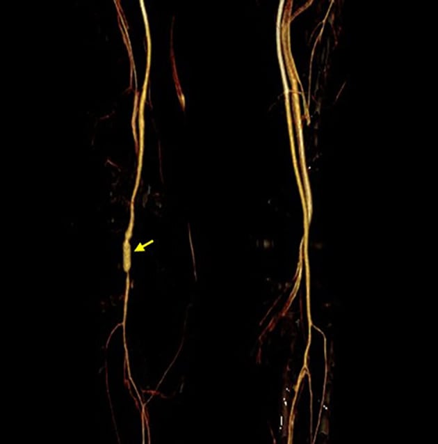
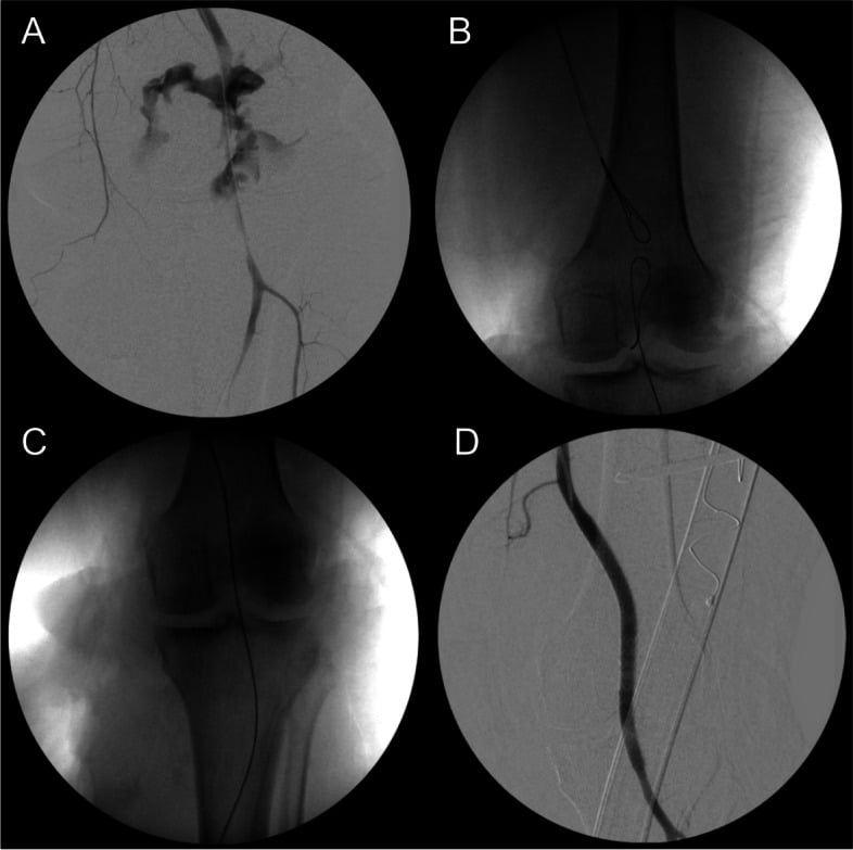
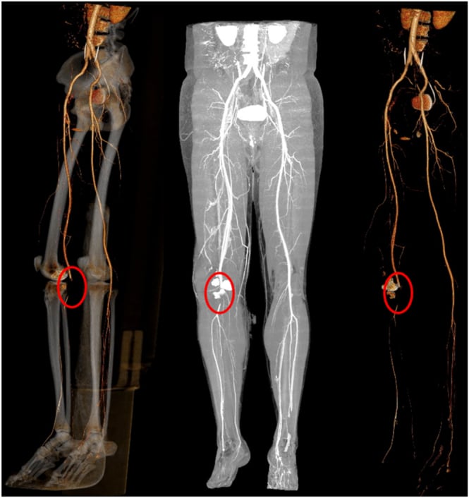
- Knee dislocation and high-energy tibial plateau fracture, with popliteal artery risk
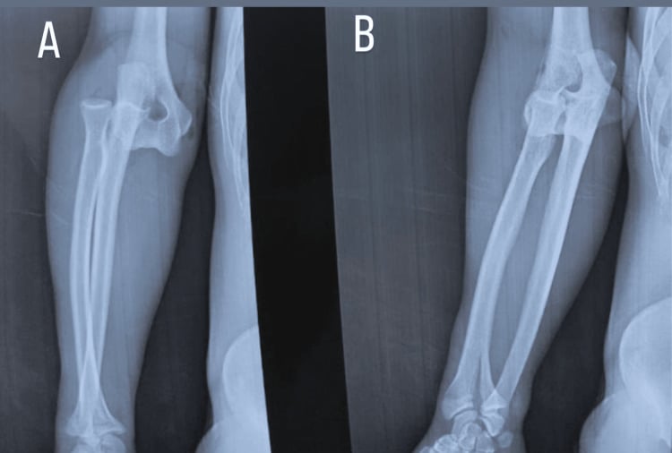
- Elbow fracture-dislocation or paediatric supracondylar fracture, with brachial artery risk
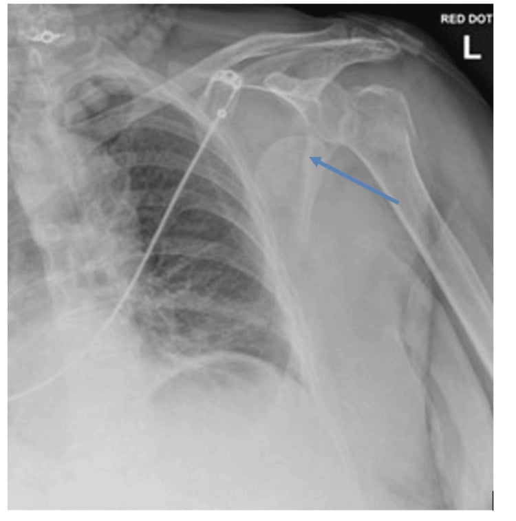
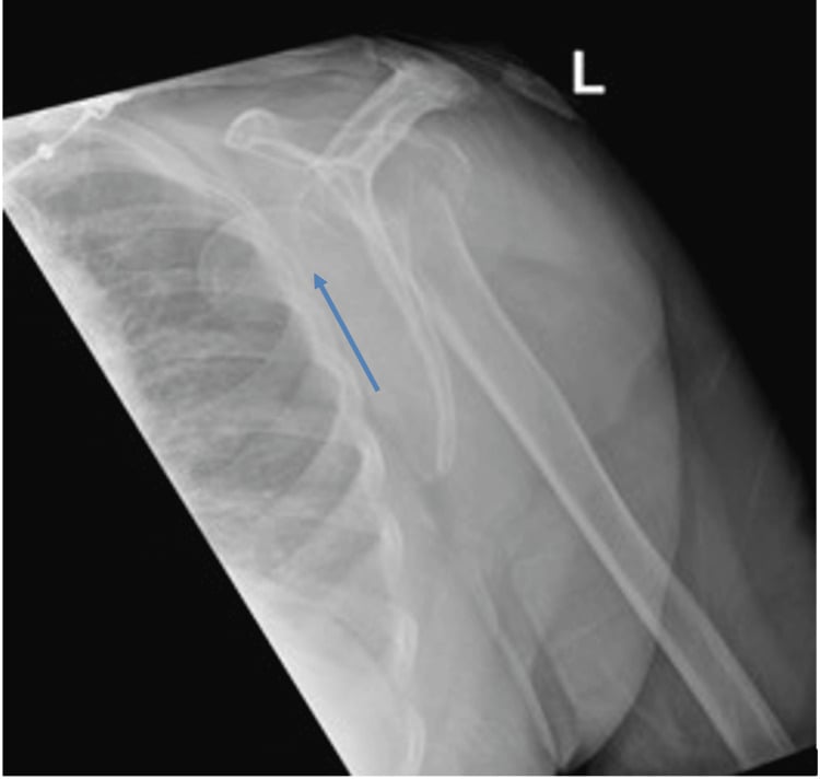
- Shoulder dislocation or proximal humeral fracture, especially in older patients, with axillary artery risk
- Pelvic, acetabular, femoral and tibial trauma with major haemorrhage or limb ischaemia
- Open fracture, crush, blast or ballistic injury with arterial disruption
- Iatrogenic injury during fixation, arthroplasty, deformity correction or revision surgery
Treat vascular injury as a sequence problem: resuscitate, control haemorrhage, restore or preserve perfusion, stabilise the skeleton when it protects the repair, and monitor for reperfusion and compartment complications.
Anatomy and Biomechanics
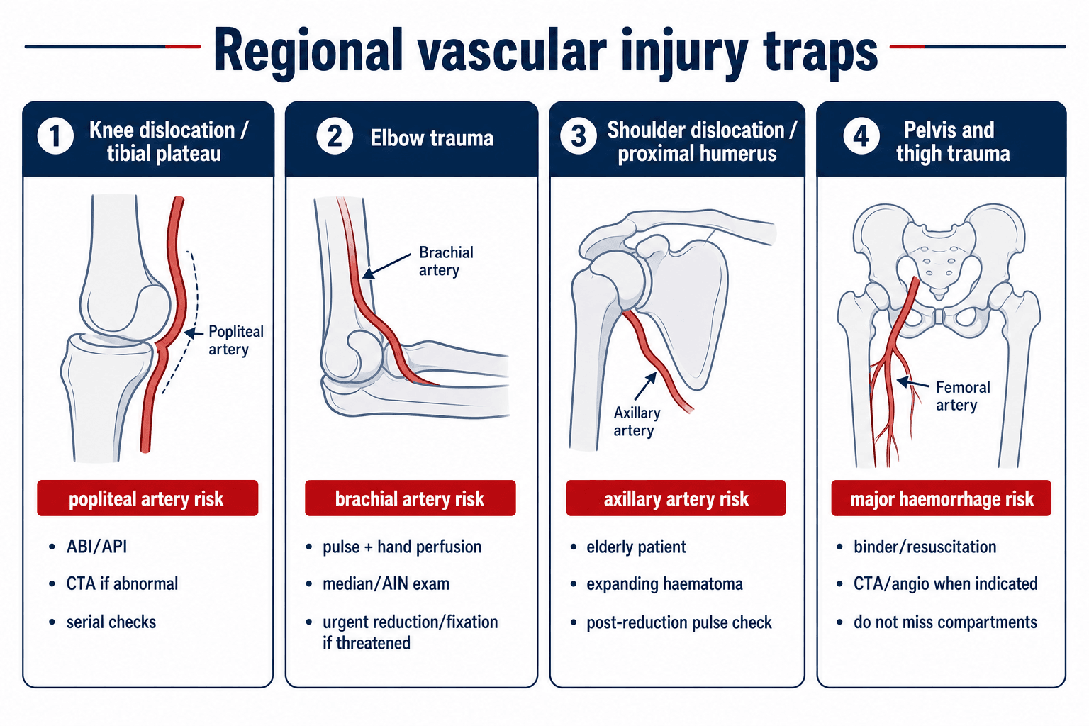
How fracture-dislocations injure vessels. Vessels are injured by stretch, compression, transection, intimal disruption, thrombosis, spasm or entrapment. A dislocation can tear the artery at the moment of injury, while a fracture spike can lacerate or tent it. Reduction may restore flow, but it can also reveal an intimal injury that later thromboses.
Tethered vessels are vulnerable. By region:
- The popliteal artery is relatively tethered at the adductor hiatus and soleus arch, so knee dislocation and plateau fracture patterns can stretch or disrupt it
- The brachial artery crosses the elbow anteriorly and is vulnerable in supracondylar fracture, elbow dislocation and distal humerus trauma
- The axillary artery is at risk in shoulder dislocation, proximal humeral fracture and traction injuries around the shoulder girdle
- The femoral and iliac vessels are at risk in pelvic, acetabular and proximal femoral trauma, especially when haemorrhage is the dominant problem
Why a pulse can mislead. Palpable pulses may persist through collateral flow, a partial injury, resolving vasospasm, or an intimal flap that has not yet thrombosed. A normal pulse is reassuring only when it fits with a normal examination, a normal ABI/API and a low-risk mechanism.
Mechanism of Injury
The mechanism shifts both the probability of vascular injury and the kind of lesion to expect, and each has its own orthopaedic implication.
- Likely Vessel Problem
- Stretch, intimal tear, thrombosis or complete rupture.
- Orthopaedic Implication
- Reduce urgently if perfusion is threatened; reassess pulse and Doppler after reduction.
- Likely Vessel Problem
- Laceration by spike, segmental vessel injury, crush or thrombosis.
- Orthopaedic Implication
- CTA or direct vascular assessment if abnormal examination or high-risk pattern.
- Likely Vessel Problem
- Transection, partial laceration, contamination and compartment risk.
- Orthopaedic Implication
- Control bleeding, antibiotics, debridement, fixation and vascular plan.
- Likely Vessel Problem
- Vascular disruption plus muscle necrosis and reperfusion injury.
- Orthopaedic Implication
- Low threshold for fasciotomy and metabolic monitoring.
- Likely Vessel Problem
- Axillary artery injury from atherosclerotic or fragile vessel stretch.
- Orthopaedic Implication
- Check pulse before and after reduction; consider CTA with haematoma or pulse change.
- Likely Vessel Problem
- Drill, screw, retractor, saw, osteotome or implant-related injury.
- Orthopaedic Implication
- Recognise immediately; control bleeding; call vascular help early.
Classification Systems
Classify vascular injury in terms that change treatment: by the clinical signs, by the arterial lesion and by the orthopaedic context.
- Examples
- Active arterial bleeding, expanding or pulsatile haematoma, bruit, thrill, absent pulse, distal ischaemia.
- Action
- Urgent vascular control; image only if stable and imaging will not delay needed treatment.
- Examples
- History of bleeding, small stable haematoma, pulse asymmetry, neurological deficit, proximity injury.
- Action
- ABI/API and selective imaging; serial examination.
- Examples
- Warm limb, symmetric pulses, normal Doppler, normal ABI/API and reliable clinical setting.
- Action
- Observation with documented serial checks.
Clinical Presentation
The obvious case. A bleeding or pulseless limb after a major fracture or dislocation: active external bleeding or a rapidly expanding haematoma, a cold, pale or mottled distal limb, absent or asymmetric pulses, an absent Doppler signal, and severe pain, paraesthesia or motor weakness, in the setting of a major fracture-dislocation, an open wound or a crush mechanism.
The subtle case is the dangerous one. Examples include:
- A knee dislocation that has reduced before arrival
- A tibial plateau fracture with symmetric pulses but a high-risk mechanism
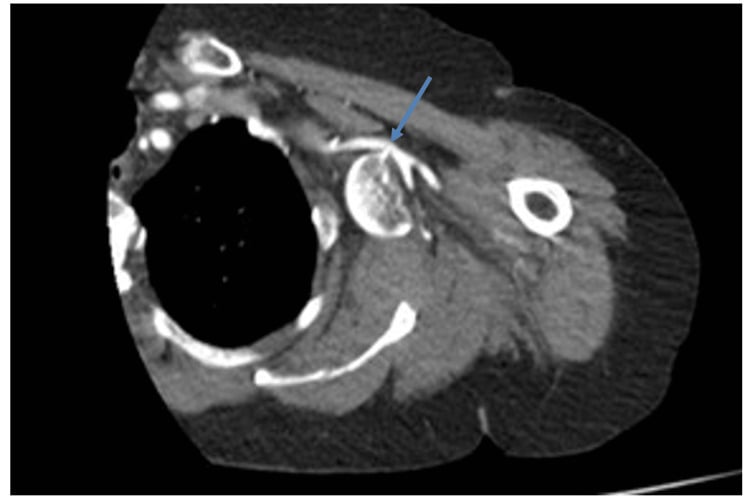
- A shoulder dislocation in an older patient with a small pulse difference
- An elbow fracture with a pink hand but an abnormal Doppler or a neurological deficit
- A ballistic wound near a major vessel with no hard signs
- Delayed thrombosis after an initially normal examination
Delayed presentation. A missed injury can surface later as increasing pain or swelling, a pseudoaneurysm, an arteriovenous fistula, delayed thrombosis or compartment syndrome. It can also present as claudication or reduced exercise tolerance, or as wound breakdown or failed fracture healing from poor perfusion.
Physical Examination
Examine repeatedly, and time it. Document the findings before reduction, after reduction, after splinting or fixation, after vascular repair and during postoperative observation.
Look. Record the colour, temperature and capillary refill of the limb, any deformity or dislocation, an open wound or active bleeding, and an expanding haematoma. Look at the skin for tension, blistering or threatened areas, at the muscle for swelling and compartment tension, and at the whole patient for polytrauma or shock.
Feel. Palpate the radial, ulnar, brachial, dorsalis pedis and posterior tibial pulses as relevant, and compare both limbs. Palpate a haematoma gently so as not to provoke bleeding, and feel for a temperature gradient from proximal to distal. Assess compartment firmness, but do not rely on firmness alone.
Move, and the nerves. Document motor function by named nerves and sensation by territory, and look for deterioration after reduction or revascularisation. Remember that a neurological deficit can be vascular, nerve, compartment or fracture-related.
- How To Use It
- Check distal arterial signal and compare sides.
- Interpretation
- Absent or changing signal is concerning even if the limb looks warm.
- How To Use It
- Doppler systolic pressure injured limb divided by uninjured limb or brachial pressure.
- Interpretation
- Screening threshold 0.9; how the result directs care is set out under the management algorithm.
- How To Use It
- Adjunct in fingers or toes when available.
- Interpretation
- A waveform can support perfusion assessment but does not replace Doppler and ABI/API.
- How To Use It
- Pain, passive stretch pain, paraesthesia, analgesia requirement and tense compartments.
- Interpretation
- Treat clinically clear compartment syndrome urgently.
Imaging and Investigations
Plain radiographs. Order the films needed to define and reduce the fracture or dislocation, and do not let routine imaging delay reduction of a limb-threatening dislocation. They show the direction of dislocation and the quality of reduction, the fracture-dislocation pattern, the tibial plateau, distal femoral, proximal tibial or elbow injuries that carry vascular risk, and open-fracture contamination and bone loss. They also inform the fixation strategy if external fixation is needed.
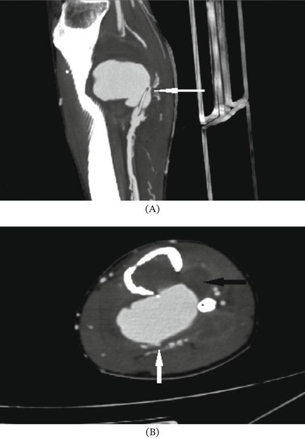
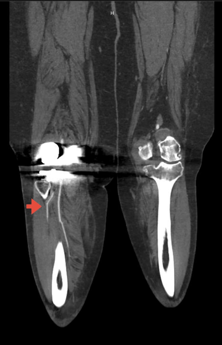
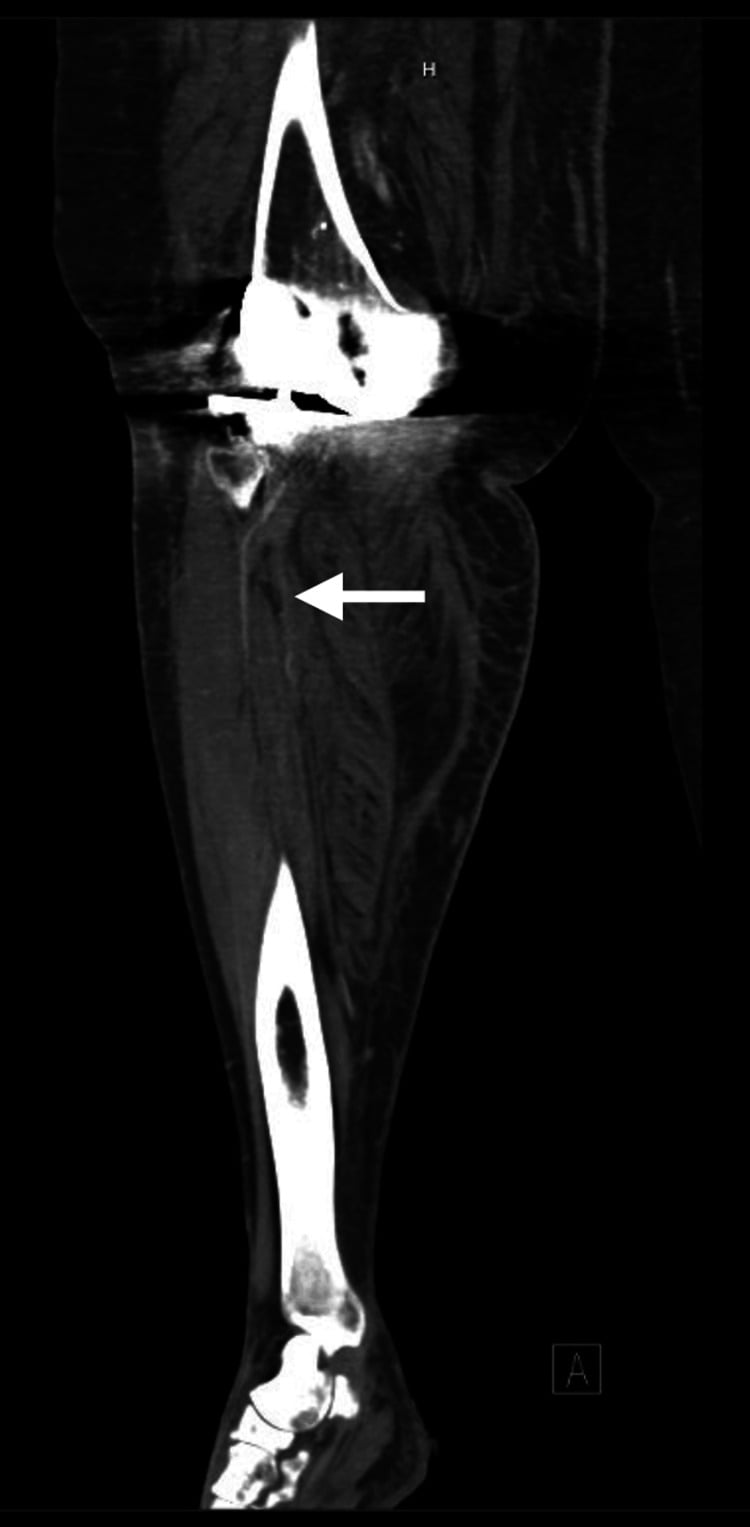
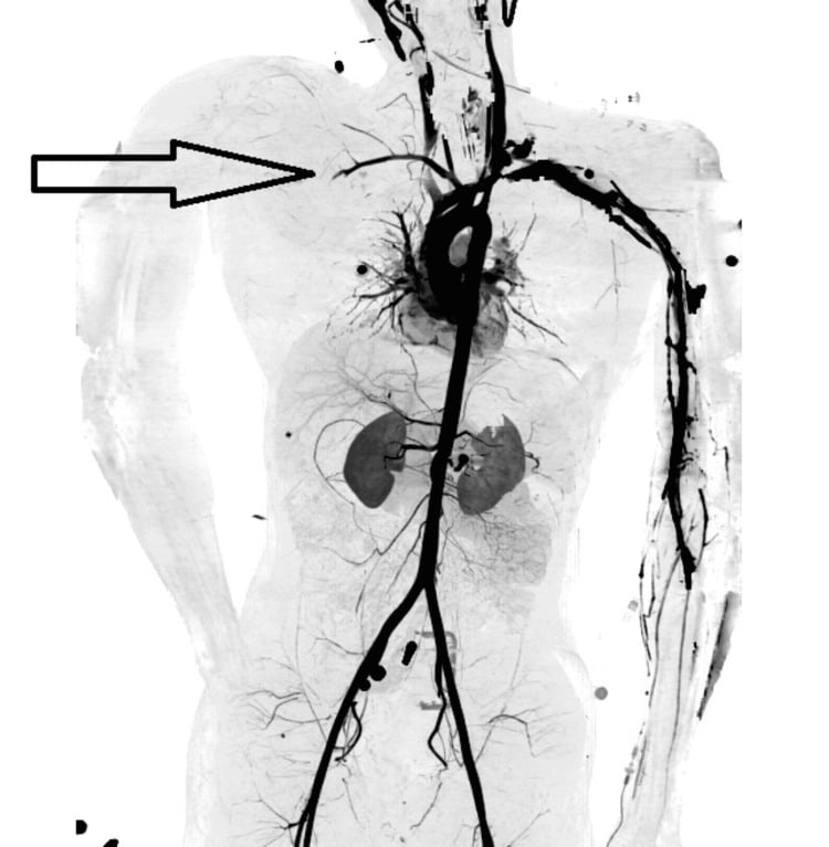
CT angiography. CTA is the usual first-line vascular imaging test when imaging is needed and the patient is stable enough. It defines:
- Occlusion, transection, active extravasation or pseudoaneurysm
- The length and level of the arterial injury
- Its relationship to the fracture fragments
- The distal runoff
- The surgical approach and graft planning
CTA is not a delay tool. It is used when the limb is stable enough for imaging and the result will guide treatment. Hard signs with an obvious operative target can proceed directly to theatre.
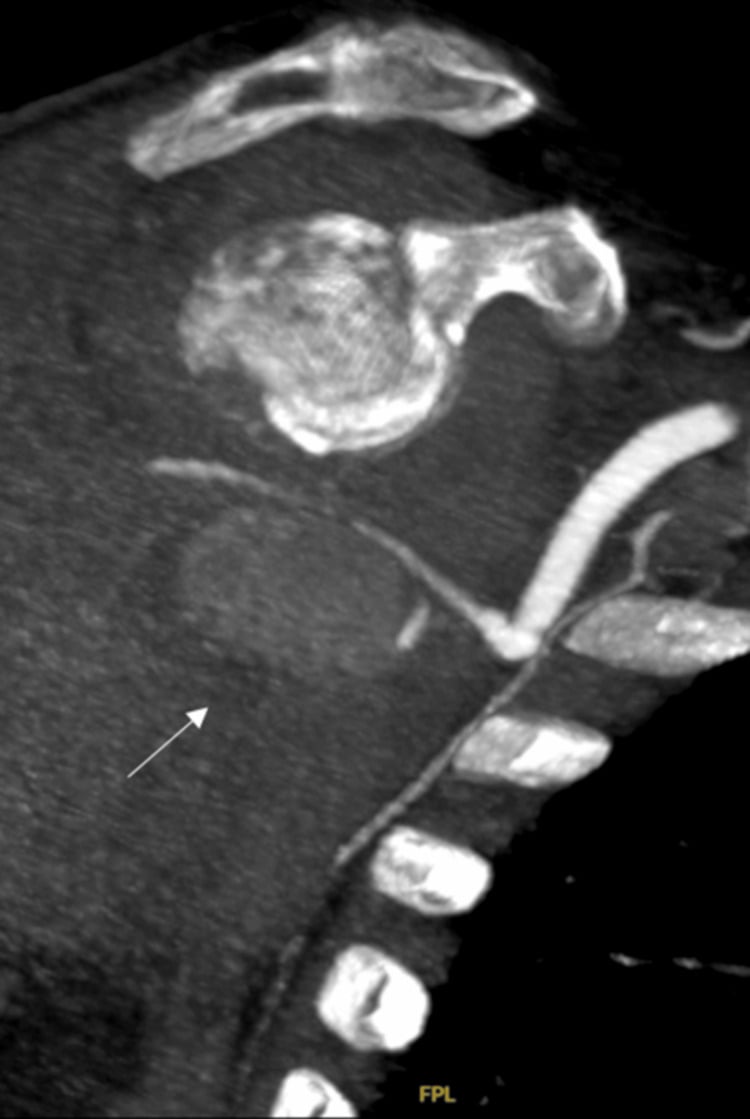
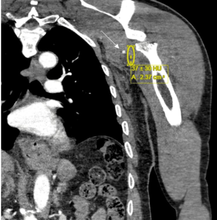
Duplex ultrasound. Duplex can be useful in selected stable patients, for postoperative surveillance, or where rapid expert scanning is available. It is less useful when wounds, dressings, splints, body habitus, pain or time pressure make the study unreliable.
Laboratory tests. The useful ones are:
- Full blood count and coagulation profile
- Group and crossmatch
- Lactate, pH and base deficit in major trauma
- Creatine kinase, potassium and renal function when there is crush, prolonged ischaemia or reperfusion risk
- Serial haemoglobin when haemorrhage is suspected
Differential Diagnosis
Not every cold or painful limb has the same cause, and the clues below tell them apart.
- Clue
- Hard signs, abnormal ABI/API, CTA lesion, absent Doppler.
- Management Implication
- Urgent vascular plan.
- Clue
- Often transient, especially in children or after manipulation.
- Management Implication
- Observe only after senior review and reliable perfusion.
- Clue
- Pain on passive stretch, rising analgesia, tense compartments, neurological symptoms.
- Management Implication
- Fasciotomy; pulses may remain present.
- Clue
- Motor or sensory deficit without perfusion abnormality.
- Management Implication
- Document and reassess; explore if associated open injury or transection suspected.
- Clue
- Bilateral cool limbs, systemic hypotension.
- Management Implication
- Resuscitate; still examine the injured limb separately.
- Clue
- Older patient, calcified vessels, baseline pulse difference.
- Management Implication
- Compare sides, use Doppler/CTA, avoid false reassurance.
Management Algorithm
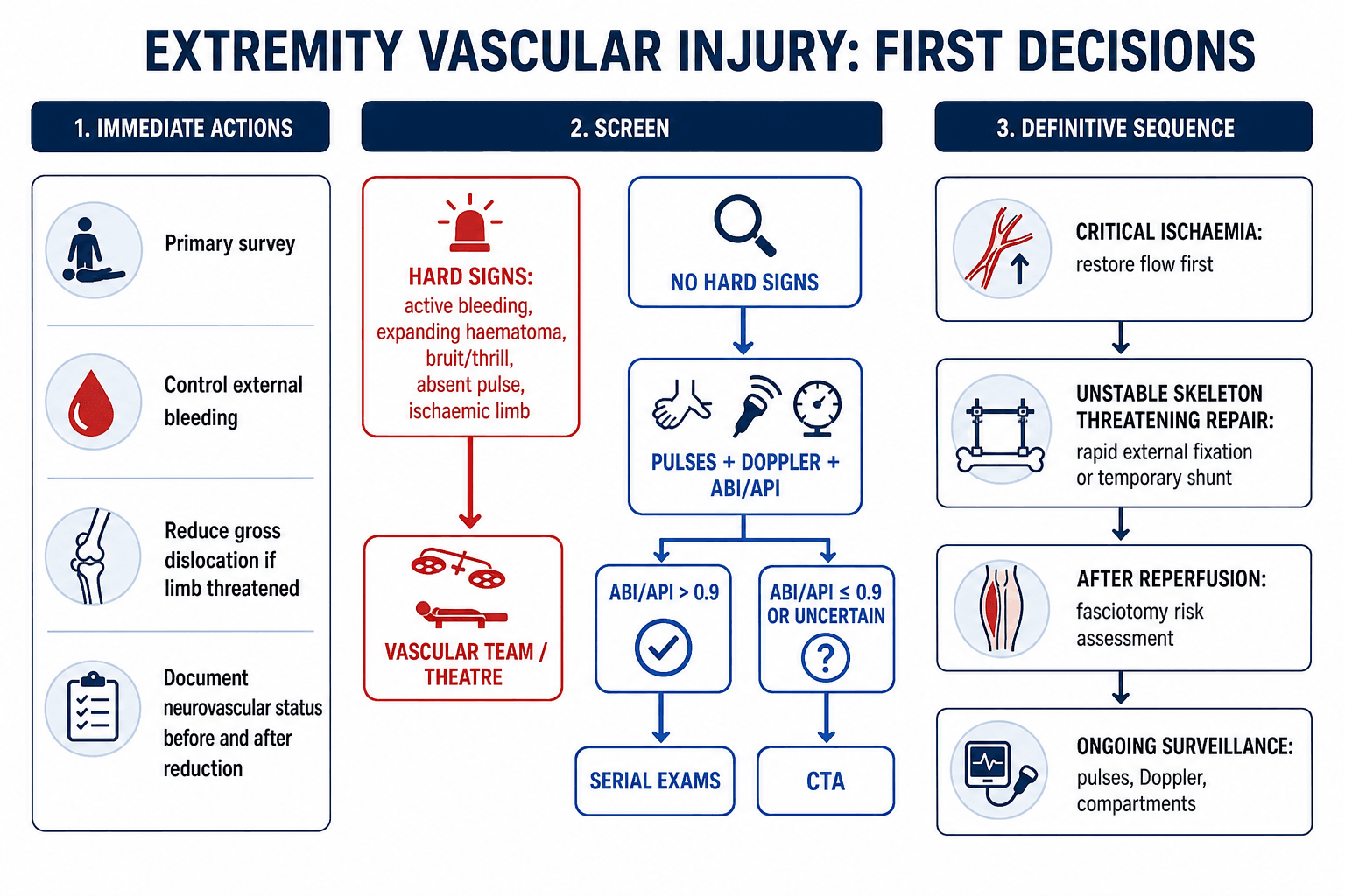
Management turns on physiology, perfusion, fracture stability, time since ischaemia and team availability. An unstable patient (shock, uncontrolled haemorrhage, multiple trauma or a threatened airway) needs the primary survey, haemorrhage control and damage-control decision-making. An ischaemic limb (cold, pale, pulseless and painful, with an absent Doppler or motor or sensory deterioration) needs urgent vascular involvement and a reperfusion pathway, and a gross knee, ankle, elbow or shoulder deformity with threatened perfusion is reduced urgently, with neurovascular status documented before and after.
- Primary survey and haemorrhage control.
- Remove constrictive dressings or splints if compromising perfusion.
- Document neurovascular status.
- Reduce gross dislocation if the limb is threatened.
- Reassess and document neurovascular status.
- Identify hard signs.
- If hard signs are present, involve vascular surgery and proceed to control or repair.
- If no hard signs, use ABI/API and CTA pathway when indicated.
- Choose vessel-first, bone-first or temporary shunt sequence.
- Assess fasciotomy need after ischaemia, repair, shunting or reperfusion.
Surgical Technique
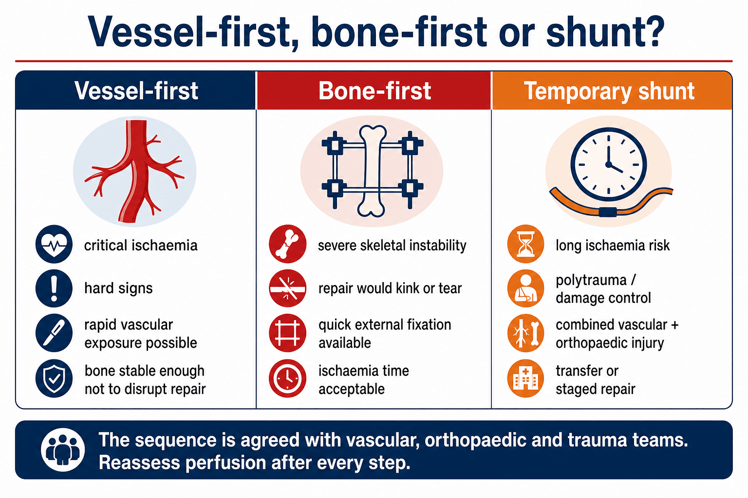
Reduce first when flow is threatened. Reduce dislocations urgently when perfusion is threatened, and splint in a stable position that does not compromise flow.
Fix fast, and out of the way. Use external fixation when skeletal instability risks the vascular repair or the soft tissues. Keep it rapid, place it outside planned vascular or soft-tissue incisions where possible, and reassess Doppler and compartments afterwards.
The vein is a decision too. The artery gets the attention, but a concomitant major venous injury is a genuine decision point and a frequent omission. A divided major outflow vein (popliteal, femoral) causes venous hypertension and limb oedema, which worsens compartment syndrome and can compromise the arterial repair, because poor outflow promotes arterial thrombosis.
Repair or ligate. In the stable patient with a salvageable limb, repair a major vein by lateral suture or interposition graft; this reduces oedema and compartment risk and supports arterial patency, particularly for the popliteal vein. In the unstable or polytrauma patient, or with a difficult exposure, ligating even a major vein is a legitimate life-saving damage-control shortcut. Expect marked limb swelling: ligation mandates fasciotomy, with leg elevation and compression afterwards. Small, expendable veins are simply ligated.
Which first. When both are injured, many surgeons restore arterial inflow first, by shunt or repair, to limit ischaemia, and then address the vein.
Regional Injury Patterns
Each region has its own vessel at risk and its own checks, and these are the patterns to look for actively.
- Vessel Concern
- Popliteal artery intimal tear, thrombosis or rupture.
- Practical Checks
- Reduce, document pulse/Doppler before and after, ABI/API, CTA when abnormal or uncertain.
- Vessel Concern
- Knee-dislocation equivalent mechanism can injure popliteal vessels.
- Practical Checks
- ABI/API screening even with symmetric pulses is used in many trauma pathways.
- Vessel Concern
- Brachial artery injury or entrapment.
- Practical Checks
- Check radial/ulnar pulses, hand perfusion, median/AIN function and post-reduction status.
- Vessel Concern
- Axillary artery injury, especially with haematoma or pulse change.
- Practical Checks
- Check pulse before and after reduction; CTA if abnormal or expanding haematoma.
- Vessel Concern
- Major haemorrhage from pelvic vessels more than isolated limb ischaemia.
- Practical Checks
- Binder, resuscitation, pelvic haemorrhage pathway, CTA/angio or packing as indicated.
- Vessel Concern
- Anterior tibial, posterior tibial or peroneal artery injury with compartment risk.
- Practical Checks
- Doppler, ABI/API, CTA if abnormal; low fasciotomy threshold.
Fasciotomy and Reperfusion
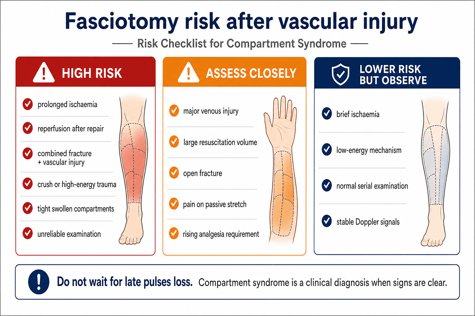
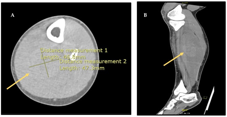
When it happens. Compartment syndrome can occur before repair, after reduction, after vascular repair, after shunting or during resuscitation, and it can occur despite restored pulses.
Do not wait for late findings. Paralysis, pulselessness and pallor are late, and should not be the trigger for fasciotomy. The early pattern that matters is escalating pain, pain on passive stretch, an increasing analgesic requirement, paraesthesia and tense compartments.
Part of the operation. When the risk is high, fasciotomy belongs to the vascular-injury operation rather than following it as an afterthought. The indications include:
- Established compartment syndrome
- Prolonged ischaemia
- Reperfusion after arterial repair
- Combined arterial and venous injury
- High-energy fracture, crush or open injury
- Tense compartments or an unreliable examination
- Need for major resuscitation
Prophylactic fasciotomy. It is considered when the risk of missing compartment syndrome is higher than the morbidity of fasciotomy. This is common after prolonged ischaemia, reperfusion, combined fracture and vascular injury, crush injury or an unreliable examination.
Documentation. Record, with times:
- Time of injury and estimated ischaemia duration
- Time of reduction, shunt and repair
- Pulse, Doppler and ABI/API findings
- Compartment findings and analgesic requirement
- The reason for fasciotomy, or the reason for observation
The warm-ischaemia clock. Restoring flow to a long-ischaemic limb is not purely good news: the systemic reperfusion load can be lethal. Skeletal muscle tolerates roughly six hours of warm ischaemia before irreversible necrosis, and beyond this, reperfusion washes the products of dead muscle into the circulation.
Reperfusion syndrome. Historically the "myonephropathic-metabolic syndrome": the sudden release of potassium, myoglobin, hydrogen ions and lactate causes hyperkalaemia (arrhythmia or arrest), myoglobinuric acute kidney injury and metabolic acidosis. The hit can come the moment the clamp or shunt is released.
Anticipate it. Warn anaesthesia before reperfusion. Give aggressive IV fluids, monitor and treat hyperkalaemia, monitor creatine kinase and renal function, and maintain a brisk urine output, with consideration of urinary alkalinisation. Correct acidosis, and perform fasciotomy to relieve the muscle oedema.
Reperfusing an unsalvageable, necrotic limb can kill the patient. In the moribund or severely physiologically deranged patient with a clearly non-viable limb (prolonged ischaemia, dead muscle), primary amputation is the safer choice than revascularisation. The whole-patient physiology, not just the artery, drives the decision.
Complications
The complications to anticipate, with why each occurs and how to prevent or answer it.
- Why It Occurs
- Normal pulses, spontaneous reduction, incomplete examination.
- Prevention or Response
- Repeat examination, ABI/API, CTA when abnormal or uncertain.
- Why It Occurs
- Intimal injury, poor runoff, kinking, compression by fracture or swelling.
- Prevention or Response
- Protect repair with alignment, monitor Doppler, urgent vascular reassessment if signal changes.
- Why It Occurs
- Ischaemia, reperfusion, crush, fracture bleeding and resuscitation swelling.
- Prevention or Response
- Early fasciotomy when clinical or high risk.
- Why It Occurs
- Open fracture, contamination, devitalised tissue.
- Prevention or Response
- Debridement, soft tissue cover, antibiotics and avoid synthetic graft when possible.
- Why It Occurs
- Muscle necrosis, infection, severe bone/soft tissue loss or failed repair.
- Prevention or Response
- Realistic counselling and serial viability assessment.
- Why It Occurs
- Nerve injury, compartment syndrome, stiffness, nonunion and scarring.
- Prevention or Response
- Early multidisciplinary rehabilitation and surveillance.
Postoperative Care
Active surveillance. Postoperative care is active surveillance rather than routine observation. Monitor:
- Neurovascular status, hourly or by protocol initially
- The Doppler signal, documented
- Compartments and analgesic requirement
- Wounds and fasciotomy dressings
- The graft or repair, according to the vascular plan
- Renal and metabolic function after crush or reperfusion
Immobilisation and fixation. External fixator pin sites must not compromise vascular access or planned flaps, and splints should not compress the repair or the compartments. The timing of definitive fixation depends on physiology, perfusion, soft tissues and infection risk, and the location of the vascular graft affects the surgical approach and reoperation planning.
Rehabilitation. Recovery depends on nerve, muscle, bone and soft tissue, and a perfused limb may still be weak, painful or functionally limited. The early goals are protection of the repair, oedema control, joint motion where safe, wound care and realistic patient counselling.
Outcomes and Prognosis
Outcome depends less on the vessel alone than on the whole limb. The prognostic factors are:
- Warm ischaemia time
- Blunt versus penetrating mechanism
- Popliteal or multi-level tibial vessel injury
- Associated fracture-dislocation or open fracture
- Nerve injury
- Muscle viability
- Need for fasciotomy
- Infection and soft-tissue cover
- Patient physiology and rehabilitation capacity
Successful revascularisation is necessary but not sufficient. Limb salvage still depends on viable muscle, stable bone, durable soft tissue, nerve function and absence of infection.
Guidelines, Registries & Global Practice
Extremity vascular injury complicates roughly 1 to 3 percent of major limb trauma in civilian series, rising sharply in penetrating and military cohorts. The popliteal and superficial femoral arteries dominate lower-limb injury, and the brachial artery dominates the upper limb. Outcomes are driven less by the artery and more by ischaemia time, mechanism (blunt worse than penetrating) and associated musculoskeletal and nerve injury.
Side-by-side guidance
- Core Position
- Hard signs go to operative control; selective imaging otherwise.
- Practical Emphasis
- Physical examination plus ABI for triage; CTA as imaging of choice; tourniquets and shunts as adjuncts.
- Core Position
- Damage-control and restoration of perfusion in the unstable patient.
- Practical Emphasis
- Shunting and external fixation as part of staged limb salvage.
- Core Position
- Skeletal stabilisation must protect the vascular repair.
- Practical Emphasis
- Rapid external fixation before or alongside revascularisation when the skeleton is unstable.
- Core Position
- Combined orthoplastic and vascular care in specialist centres.
- Practical Emphasis
- Early senior decision-making, fix-and-flap planning and timely transfer.
- Core Position
- Time-to-reperfusion is the key modifiable factor.
- Practical Emphasis
- Minimise warm ischaemia; low threshold for fasciotomy after reperfusion.
Registry and large-cohort data (multicentre civilian shunt series and the PROOVIT vascular-injury registry) consistently show that severely injured shunted limbs achieve salvage rates comparable to or better than non-shunted limbs despite greater injury severity, and that blunt mechanism, popliteal location and pulseless presentation predict eventual amputation.
High- versus limited-resource practice
In high-resource trauma networks, definitive CTA, vascular surgery, interventional radiology, intensive care and orthoplastic reconstruction are available in one centre, allowing single-stage vessel-and-bone management. In limited-resource or pre-transfer settings, the priorities are haemorrhage control, reduction of limb-threatening dislocations, documented serial neurovascular checks, early senior consultation and transfer planning. A temporary intravascular shunt plus a spanning external fixator is a powerful damage-control combination that restores perfusion and stabilises the skeleton while definitive vascular and orthopaedic reconstruction is arranged or the patient is transferred.
Controversies and Areas of Uncertainty
Several questions in this field have no single right answer and are common viva ground.
- The Debate
- Observe a well-perfused but pulseless hand after reduction versus explore.
- Pragmatic Position
- Reduce and stabilise first; a perfused hand may be observed closely, but persistent pulselessness with poor perfusion or median/AIN deficit favours exploration in many protocols.
- The Debate
- Whether to shunt routinely in combined orthopaedic-vascular injury.
- Pragmatic Position
- No randomised data; cohort evidence links shunting to less compartment syndrome. Shunt when bone fixation will delay flow or physiology is hostile.
- The Debate
- Routine fasciotomy after revascularisation versus close monitoring.
- Pragmatic Position
- Low threshold after prolonged ischaemia, combined arterial and venous injury or unreliable examination; selective monitoring when ischaemia time is short and examination reliable.
- The Debate
- Whether all Schatzker 4-6 fractures with symmetric pulses need ABI.
- Pragmatic Position
- Single-centre data show ABI screening misses no injuries; many trauma services now screen routinely.
- The Debate
- Stenting or embolisation versus open repair in extremity arterial injury.
- Pragmatic Position
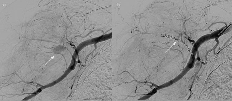
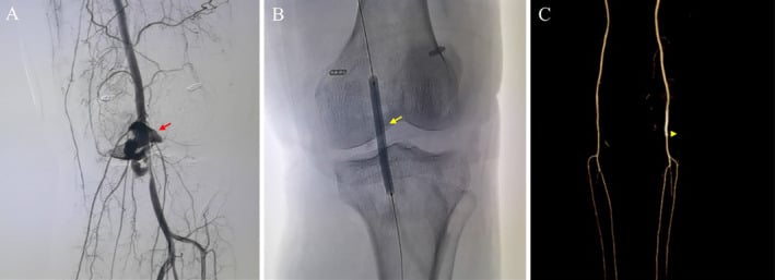
- Endovascular is useful for selected proximal, pseudoaneurysm or AV-fistula lesions; open repair remains standard for most acute limb-threatening transection.
Common Pitfalls
- Calling the limb safe because a pulse is palpable once.
- Forgetting that a knee dislocation may reduce before presentation.
- Delaying reduction of a threatened limb for routine imaging.
- Sending a patient with hard signs for CTA without vascular agreement.
- Performing prolonged definitive fixation while the foot is ischaemic.
- Repairing the artery without stabilising a skeleton that will disrupt the repair.
- Failing to consider a temporary shunt in a damage-control situation.
- Waiting for pulselessness before diagnosing compartment syndrome.
- Forgetting to document serial neurovascular checks.
- Assuming limb salvage means useful function.
MCQ Practice Points
Q: What are hard signs of extremity vascular injury? A: Active arterial bleeding, expanding haematoma, bruit, thrill, absent pulse and distal ischaemia are hard signs. They require urgent vascular control rather than routine delayed imaging.
Q: What does an ABI/API above 0.9 mean after extremity trauma? A: If the examination is normal and reliable, it supports serial observation. An ABI/API of 0.9 or less, uncertainty or a discordant high-risk mechanism prompts CTA or vascular assessment.
Q: Why is a spontaneously reduced knee dislocation still dangerous? A: Popliteal artery injury can be occult initially, so the patient still needs vascular screening with documented pulses, Doppler, ABI/API and serial examination.
Q: What is the role of a temporary vascular shunt? A: It restores perfusion during damage control, transfer, skeletal stabilisation or staged repair. It is a bridge to definitive vascular reconstruction, not definitive treatment.
Q: Can compartment syndrome occur with pulses present after vascular repair? A: Yes. Reperfusion after vascular repair is a major risk setting, and compartment syndrome remains a clinical diagnosis when signs are clear.
Exam Cheat Sheet
First Actions
- Primary survey and haemorrhage control
- Document neurovascular status
- Reduce gross dislocation if limb threatened
- Repeat examination after reduction
- Hard signs go to urgent vascular pathway
Screening
- Pulses alone are not enough
- Use Doppler and ABI/API
- Above 0.9 plus normal reliable exam supports serial observation
- 0.9 or less, uncertainty or discordance prompts CTA or vascular review
- CTA is first-line imaging when stable enough
Sequence
- Critical ischaemia favours vessel-first
- Unstable skeleton threatening repair may need rapid external fixation
- Temporary shunt restores flow during staged care
- Definitive fixation follows physiology, perfusion and soft tissue readiness
- Reassess perfusion after every step
Fasciotomy
- Established compartment syndrome is an emergency
- High risk after prolonged ischaemia and reperfusion
- Combined fracture and vascular injury lowers threshold
- Do not wait for pulselessness
- Document timing and serial findings
Applied Scenarios
Practise clinical reasoning and management decisions out loud
“A patient reports a knee deformity that reduced before arrival. The limb is warm and pulses are palpable.”
“A patient has an ankle fracture-dislocation with a cool foot and absent Doppler signal.”
“A patient has a distal femur fracture, absent distal pulses and CTA shows popliteal artery occlusion. The fracture is very unstable.”
“An older patient has shoulder dislocation with axillary artery occlusion. Flow is restored after repair, but the arm becomes tense and analgesic requirement rises.”
Evidence Base
Penetrating Lower-Extremity Arterial Trauma: EAST Practice Management Guideline
- Forty-three articles screened; 20 used to construct the guideline updating the 2002 EAST recommendations.
- Physical examination and ankle-brachial index allow expedited triage of penetrating lower-limb arterial injury.
- CTA has become the diagnostic study of choice when imaging is required.
- Tourniquets and temporary intravascular shunts are recognised adjuncts in lower-extremity arterial trauma.
Imaging Vascular Trauma
- Systematic review of 58 articles (2000-2010); CTA was the dominant modern diagnostic modality across the vascular-trauma literature.
- CTA showed acceptable sensitivity and specificity for blunt and penetrating vascular injury within the neck and extremity.
- Duplex, MRA and transoesophageal echocardiography each had significant drawbacks as first-line tools.
CT Angiography Effectively Evaluates Extremity Vascular Trauma
- Single Level-I trauma centre series; 38 patients underwent CTA and 14 conventional arteriography for extremity vascular trauma.
- There were no false negatives or missed injuries with CTA.
- CTA supplanted catheter arteriography as the initial radiographic test, offering non-invasiveness and immediate availability.
Nonoperative Management of Occult Arterial Injuries and Physical Examination Alone
- Clinically occult arterial injuries without hard signs were safely managed nonoperatively; only 4 of 43 (9%) deteriorated and needed delayed surgery, all with good results.
- Of 287 patients managed by physical examination alone, only 4 (1.3%) required delayed surgery within the first week.
- No patient contacted at long-term follow-up reported vascular symptoms.
Reference Links
- PubMed: EAST lower-extremity arterial trauma guideline
- PubMed: CTA effectively evaluates extremity vascular trauma
- PubMed: imaging vascular trauma systematic review
- PubMed: physical examination and occult arterial injury follow-up
- PubMed: ABI and ultrasonography systematic review
- PubMed: temporary shunts systematic review
- PubMed: combined orthopaedic and vascular trauma shunting
- PubMed: civilian lower-extremity arterial trauma outcomes