Centre of Rotation of Angulation | Mechanical Axis Deviation | Osteotomy Planning
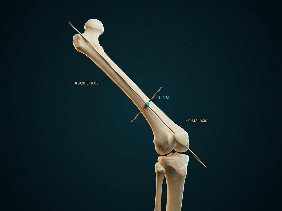
- CORA = the point where the proximal and distal axis lines cross - the apex of the deformity
- MAD = perpendicular distance from the mechanical axis to the knee centre; normally about 8 to 10 mm medial (roughly plus or minus 7 mm), not zero
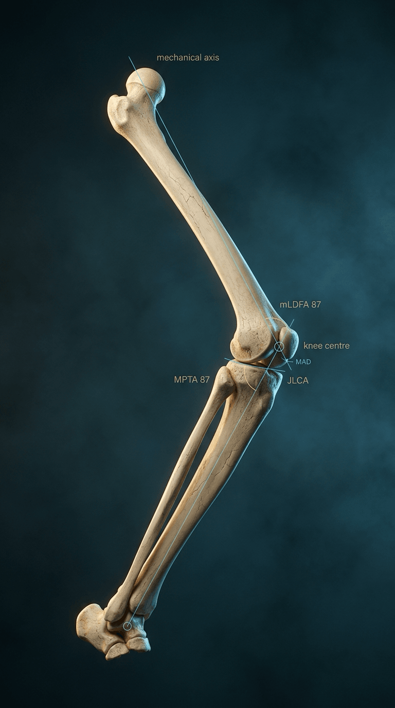
- mLDFA 87°, MPTA 87°, JLCA 0-2° - these three localise the deformity to femur, tibia or joint
- Osteotomy away from the CORA forces translation, and the amount is predictable: 2 x d x tan(half the correction)
- Predicted MPTA above 95° means a single-level tibial correction will leave an oblique joint line - consider double-level
- “aLDFA is 81°, not 87° - anatomic and mechanical femoral angles differ by the 6-7° anatomic-mechanical angle
- “A raised JLCA is joint, not bone - subtract it before sizing the wedge or you will over-correct
- “HTO obeys Rule 2: the CORA of a varus knee sits at the joint line and the cut is made below it
- “Rotation is invisible on the long-leg AP - torsion must be assessed separately
Overview and Epidemiology
Deformity analysis is the systematic evaluation of limb alignment to identify the location and magnitude of angular, translational, rotational and length deformity. The CORA (centre of rotation of angulation) method, described by Dror Paley, turns that assessment into a drawing that specifies where to cut and by how much.
Where it is used. Angular deformity correction, limb length discrepancy with deformity, malunion correction, developmental deformity and post-traumatic reconstruction all start from the same drawing. Accurate analysis prevents secondary deformities from osteotomy, guides the choice of osteotomy level and predicts the outcome of correction.
Paley's method is two questions asked in order. The malalignment test asks is there a deformity? - draw the hip-to-ankle line and measure MAD. The malorientation test asks where does it live? - measure mLDFA, MPTA, LDTA and JLCA to see which joint is facing the wrong way. Only then does the CORA construction tell you where to cut. Candidates who name an operation before finishing the second question correct the wrong bone.
Pathophysiology
Mechanical Axis and MAD
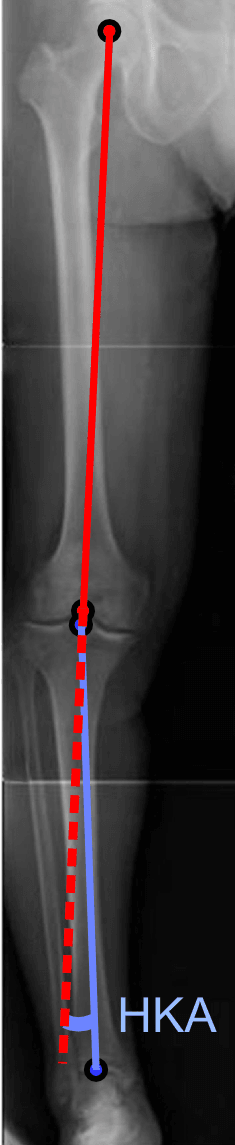
The mechanical axis (the Mikulicz line) runs from the centre of the femoral head to the centre of the ankle. It represents the weight-bearing axis of the whole limb, ignoring where the individual bones sit, and it does not pass through the middle of the knee: it lies slightly medial to it.
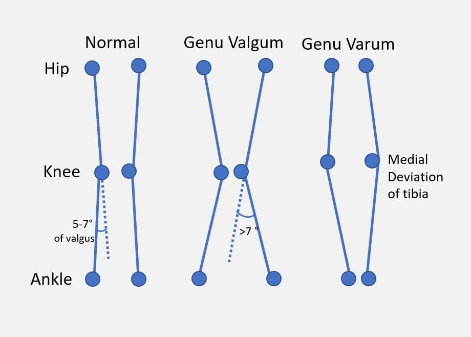
Mechanical axis deviation (MAD) is the perpendicular distance from that line to the centre of the knee. Normal is about 8 to 10 mm medial (Paley's reference figure, with a spread of roughly 7 mm either side), so up to around 15 mm medial is still within the normal range. Increasing medial deviation is varus; any appreciable lateral deviation is valgus.
Zero is not normal. A MAD of zero is slightly valgus relative to the population mean, which is the geometric reason "restore the axis to the exact centre of the knee" is not the same instruction as "restore normal alignment".
Name the side. Report MAD in millimetres as "14 mm medial", not "-14": sign conventions are not standardised between texts, and a swapped sign inverts the diagnosis.
Anatomic Axis and the Anatomic-Mechanical Angle
The anatomic axis is the line through the centre of the bone's diaphysis, the mid-shaft line you can draw on a short film. In the tibia it is effectively the same line as the mechanical axis, which is why tibial angles are quoted only once. In the femur the two are not the same line.
The femoral anatomic-mechanical angle (AMA) is about 6 to 7 degrees (Paley quotes 7°, range 5-9°). The anatomic axis enters the knee lateral to the mechanical axis, and this offset is exactly the difference between the aLDFA of 81° and the mLDFA of 87°. If only a knee-length film is available you can measure the aLDFA, but you must compare it with 81°: holding an anatomic measurement against the mechanical normal invents 6° of valgus that the patient does not have.
Joint Orientation Angles
The tables give the normal values and how each angle is drawn.
- Normal Value
- 87° (85-90°)
- How it is drawn
- Lateral angle between the femoral MECHANICAL axis and the knee joint line
- Normal Value
- 81° (79-83°)
- How it is drawn
- Same joint line, but measured off the femoral ANATOMIC (mid-shaft) axis
- Normal Value
- 87° (85-90°)
- How it is drawn
- Medial angle between the tibial mechanical axis and the tibial plateau line
- Normal Value
- 90° (85-95°)
- How it is drawn
- Lateral angle between the femoral mechanical axis and the femoral neck axis
- Normal Value
- 89° (86-92°)
- How it is drawn
- Lateral angle between the tibial mechanical axis and the ankle plafond
- Normal Value
- 0-2° (abnormal above 3°)
- How it is drawn
- Angle between the distal femoral and proximal tibial joint lines - the intra-articular contribution
- Normal Value
- 8-10 mm medial (± ~7 mm)
- How it is drawn
- Perpendicular distance from the hip-to-ankle line to the knee centre
The two knee angles are both 87°, so a knee angle that is not near 87 points to the bone it belongs to, as long as it was measured off the mechanical axis.
- Normal Value
- 83° (79-87°)
- How it is drawn
- Posterior angle between the femoral anatomic axis and the distal femoral condylar line on the lateral view
- Normal Value
- 81° (77-84°)
- How it is drawn
- Posterior angle between the tibial anatomic axis and the plateau line - the reciprocal of posterior tibial slope
- Normal Value
- 80° (78-82°)
- How it is drawn
- Anterior angle between the tibial anatomic axis and the ankle plafond on the lateral view
- Normal Value
- 7-10°
- How it is drawn
- Approximately 90° minus the PPTA; changed unintentionally by most tibial osteotomies
The sagittal plane. The sagittal angles are the half of deformity analysis most candidates leave out. A proximal tibia with a normal MPTA and a PPTA of 70° carries an 11° sagittal deformity, a posterior slope of about 20° instead of 9°, and nothing on the AP film will ever show it.
The blind spot works in reverse. A medial opening-wedge HTO hinged too far anteriorly raises the slope and drives the tibia forward on the femur, which matters enormously if the anterior cruciate ligament is already deficient. A lateral closing wedge tends to reduce the slope, which matters in the cruciate-deficient knee in the opposite direction.
CORA Determination
- Decide which bone is abnormal from the joint orientation angles - the CORA is constructed on that bone, not on the limb as a whole
- Draw the axis of the proximal segment and the axis of the distal segment. Where a segment is too short to have a visible mid-shaft line, construct its axis from the joint orientation angle instead (for example, draw a line at 87° to the plateau to obtain the proximal tibial mechanical axis)
- The point where the two lines cross is the CORA; the angle between them is the magnitude of angulation
- The CORA always lies on the bisector of the angle between the two axis lines; where the axes have been drawn correctly it coincides with the visible apex of the bend
What the construction tells you beyond the angle. If the two axis lines cross at the level of the visible bend, the deformity is purely angular and there is one CORA. If they cross at a level away from the visible bend, there is an additional translation component: the bone is both angulated and shifted. If the two lines are parallel but not collinear there is no CORA at all, because parallel lines never meet; that is a pure translation deformity, and no amount of angulation will fix it.
How many CORAs. One deformity in an oblique plane still has only one CORA. It is the multi-apical deformity - rickets, multiple hereditary exostoses, several malunions in one bone - that has more than one.
Oblique Plane Deformity
A deformity whose true apex lies between the cardinal planes shows up on both the AP and the lateral view, and on neither of them at full size.
The true magnitude is always larger than either projection. Combine them trigonometrically: the tangent of the true angle is the square root of the sum of the squared tangents of the two components. A 20° varus on the AP with a 20° procurvatum on the lateral is a true deformity of about 27°, not 20°.
The plane of the deformity is found from the ratio of the two components. Its angle from the AP plane is the arctangent of tan-lateral over tan-AP, and the correction axis must be set perpendicular to that oblique plane. Correcting 20° in the AP plane and 20° in the sagittal plane as two separate manoeuvres does not reproduce a single 27° correction in the oblique plane.
The CORA is a property of the patient - it is where the deformity actually is. The ACA (angulation correction axis) is a choice made by the surgeon - it is the hinge the bone will rotate about, drawn perpendicular to the plane of the deformity. Paley's three osteotomy rules are simply the three ways those two points and the osteotomy cut can be arranged relative to each other, and the whole of osteotomy planning is deciding where to put the ACA once the CORA is known.
Rotational (Torsional) Deformity
The CORA method handles angulation, translation and length; rotation (torsion) is the axial deformity it does not capture on the coronal film and must be assessed separately - a high-yield omission in many answers.
Normal values. Femoral anteversion is about 15 degrees in adults (higher in infancy, decreasing through growth), and tibial torsion about 15 to 20 degrees of external (lateral) torsion in adults. Thigh-foot angle is normally about 10 degrees external in a child and 15 degrees external by adolescence, with a wide normal spread, which is why the clinical profile screens and the CT decides.
The clinical rotational profile (Staheli) has three parts:
- Hip internal and external rotation (femoral version - excess internal rotation suggests increased anteversion)
- Thigh-foot angle and the transmalleolar axis (tibial torsion)
- Foot progression angle (net in- or out-toeing) and foot shape (to exclude metatarsus adductus)
Measurement. The clinical profile screens, but a CT (or MRI) rotational study is the gold standard for quantifying femoral version and tibial torsion when surgery is considered. On CT, femoral version (neck axis against the posterior condylar line) and tibial torsion (proximal tibial posterior condylar line against the transmalleolar axis) are measured on the same scan so that the two are not confused.
Malrotation after intramedullary nailing of the femur or tibia is a common iatrogenic torsional deformity; compare with the contralateral limb on CT. A rotational difference of more than about 15 degrees from the uninjured side after nailing is the usual figure quoted for symptomatic malrotation and for offering derotation.
"Miserable malalignment syndrome" is increased femoral anteversion plus external tibial torsion, producing squinting patellae, an increased Q-angle and patellofemoral pain.
Correction. Symptomatic torsion is corrected by a derotation osteotomy (femoral or tibial), planned from the version difference relative to the normal or contralateral side.
Classification Systems
Deformity is classified by plane, by source segment and by the Paley osteotomy rule that the chosen cut will obey.
By plane. Deformity is described in the plane it occupies:
- Frontal (coronal): varus / valgus - measured on the long-leg AP (mLDFA, MPTA, LDTA, MAD)
- Sagittal: procurvatum / recurvatum - measured on the lateral (PDFA, PPTA, ADTA)
- Axial (rotational): internal / external torsion - assessed clinically and on CT rotational profile
- Oblique-plane: the true apex lies between the cardinal planes; the deformity looks different on AP and lateral, and the correction axis (ACA) must be set in the oblique plane
- Translational and length: pure axis shift (no angulation) and limb-length discrepancy frequently coexist with angular deformity
By source segment. The joint orientation angles assign the deformity to the femur, the tibia, both bones, or the joint itself; the patterns and the osteotomy each implies are set out under localising the deformity below.
By Paley rule. Rules 1 to 3 describe where the osteotomy line and the correction axis sit relative to the CORA, and are set out under surgical management.
Clinical Presentation
History. Establish the aetiology of the deformity (congenital, developmental or post-traumatic), its duration and progression, the symptoms it causes - pain, instability, functional limitation - and any previous surgery.
Examination. Analyse the gait, assess limb alignment standing and supine, and record joint range of motion, ligamentous stability, limb length and the rotational profile.
Indications for correction are functional or prophylactic:
- Functional: pain related to malalignment, gait abnormality, progressive deformity, accelerated compartmental wear
- Prophylactic: to prevent arthrosis progression, improve joint preservation, and optimise alignment before or instead of arthroplasty
Investigations
Long-leg standing radiographs. A full-length hip-to-ankle AP taken standing, on a single long cassette or as a stitched composite. Supine films underestimate varus because they unload the joint, and every published normal value assumes weight-bearing.
How the film must be taken, because a badly positioned film gives a confidently wrong number:
- Patellae pointing straight forward, not the feet. This is the single positioning rule that matters: comparing 180 limbs on plain long films against EOS, Moon et al (2020) found that axial rotation of the knee is what lets error in, and that a strictly patella-forward film measured accurately even in the presence of a fixed flexion contracture
- Knee flexion is forgiving only if rotation is controlled. In the same series, coronal error correlated with knee flexion (r = 0.368) and the correlation was confined to flexed knees with patellar rotation above 3%; below that threshold flexion made no measurable difference. So record the flexion contracture and plan for it, but do not discard an otherwise well-rotated film because the knee is not straight
- Both limbs on the same exposure, so length and alignment are compared directly rather than between films
- A calibration marker of known size if any measurement in millimetres (MAD, limb length, wedge height) is going to be trusted, because long-cassette magnification is not uniform
- Foot position and blocks - if there is a limb-length discrepancy, level the pelvis with blocks of recorded height, otherwise pelvic obliquity distorts the apparent alignment of both limbs
What to measure on it. The mechanical axis (femoral head centre to ankle centre) and MAD; mLDFA, MPTA, LDTA, and the mLPFA if the hip is in question; the JLCA, the intra-articular contribution; and limb lengths segment by segment, femur and tibia separately, not just total.
The lateral view gives the sagittal joint orientation angles (PDFA, PPTA, ADTA), the posterior tibial slope (the parameter most often changed by accident during a tibial osteotomy), and any procurvatum or recurvatum. Measure patellar height (Caton-Deschamps or Insall-Salvati) before any tibial osteotomy, because opening-wedge HTO can lower the patella.
CT scanogram measures segmental length accurately, unaffected by magnification, and gives the rotational profile - femoral version and tibial torsion, both limbs on one study. Its 3D reconstruction supports virtual planning for multi-apical or oblique-plane deformity.
EOS or biplanar slot-scanning, where available, gives a standing, low-dose, magnification-free image with simultaneous AP and lateral, and 3D reconstruction including torsion - which removes the calibration and rotation problems above in a single acquisition.
Stress radiographs. Varus and valgus stress views assess ligament integrity, determine whether the deformity is reducible, and distinguish bony from ligamentous deformity.
Localising the Deformity: Femur, Tibia or Joint
The single most examined skill is using the joint orientation angles to decide WHERE the deformity sits. The same overall mechanical axis deviation can arise from very different sources, and each implies a different operation. Work through the angles before naming a procedure.
- mLDFA
- Normal (~87°)
- MPTA
- Decreased (e.g. 82°)
- JLCA
- Normal (0-2°)
- Interpretation / Osteotomy
- Tibia is the source → proximal tibial (HTO) realignment
- mLDFA
- Increased (e.g. 93°)
- MPTA
- Normal (~87°)
- JLCA
- Normal (0-2°)
- Interpretation / Osteotomy
- Femur is the source → distal femoral osteotomy
- mLDFA
- Abnormal
- MPTA
- Abnormal
- JLCA
- Normal
- Interpretation / Osteotomy
- Both bones contribute → correct at both levels, particularly in the young
- mLDFA
- Normal or near-normal
- MPTA
- Correctable, but only by over-tilting the plateau
- JLCA
- Normal
- Interpretation / Osteotomy
- The tibia cannot absorb the whole correction → split it between tibia and femur
- mLDFA
- Normal
- MPTA
- Normal
- JLCA
- Increased (greater than 3°)
- Interpretation / Osteotomy
- Deformity is in the joint (cartilage wear or laxity), NOT bone → osteotomy alone will not correct it
- mLDFA
- ~87°
- MPTA
- ~87°
- JLCA
- 0-2°
- Interpretation / Osteotomy
- MAD about 8-10 mm medial → no bony realignment indicated
Always measure the JLCA before planning. A raised JLCA means part of the apparent deformity comes from the joint itself - cartilage loss on one side, collateral laxity on the other - and not from the bone. Correct the whole apparent deformity in bone and you have corrected the intra-articular part twice, which is over-correction. Subtract the intra-articular component from the planned wedge, and remember that a JLCA that is high because of laxity may partly reduce once the limb is realigned, while a JLCA that is high because of bone loss will not.
Management
The plan follows Paley's two questions and then the construction, in a fixed order.
Step 1: Malalignment test - is there a deformity? Draw the limb mechanical axis from the femoral head centre to the ankle centre and measure MAD at the knee. Anything well outside the normal band is a deformity to be located.
Step 2: Malorientation test - measure the joint orientation angles. Measure the mLDFA off the mechanical axis, not the shaft, the MPTA, the LDTA - because a tibial deformity can be distal as easily as proximal - and the JLCA, each against its normal value.
Step 3: Localise the deformity. An abnormal mLDFA is femoral, an abnormal MPTA is tibial, both abnormal is double-level, and normal angles with a raised JLCA is intra-articular or ligamentous, which no bony osteotomy alone will correct.
Step 4: Construct the CORA on the abnormal bone. Draw the axis of the proximal segment and the axis of the distal segment of that bone - not the hip-to-knee and knee-to-ankle limb axes, which cross at the knee whatever the deformity and will send you to the wrong level. Their intersection is the CORA and the angle between them is the magnitude to be corrected. Repeat on the lateral view; if there is a component in both, combine them into the true oblique-plane magnitude before choosing a wedge.
Step 5: Choose the osteotomy level and the correction axis. Put the cut and the correction axis at the CORA if the anatomy allows (Rule 1). If the CORA is intra-articular or too close to the joint to fix, keep the correction axis at the CORA and accept a planned translation at the cut (Rule 2), sized by the formula given with the rules below. Leave enough metaphyseal bone for fixation, and stay out of the joint.
Step 6: Set the correction target. For a neutral limb, restore MAD to the normal band. For medial-compartment OA (varus HTO) the target is deliberate overcorrection into slight valgus: the classic target is the Fujisawa point, the weight-bearing line crossing at about 62% of the plateau width measured from the medial edge, and Coventry's survival data supports at least 8° of valgus tibiofemoral angle. For lateral-compartment OA (valgus DFO) the target is neutral to only very slight varus, never a mirror-image overcorrection.
Check the predicted MPTA before you commit. If the planned correction pushes it beyond 95°, the joint line will be left oblique and the correction should be split between tibia and femur.
Surgical Management
Paley's Osteotomy Rules
All three rules describe the same three ingredients in different arrangements: the CORA (where the deformity is), the osteotomy line (where you cut) and the ACA (the axis the distal fragment rotates about). Rule 1 puts all three together. Rule 2 keeps the ACA on the CORA but moves the cut. Rule 3 moves both away, and is the one that leaves the patient malaligned.
Rule 1: osteotomy line and correction axis both at the CORA. The cut passes through the CORA and the fragment rotates about an axis at the CORA. The correction is pure angulation: the axes realign, the bone ends stay in contact, and no translation is created or needed.
Why it is the ideal. There is no secondary deformity, bone contact is maintained so union is predictable and fixation straightforward, and limb length is essentially unchanged. It is the reason a diaphyseal malunion is easier to plan than a periarticular one, and a focal dome osteotomy centred on the CORA is its physical embodiment: the curved cut lets the fragment rotate about the CORA with broad bone contact throughout.
Why it is often not practical. The CORA of a varus arthritic knee lies at the joint line, and the CORA of a distal femoral valgus malunion may sit within the condyles. Obeying Rule 1 there would require an intra-articular or unfixable periarticular cut.
Confirming the Correction on the Table
Once planning is complete, the correction is executed and confirmed intra-operatively. A temporary external fixator (rail fixator with swivel clamps) or a guide-wire construct holds the planned correction while an alignment rod - a cautery cord or long metal rod from femoral head centre to ankle centre - is screened across the knee under the image intensifier to confirm the mechanical axis passes through the desired point before definitive internal fixation (plate or nail) is applied. A straight-looking bone is not the endpoint; the axis passing through the planned point is.
Opening versus Closing Wedge
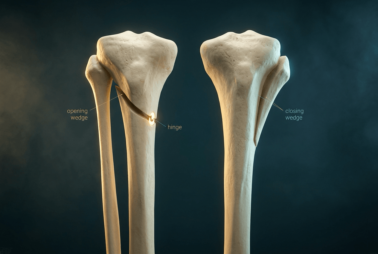
Locating the CORA decides where to cut; the next decision is how. Opening and closing wedges are not interchangeable - they differ in limb length, union behaviour, the structure at risk and the sagittal side-effect.
- Opening wedge
- Cut on the CONCAVE side and distract it open
- Closing wedge
- Remove a wedge from the CONVEX side and close it
- Opening wedge
- Lengthens
- Closing wedge
- Shortens
- Opening wedge
- Needs graft or substitute to fill the gap; slower union
- Closing wedge
- Bone-on-bone, no graft; faster and more inherently stable union
- Opening wedge
- Single cut, easier fine-tuning, but relies on an intact far hinge
- Closing wedge
- Two cuts, less adjustable but inherently stable
- Opening wedge
- Medial opening-wedge HTO increases posterior tibial slope and can cause patella baja
- Closing wedge
- Lateral closing-wedge HTO risks the common peroneal nerve and usually needs a proximal fibular osteotomy or division
Wedge magnitude (rule of thumb). Each degree of coronal correction shifts the weight-bearing line roughly 3 to 4 mm across the tibial plateau (Dugdale), and any joint-space opening from lateral laxity must be subtracted to avoid over-correction. The opening-wedge gap height is approximately the osteotomy width multiplied by the tangent of the correction angle. Choose the wedge by the desired length change and the cortex you wish to preserve.
Acute versus Gradual Correction
The CORA construction says where the deformity is; it does not say how fast the soft tissues will tolerate having it removed. That is a separate decision, and it is the one that determines whether the correction is done with a plate in a single sitting or with a frame over weeks.
Acute correction, internally fixed suits a single-apex deformity of modest size in good soft tissue, with a healthy limb length. It is one operation, no frame, and the correction is set at the moment of fixation - which is also its weakness, because a planning error is now built in.
Gradual correction with a circular frame or hexapod suits large corrections, multi-apical deformity, stiff or scarred soft tissue, an associated limb-length discrepancy, and any correction where nerve or vessel stretch is a genuine concern. Its real advantage is that the correction is adjustable after the operation, so imperfect planning can be recovered.
The numbers that govern distraction.
- Value
- 5-7 days (shorter in young children, longer in adults and poor biology)
- Why
- Lets the regenerate begin to organise before it is pulled
- Value
- About 1 mm/day
- Why
- Faster risks a thin or absent regenerate; slower risks premature consolidation
- Value
- 0.25 mm four times a day
- Why
- The same daily rate delivered in smaller increments makes a better regenerate than one large turn
- Value
- Roughly 30-45 days of frame time per centimetre gained
- Why
- The figure used to counsel patients about total treatment length
Why the pace matters clinically. Nerve is the limiting tissue: acute correction of a large angular deformity - particularly varus taken into valgus at the proximal tibia, where the peroneal nerve is tethered at the fibular neck - stretches the lateral structures over a short interval and produces palsy. Gradual correction distributes the same lengthening over weeks and is the standard answer when the correction is large.
What to watch during a frame correction. Joint subluxation during correction or lengthening (the knee and ankle can be dragged out of position by the soft tissues), equinus contracture during tibial lengthening, premature consolidation, delayed consolidation and pin-site infection, which is near-universal at some level and is managed by pin-site care and oral antibiotics before it becomes a ring sequestrum.
Correcting the Growing Skeleton
In a child with a physis still open, the deformity can be corrected by steering growth rather than by cutting bone - and the analysis that decides it is the same analysis.
Guided growth (temporary hemiepiphysiodesis) with a tension-band plate or staples tethers one side of the physis and lets the other side catch up. It is low-morbidity, does not require a frame, and is reversible by removing the implant.
It requires two things: enough growth remaining to deliver the correction, and a deformity that is in the bone. A high JLCA means the malalignment is intra-articular or ligamentous, and no amount of physeal tethering will address it. The rate of correction is roughly a degree a month around the knee, so the remaining growth has to be checked against the size of the deformity before promising a result.
Rebound after implant removal is common, particularly in the younger child and after large corrections; slight overcorrection and continued surveillance until maturity are the usual responses.
When guided growth cannot work - the physis is closed, the deformity is too large for the growth remaining, or the apex is diaphyseal rather than physeal - the plan reverts to osteotomy, planned exactly as above.
Multi-apical disease deserves naming. Rickets and other metabolic bone disease, multiple hereditary exostoses and skeletal dysplasia produce several CORAs in one bone, and correcting only the most obvious one leaves the limb bent. Treat the metabolic cause first where there is one, because operating on unhealed rickets invites recurrence.
Complications
Planning errors, and what each one produces
- What the patient ends up with
- CORA placed at the knee, osteotomy at the wrong level
- What the patient ends up with
- Six degrees of femoral valgus diagnosed that does not exist
- What the patient ends up with
- The intra-articular component corrected in bone as well - over-correction
- What the patient ends up with
- Straight bone, residual mechanical axis deviation
- What the patient ends up with
- Sagittal deformity or slope change missed entirely
- What the patient ends up with
- Coronal deformity corrected, torsional deformity left or worsened
- What the patient ends up with
- Oblique joint line, shear across the plateau, poorer durability
Intraoperative and early complications
Lateral hinge fracture in medial opening-wedge HTO is the commonest technical complication - 26 of 104 knees (25%) in Takeuchi's series. His classification is the one used: type I reaches just proximal to or within the proximal tibiofibular joint and behaves benignly (no complications in 19 patients); type II runs distal to the tibiofibular joint and is unstable - 2 of 5 went on to delayed union with 3° and 7° of correction loss; type III is an intra-articular lateral plateau fracture. Type II and III need protected weight-bearing until callus is visible, and may need a lateral buttress screw or plate.
Protecting the hinge. Aim the cut at a point just distal to the tip of the fibular head, leave a hinge of intact lateral cortex around 1 cm wide, protect it with a K-wire before opening the gap, and open slowly.
Common peroneal nerve injury. The nerve winds around the fibular neck, so it is at risk during a lateral closing-wedge HTO, during any fibular osteotomy or division, and from acute correction of varus into valgus, which lengthens the lateral side of the limb. Cut the fibula either within about 1 cm of the proximal tibiofibular joint or at the junction of the middle and distal thirds, and stay out of the neck. A foot drop after correction is a stretch injury until proven otherwise: release the correction rather than observe it.
Popliteal neurovascular injury during the posterior cortical cut: flex the knee to relax the neurovascular bundle, place a retractor on the posterior cortex, and never complete the posterior cut blind.
Compartment syndrome after proximal tibial osteotomy, particularly with a closing wedge and fibular osteotomy, and infection and thromboembolism, as for any periarticular osteotomy.
Deformity-specific and late complications
- Under-correction is the commonest cause of failure after HTO for medial OA and is worse in the overweight patient
- Over-correction, especially in a valgus knee taken past neutral into varus, sharply loads the opposite compartment
- Secondary translation deformity from a Rule 3 correction: the angle is right, the axis is not
- Joint line obliquity, either left uncorrected or created by forcing the whole correction through one bone
- Unintended slope change, in the directions set out under the sagittal plane above
- Patella baja after opening-wedge HTO, which complicates a later arthroplasty
- Delayed union or non-union at the gap of an opening wedge, in proportion to gap size and smoking
- Loss of correction, usually through hinge collapse or premature loading
Postoperative Care
Rehabilitation protects the osteotomy hinge and fixation while restoring motion.
Weight-bearing. Opening-wedge osteotomies, a tension construct relying on the intact far hinge, are usually protected with toe-touch or partial weight-bearing for about 6 weeks; closing-wedge and compressed constructs, being more inherently stable, allow earlier loading at the surgeon's discretion. Premature full load can fracture or collapse the far cortex and lose the correction.
Motion. Early knee range of motion prevents stiffness, with quadriceps activation and patellar mobilisation, especially after opening-wedge HTO where patella baja can develop.
The gap and the frame. Opening wedges are filled with autograft, allograft or substitute and monitored for delayed union at the gap. Gradual correction needs pin-site care, strut or ring adjustments to the planned schedule, and serial alignment radiographs.
Imaging and the rest. Check correction and union with standing long-leg films, and confirm the mechanical axis has reached the planned target rather than just "looks straight". Thromboprophylaxis is per unit protocol, and temporary external fixation is removed once internal fixation or union is secure.
Outcomes and Prognosis
Alignment is the dominant survival factor. After valgus tibial osteotomy, achieving at least about 8° valgus (slight overcorrection) gave about 90% 5-year and at least 65% 10-year survival; under-correction in an overweight patient dropped 10-year survival to about 19% (Coventry 1993).
Disease modification. Frontal-plane malalignment multiplies the odds of compartmental OA progression 4-5 fold (Sharma 2001), so restoring the mechanical axis genuinely slows medial (or lateral) wear, not just symptoms.
Valgus correction is different. Distal femoral osteotomy should aim for neutral, since over-correction into varus sharply overloads the medial compartment (Wu 2022).
Conversion to arthroplasty. A prior osteotomy does not preclude later TKA but can modestly increase technical difficulty; osteotomy is best viewed as an arthroplasty-sparing, time-buying procedure in the young active patient.
Prognostic modifiers. Obesity, smoking, large corrections, advanced cartilage loss and inaccurate planning all worsen outcome; accurate CORA-based planning and adequate (not excessive) correction optimise it.
Guidelines, Registries & Global Practice
Deformity analysis using the Paley CORA framework is the internationally accepted standard and is examined in essentially every national orthopaedic curriculum. Practice differs mainly in imaging access, fixation philosophy and resource availability rather than in the underlying geometry, which does not vary by country.
Global Epidemiology and Burden
- Symptomatic knee osteoarthritis affects an estimated 250 to 300 million people worldwide, and frontal-plane malalignment is one of the strongest modifiable mechanical risk factors for progression (Sharma et al, JAMA 2001).
- Realignment osteotomy volume is rising in younger, active patients (typically under 60) as an arthroplasty-sparing option, with the highest relative use in regions where joint preservation is prioritised.
- Post-traumatic and developmental (rickets, Blount disease, physeal injury, skeletal dysplasia) deformities form a larger share of the caseload in limited-resource and high-prevalence settings.
Society Guidance and Frameworks (side by side)
- Position on realignment around the knee
- Osteotomy is an accepted option for unicompartmental OA with malalignment in younger, active patients; emphasises full-length weight-bearing alignment assessment
- Position on realignment around the knee
- Recognises osteotomy as a joint-preserving alternative to arthroplasty in selected younger patients; alignment correction central to decision-making
- Position on realignment around the knee
- Provides the operative technique standards and the Paley malalignment test / CORA planning method as the reference for osteotomy execution
- Position on realignment around the knee
- Consensus supports osteotomy for early compartmental OA with correctable malalignment; target alignment individualised, avoiding routine large overcorrection
The named-society positions above are guideline-level statements rather than primary trial data.
Registry and Outcome Notes
- National arthroplasty registries (NJR UK, AJRR US, AOANJRR Australia, SHAR Sweden, NZJR New Zealand) record prior osteotomy as a factor in subsequent knee arthroplasty; prior osteotomy does not preclude later conversion but can modestly increase technical complexity.
- Long-term cohort data (Coventry et al, JBJS 1993) anchor the principle that adequate, slightly over-corrected alignment is the dominant survival factor after valgus tibial osteotomy.
High- vs Limited-Resource Practice Variation
- Typical workflow
- Long-leg weight-bearing or EOS imaging, digital templating (TraumaCad, Bone Ninja, mediCAD), patient-specific cutting guides and locking-plate fixation; CT for rotational and 3D analysis
- Typical workflow
- Stitched/composite plain films or clinical alignment assessment, manual acetate templating, circular (Ilizarov/Taylor Spatial Frame) or monolateral external fixators that allow gradual, adjustable correction without advanced imaging
Gradual correction with circular frames remains a globally important technique because it tolerates planning imprecision, corrects multi-apical and large deformities, and manages associated limb-length discrepancy and soft-tissue contracture.
Controversies & Areas of Uncertainty
Target alignment - neutral versus constitutional varus. The traditional instruction is to overcorrect a varus knee into slight valgus, to the Fujisawa point supported by Coventry's survival data. The counter-argument from constitutional varus is that a proportion of people are natively varus and have always been, and that driving them to mechanical neutral is neither necessary nor comfortable. There is no trial that settles it; correction is individualised to native alignment, cartilage state and activity rather than to a fixed formula. The valgus knee is the other way round: distal femoral osteotomy should generally aim only for neutral or very slight varus, because the biomechanical data show that pushing into varus may accelerate medial OA.
Single-level versus double-level correction. There is a working threshold, and it is a predicted post-correction MPTA above 95°: beyond that the plateau is tilted enough that the joint line becomes oblique. Akamatsu compared 34 knees corrected by opening-wedge HTO alone with 34 by double-level osteotomy, all with a predicted MPTA above 95°: joint line obliquity rose from 1.4° to 6.3° after HTO alone but stayed at 1.0° to 1.3° after double-level correction. Joint-line obliquity left uncorrected (a single-level fix of a double-level deformity) is associated with shear and less durable results, which is the rationale for double-level osteotomy in the young.
The ceiling on that result matters. At two years the Knee Society and KOOS scores did not differ between the groups, and only the Lysholm score favoured double-level correction. The geometric case is therefore proven and the clinical case, at two years, is not; the argument for operating at two levels in a young patient rests on a durability advantage that no randomised trial has yet demonstrated.
Acute internal fixation versus gradual external correction. Acute correction with plates is faster and avoids prolonged frame time but is less forgiving of planning error; gradual correction with circular frames allows fine adjustment and large or multi-apical correction but carries pin-site and patient-tolerance burden. The choice is driven by deformity magnitude, soft tissue and resources, not by a single right answer.
Sagittal-plane consequences. Medial opening-wedge HTO tends to increase posterior tibial slope and can alter patellar height; these secondary effects are real and must be planned for, but the precise magnitude and clinical significance remain debated.
MCQ Practice Points
Q: What is the CORA and why is an osteotomy at it ideal? A: The centre of rotation of angulation - the point where the proximal and distal segment axes of the deformed bone intersect, which is the apex of the deformity. Cutting there, and hinging there, gives pure angular correction with no translation (Paley Rule 1).
Q: What are the normal values of the mLDFA, aLDFA and MPTA? A: mLDFA 87° (85-90°), aLDFA 81° (79-83°), MPTA 87° (85-90°). The 6 to 7 degree gap between the two femoral values is the anatomic-mechanical angle; measuring off the shaft and comparing with 87 invents valgus that is not there.
Q: Where does the mechanical axis normally cross the knee? A: About 8 to 10 mm MEDIAL to the knee centre, with a spread of roughly 7 mm - not through the centre. A mechanical axis restored to the exact midpoint of the knee is therefore very slightly valgus relative to the population mean.
Q: A varus knee has a normal mLDFA and MPTA but a JLCA of 6°. Where is the deformity? A: In the joint - intra-articular cartilage loss or collateral laxity, not the bone. A bony osteotomy alone will not correct it, and failing to subtract the intra-articular component before sizing the wedge produces over-correction.
Q: Why is a high tibial osteotomy a Paley Rule 2 osteotomy? A: Because the CORA of a varus knee sits at the joint line while the cut is made below it. The correction axis stays at the CORA, so the shaft has to translate laterally as the angle is corrected. That translation is planned geometry, not a complication.
Q: You correct 10° at an osteotomy 3 cm from the CORA. How much translation is required? A: About 5 mm. Translation = 2 x d x tan(half the correction) = 2 x 30 mm x tan 5° = 5.2 mm. Double the distance and you double the translation, which is why the cut is made as close to the apex as fixation allows.
Q: What number tells you to correct at two levels rather than one? A: A predicted post-correction MPTA above 95°. Beyond that the plateau is tilted enough to leave an oblique joint line - obliquity rose from 1.4° to 6.3° after single-level HTO in that group, and stayed unchanged after double-level correction.
Q: What is the alignment target for an HTO in medial-compartment OA, and how does it differ for a valgus DFO? A: Deliberate slight overcorrection into valgus for varus-OA HTO - the Fujisawa point at about 62% of plateau width from medial, with Coventry's survival data supporting at least 8° of valgus. A valgus distal femoral osteotomy aims only for neutral, because overcorrection into varus sharply overloads the medial compartment.
Q: A malunion shows 20° varus on the AP and 20° procurvatum on the lateral. What is the true deformity? A: About 27°, in an oblique plane roughly midway between the two. The tangent of the true angle is the square root of the sum of the squared tangents of the components - so it is always bigger than either view and always smaller than their sum.
Exam Viva Scenarios
Practise clinical reasoning and management decisions out loud
“You are shown a long-leg standing radiograph of a 55-year-old with medial compartment osteoarthritis. The mLDFA is 87°, the MPTA is 82°. Analyse this deformity.”
“A young adult has valgus malalignment with an mLDFA of 93° and an MPTA of 93°. How do you analyse and plan the correction?”
“Explain Paley's three osteotomy rules.”
“A 28-year-old has a mid-diaphyseal tibial malunion two years after a nailed fracture. The AP shows 20° of varus and the lateral shows 20° of procurvatum. He also complains that his foot points inwards. How do you analyse and correct this?”
“A 9-year-old is referred with progressive bilateral genu varum. How do you assess and treat?”
Normal Joint Orientation Angles
- mLDFA: 87° (85-90°) - mechanical lateral distal femoral angle
- aLDFA: 81° (79-83°) - the ANATOMIC version, 6-7° smaller
- MPTA: 87° (85-90°); LDTA: 89° (86-92°); mLPFA: 90° (85-95°)
- JLCA: 0-2°, abnormal above 3°
- MAD: about 8-10 mm MEDIAL to the knee centre, not zero
Sagittal Angles
- PDFA: 83° (79-87°)
- PPTA: 81° (77-84°)
- ADTA: 80° (78-82°)
- Posterior tibial slope: 7-10°, roughly 90 minus PPTA
CORA Determination
- Draw the proximal and distal axes OF THE ABNORMAL BONE, not of the limb
- Intersection = CORA; the angle between them = the magnitude
- Oblique-plane deformity still has ONE CORA; multi-apical disease has several
- Axes parallel but offset = no CORA at all, a pure translation deformity
Paley Osteotomy Rules
- Rule 1: cut AND correction axis at the CORA = pure angulation
- Rule 2: correction axis at the CORA, cut elsewhere = obligatory translation
- Rule 3: both away from the CORA = axes parallel but offset, residual malalignment
- Translation owed = 2 x d x tan(half the correction)
- HTO is a Rule 2 osteotomy
Localising the Deformity
- Abnormal mLDFA = femoral
- Abnormal MPTA = tibial
- Both abnormal, or predicted MPTA above 95° = double-level
- Normal angles with raised JLCA = intra-articular, not bone
HTO Planning for Varus OA
- Overcorrect into slight valgus - Fujisawa point at about 62% of plateau width
- CORA at the joint line, cut below it, shaft translates laterally
- Each degree of correction shifts the weight-bearing line 3-4 mm (Dugdale)
- Leave a 1 cm lateral hinge; hinge fracture occurs in about 25% (Takeuchi)
- Opening wedge increases posterior slope and can cause patella baja
DFO Planning for Valgus
- Target neutral to only slight varus - never mirror the HTO overcorrection
- CORA at the distal femur
- Lateral opening or medial closing wedge
- Check the sagittal and rotational planes before committing
Gradual Correction Numbers
- Latency 5-7 days before distraction
- Rate about 1 mm/day, given as 0.25 mm four times daily
- Consolidation index roughly 30-45 days per centimetre
- Favoured for large, multi-apical, rotational or shortened deformity
Evidence Base
Mechanical Axis Deviation - The CORA Concept (Landmark)
- Defined the apex of deformity using individual mechanical axis lines of each bone segment and joint reference lines of hip, knee and ankle
- Osteotomy at the apex (CORA) requires angulation only; an osteotomy proximal or distal to the apex additionally requires translation to correct accurately
- Frontal-plane angular deformity produces both mechanical axis deviation and malorientation of adjacent joints
Preoperative Planning for High Tibial Osteotomy
- Each degree of tibiofemoral angulation shifts the weight-bearing line 3 to 4 mm across the tibial plateau
- Every 1 mm of lateral tibiofemoral joint separation adds roughly 1 degree of varus, which must be subtracted to avoid overcorrection
- Provided a reproducible algorithm to calculate wedge size based on tibial and femoral length
Normal Axial Alignment of the Lower Extremity (Reference Values)
- In 25 normal young men the knee sat in a mean 1.1 to 1.5 degrees of varus between the tibial and femoral mechanical axes
- The knee transverse axis was a mean 2.6 to 3.0 degrees from perpendicular to the tibial mechanical axis
- The femoral anatomic axis does NOT pass through the centre of the knee, confirming the anatomic-mechanical offset
Malalignment Drives Knee OA Progression (Landmark Cohort)
- Varus alignment gave a 4-fold increase in odds of medial OA progression (adjusted OR 4.09, 95% CI 2.20-7.62) over 18 months
- Valgus alignment gave a near 5-fold increase in odds of lateral OA progression (adjusted OR 4.89, 95% CI 2.13-11.20)
- Greater malalignment severity correlated with greater joint-space loss and functional decline
Long-Term Survivorship After Valgus Tibial Osteotomy
- 87 valgus tibial osteotomies, median follow-up 10 years, with arthroplasty as the failure endpoint
- Valgus angulation of at least 8 degrees at 1 year gave a 90 percent 5-year and at least 65 percent 10-year survival
- Inadequate correction (under 8 degrees valgus) in an overweight patient dropped 10-year survival to 19 percent
Correction Angle in Distal Femoral Osteotomy for Valgus
- Compartment stresses were balanced near a neutral hip-knee-ankle angle of 178 to 180 degrees
- Correction into varus sharply raised medial compartment stress (90.9 percent increase) versus lateral unloading (19.3 percent)
- Optimal valgus-knee target is neutral to only slight varus (0 to 2 degrees); overcorrection risks medial OA
How Much Translation an Off-CORA Osteotomy Owes
- Translation required = 2(tan a x d), where a is 50 percent of the desired angular correction and d is the distance of the osteotomy from the CORA
- Simulated osteotomies at the distal femur, proximal tibia and distal tibia in nine limb scanograms all correlated strongly with the algorithm (correlation coefficients 0.93 to 0.99, p less than 0.0001)
- Translational deformity increases with distance from the CORA, so cutting as close to the apex as fixation allows minimises the correction owed
Joint Line Obliquity After Single- Versus Double-Level Correction
- 34 knees with opening-wedge HTO alone versus 34 with double-level osteotomy, all with a predicted MPTA above 95 degrees, matched for age, BMI and hip-knee-ankle angle
- Joint line obliquity rose from 1.4 to 6.3 degrees after HTO alone but was unchanged after double-level osteotomy (1.0 to 1.3 degrees)
- Knee Society and KOOS scores did not differ at 2 years and second-look cartilage findings were similar; only the Lysholm score favoured double-level correction, so the demonstrated advantage is geometric rather than clinical at this follow-up
Lateral Hinge Fracture After Opening-Wedge HTO
- Lateral cortex fractures in 26 of 104 knees (25 percent): 19 type I, 5 type II, 2 type III
- Type I (at or just proximal to the proximal tibiofibular joint) caused no complications in any of the 19 patients and took the standard rehabilitation protocol
- Type II (distal to the proximal tibiofibular joint) gave delayed union in 2 of 5 patients with correction loss of 3 and 7 degrees, so type II and intra-articular type III fractures need protected weight-bearing
Positioning Error on the Long-Leg Radiograph
- 180 lower limbs imaged both on full-length weight-bearing radiographs and with EOS, comparing measured mechanical tibiofemoral angle between the two
- Error correlated with sagittal-plane rotation (knee flexion; r = 0.368, p less than 0.001) while axial-plane rotation alone did not correlate
- The flexion effect was confined to limbs with patellar rotation above 3 percent; with a strict patella-forward position the measurement was accurate even with a fixed flexion contracture