Cheiralgia Paresthetica
- Wartenberg's Syndrome is purely sensory (Pain/Paresthesia in dorsal webspace).
- It is often caused by tight handcuffs, watches, or bracelets ('Handcuff Neuropathy').
- The nerve is compressed between the Brachioradialis and ECRL tendons during pronation.
- Finkelstein's test can be positive in BOTH Wartenberg's and De Quervain's.
- Differentiation relies on Tinel's sign and sensory testing.
- Treatment is largely non-operative; surgery often leads to painful neuromas.
- “Do NOT confuse Wartenberg's Syndrome (Radial) with Wartenberg's Sign (Ulnar).
- “The Superficial Radial Nerve is purely sensory and superficially located, making it vulnerable to external compression.
- “Surgical release has a high rate of failure or recurrence due to scar tissue.
Overview
Wartenberg's syndrome, or cheiralgia paraesthetica, is an entrapment neuropathy of the superficial branch of the radial nerve (SBRN) in the distal forearm. It presents with pain, numbness and paraesthesia over the dorsoradial aspect of the hand.
Two confusions to avoid. The syndrome is distinct from Wartenberg's sign, which belongs to a different nerve. It must also be carefully differentiated from De Quervain's tenosynovitis, as the two often coexist or mimic each other.
Wartenberg's syndrome is a compression neuropathy of the superficial radial nerve: dorsal hand pain and paraesthesia, Tinel's positive over the nerve, and no motor weakness.
Wartenberg's sign belongs to the ulnar nerve. The little finger sits abducted and cannot be adducted, because a weak third palmar interosseous (ulnar nerve) leaves extensor digiti minimi (radial nerve) unopposed. It is seen in ulnar nerve palsy.
Pathophysiology and Mechanisms
The course. The SBRN leaves the main radial nerve at the elbow, at the level of the radiocapitellar joint, and runs down the forearm deep to the brachioradialis muscle belly. At the junction of the middle and distal thirds of the forearm it pierces the deep fascia between the brachioradialis and ECRL tendons, becoming subcutaneous approximately 9cm proximal to the radial styloid. Distally it runs superficial to the anatomical snuffbox to supply the dorsum of the hand.
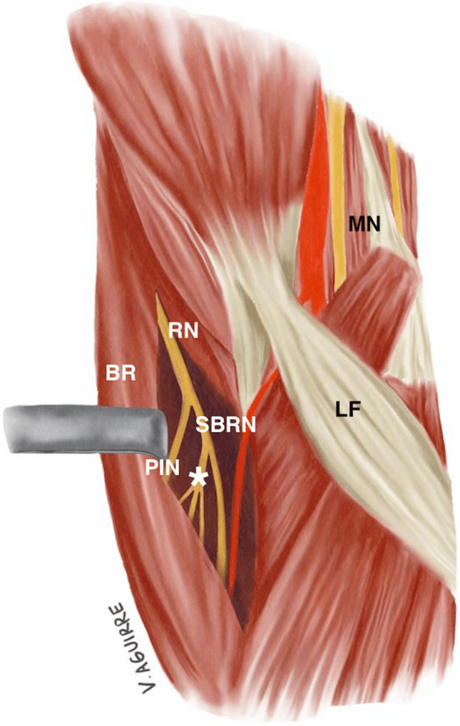
The compression site. The fascial band connecting brachioradialis and ECRL is where the nerve exits, and it is the most common site of compression, unlike carpal tunnel syndrome, which is at the wrist. Two things happen to the nerve there:
- Scissoring - during pronation the ECRL crosses under the brachioradialis, effectively scissoring the nerve between the tendons
- External compression - because the nerve becomes subcutaneous here, it is easily compressed by watches, handcuffs or casts
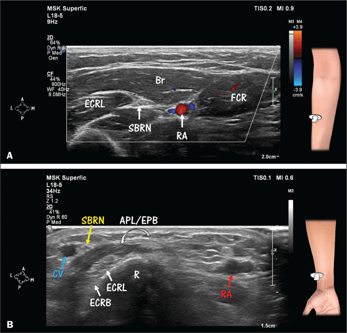
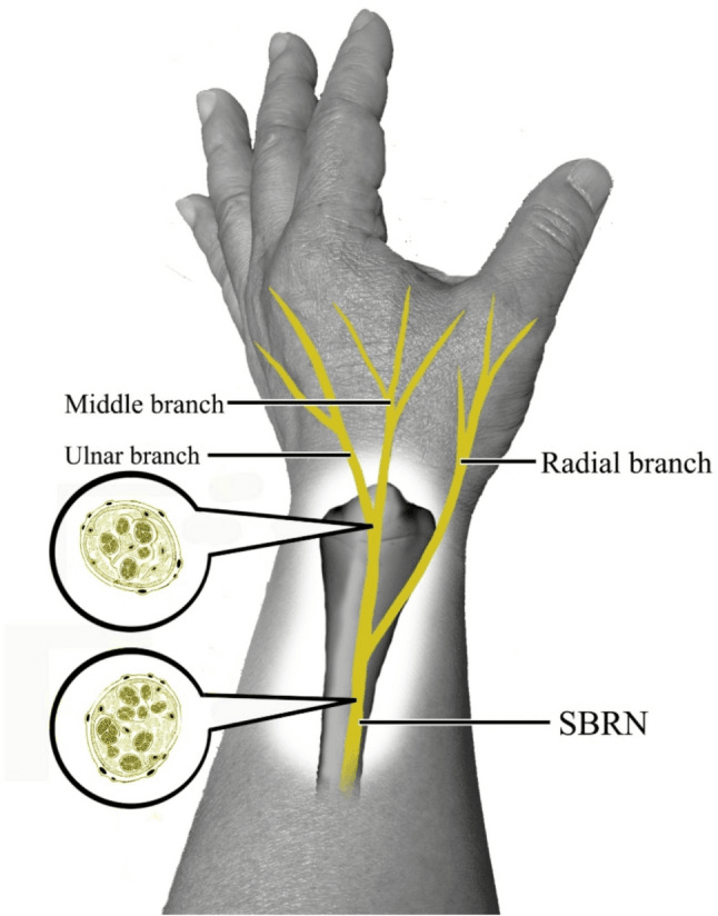
The Cephalic Vein Relationship: Injury During Venous Access
Why venepuncture is a genuine cause. As the SBRN becomes subcutaneous over the distal dorsoradial forearm and wrist, its terminal branches run immediately alongside, and repeatedly cross, the cephalic vein in the subcutaneous fat. Nerve and vein are millimetres apart in the same superficial plane, so a procedure aimed at the vein readily catches the nerve. Cheiralgia paraesthetica can therefore follow a routine blood draw, a drip or vein surgery, which makes this a high-yield, ward-relevant mechanism.
- Mechanism
- Needle spears or tethers a branch lying next to the cephalic vein
- Prevention
- Avoid the dorsoradial wrist; heed sudden radiating pain and withdraw
- Mechanism
- Open dissection near the radial styloid divides or scars branches
- Prevention
- Identify and protect the nerve; dissect under direct vision
- Mechanism
- The same branches cross the compartment and are cut/scarred
- Prevention
- Find and protect the SBRN before opening the sheath
The warning sign. Contact with the nerve during cannulation produces a sudden electric shock radiating to the thumb and dorsal hand. Radiating pain on needling the "intern's vein" is the cue to stop and reposition immediately rather than push on. A patient who develops dorsoradial burning and numbness after a wrist venepuncture or cannula has cheiralgia paraesthetica until proven otherwise.
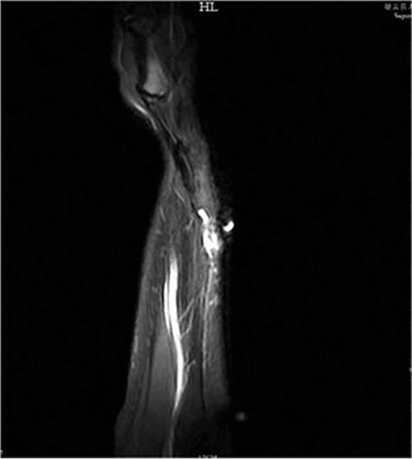
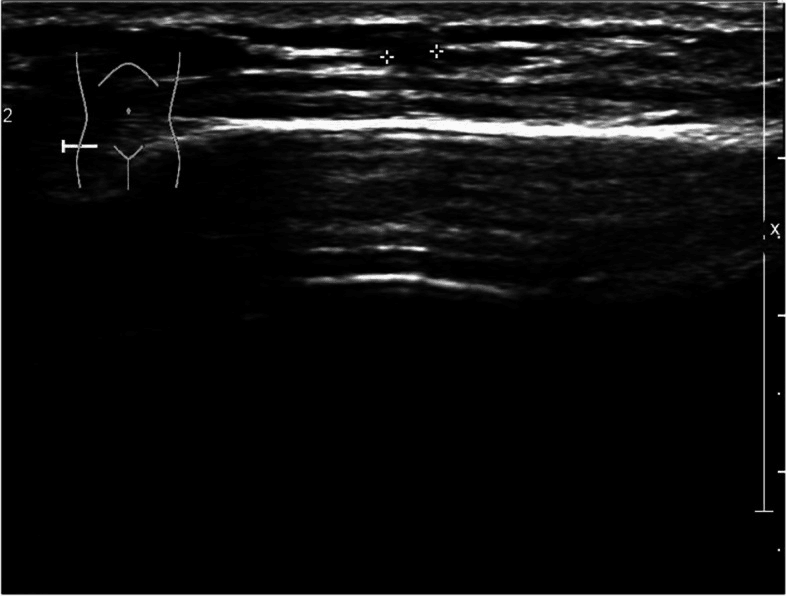
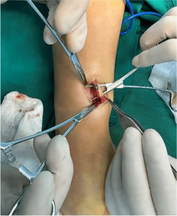
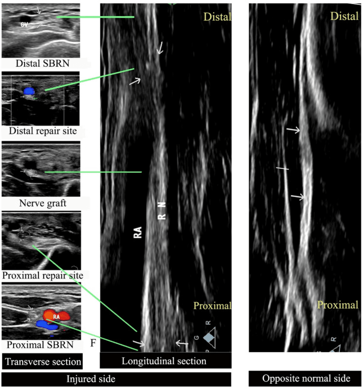
Compression by the vein itself. Cephalic-vein thrombophlebitis after venous injury can crowd and directly compress the adjacent SBRN at the distal radial forearm, another avoidable iatrogenic mechanism after venous access. Ultrasound allows this vascular compression to be distinguished from primary fascial entrapment. The three images below come from one such case.
Classification Systems
Aetiological types. The causes fall into four groups:
- Compressive - external: watch, cast, handcuffs
- Dynamic - repetitive pronation and supination (scissoring)
- Traumatic - direct blow, or venepuncture injury, since the cephalic vein is close
- Scarring - after De Quervain's release (neuroma)
Iatrogenic injury is a significant cause of litigation.
Dellon's classification (neuroma). Severity is graded by what accompanies the pain, and Type III indicates more severe axonal damage:
- Type I - pain in the nerve distribution
- Type II - pain and Tinel's sign
- Type III - pain, Tinel's sign and loss of static two-point discrimination
Clinical Assessment
History. Burning, shooting pain over the dorsum of the thumb and index finger, aggravated by wrist movement, tight sleeves and watches. There are no night symptoms, unlike carpal tunnel syndrome, and symptoms are often purely sensory, with no motor weakness. Ask about handcuffs or a new watch.
Examination. Tinel's sign is the most reliable sign, and Finkelstein's test is the trap. Compare sensation with the contralateral side, which is crucial.
- Tinel's sign - positive over the nerve, approximately 9cm proximal to the radial styloid
- Finkelstein's test - often positive, because traction on the nerve causes pain; palpate the tendons (De Quervain's) against the nerve (Wartenberg's) to tell them apart
- Dellon's test - wrist hyperextension with thumb adduction stretches the nerve
- Hyperpronation test - sustained forearm hyperpronation reproduces symptoms (Dellon and Mackinnon)
- Sensation - reduced over the dorsal first web space
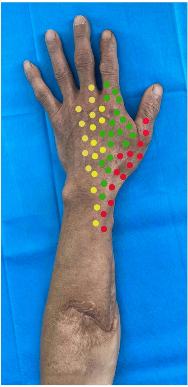
Mapping the territory. Testing sensation branch by branch across the dorsoradial hand maps the SBRN territory and helps separate Wartenberg's syndrome from C6 radiculopathy, lateral antebrachial cutaneous neuropathy and ulnar-nerve disease.
The differential. The two tendon conditions it is confused with are set against it below.
- Wartenberg's Syndrome
- Nerve Compression (SRN)
- De Quervain's
- Tenosynovitis (1st Comp)
- Intersection Syndrome
- Tenosynovitis (1st/2nd Cross)
- Wartenberg's Syndrome
- Dorsal Radial Forearm
- De Quervain's
- Radial Styloid
- Intersection Syndrome
- 4cm Proximal to Styloid
- Wartenberg's Syndrome
- Burning, Electric
- De Quervain's
- Aching, Mechanical
- Intersection Syndrome
- Crepitus, Squeaky
- Wartenberg's Syndrome
- Positive (Traction)
- De Quervain's
- Positive (Mechanical)
- Intersection Syndrome
- Negative
- Wartenberg's Syndrome
- Positive
- De Quervain's
- Negative
- Intersection Syndrome
- Negative
Imaging and Electrodiagnostics
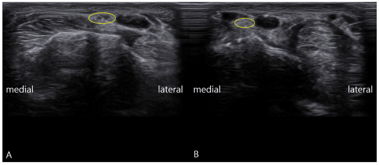
Ultrasound is excellent for visualising the nerve. It shows swelling of the nerve proximal to the fascia and a "notch" sign, can observe the nerve being compressed during pronation, and can exclude a ganglion cyst compressing the nerve. Always look for an occult ganglion if the history is unclear.
Comparing sides. Side-to-side comparison of the symptomatic and contralateral nerves supports a focal neuropathy when electrodiagnostic testing is normal. Scanning can also localise a high-Wartenberg compression in the proximal forearm, separate from the usual distal fascial emergence point.
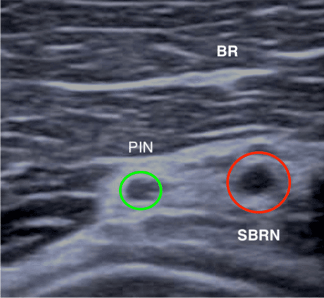
MRI is for excluding other pathology, such as a scaphoid fracture or tumour, and is better for deep lesions or bone. It may show neuritis as high signal but is less sensitive than ultrasound for small superficial nerves.
Nerve conduction studies. The radial sensory nerve action potential (SNAP) may be reduced or absent; compare with the contralateral side, where an amplitude drop greater than 50% is significant. The studies are often normal in dynamic cases, so a normal NCS does not exclude the diagnosis: clinical diagnosis dominates.
The Diagnostic Nerve Block: Confirming the SBRN Is the Pain Generator
A selective nerve block is the way to confirm the site before offering surgery, and the single most useful test before any irreversible operation on this nerve.
Why it matters here. Wartenberg's is a clinical diagnosis with notoriously unreliable confirmatory tests: conduction studies are often normal, especially in dynamic compression, and Finkelstein's test can be falsely positive because it also fires in De Quervain's. Decompression has a high failure rate and neurectomy trades pain for permanent numbness, so before either the superficial radial nerve must be proved to be the pain source.
The block. A small volume of local anaesthetic is placed around, never into, the nerve at the suspected level, ideally under ultrasound guidance because the nerve is tiny and subcutaneous. Substantial temporary relief with the expected sensory change supports the SBRN as a major pain generator. It does not by itself prove that decompression will succeed.
Nerve or tendon. Selective first-dorsal-compartment and perineural blocks can help separate coexisting tendon-sheath and nerve pain. Partial, absent or discordant relief should prompt reassessment for mixed pathology, C6 radiculopathy, thumb CMC arthritis or technical block failure.
Before neurectomy. A concordant block can confirm the intended sensory territory and let the patient experience the expected numbness. The irreversible decision still requires a secure diagnosis and explicit counselling about neuroma and persistent-pain risk.
A negative or only partial response to a well-placed selective SBRN block is a warning sign: the superficial radial nerve is probably not the (whole) problem. Operating on a nerve that a block did not silence is the classic route to a "failed", or worse, "more painful" decompression.
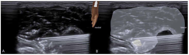
Management Algorithm
Remove the cause. Non-operative care is the mainstay, and removing the external compression is the single most important step: stop wearing watches and bracelets, and loosen casts. The rest of conservative care supports it:
- Splinting - thumb spica or wrist splint to reduce excursion
- Desensitisation - massage, textures
- Corticosteroid injection - around the nerve, avoiding intraneural injection; can be curative
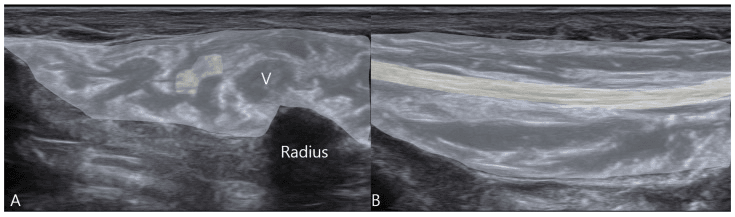
Surgical decompression. Surgery is indicated for failure of conservative care beyond 6 months and persistent pain, and should only be offered if the diagnosis is certain and blocks have confirmed it. The outcome is variable, with a high risk of dissatisfaction, so managing the patient's expectations is the key.
Surgical Technique
Neurolysis. The release is simple, but locating the nerve in scar tissue can be hard.
- Incision - longitudinal over the course of the nerve, 8-10cm proximal to the styloid
- Identify the nerve emerging between brachioradialis and ECRL
- Release - divide the deep fascia binding the two tendons
- Trace the nerve distally, avoiding its branches
- Protect - handle the nerve gently, with vessel loops
- Check that no scissoring remains in pronation
Neuroma. A painful neuroma from previous injury is a salvage situation, and outcomes of neuroma surgery are notoriously unpredictable. The options are burying the nerve end in muscle or bone, capping it, or neurectomy, which accepts numbness to cure pain.
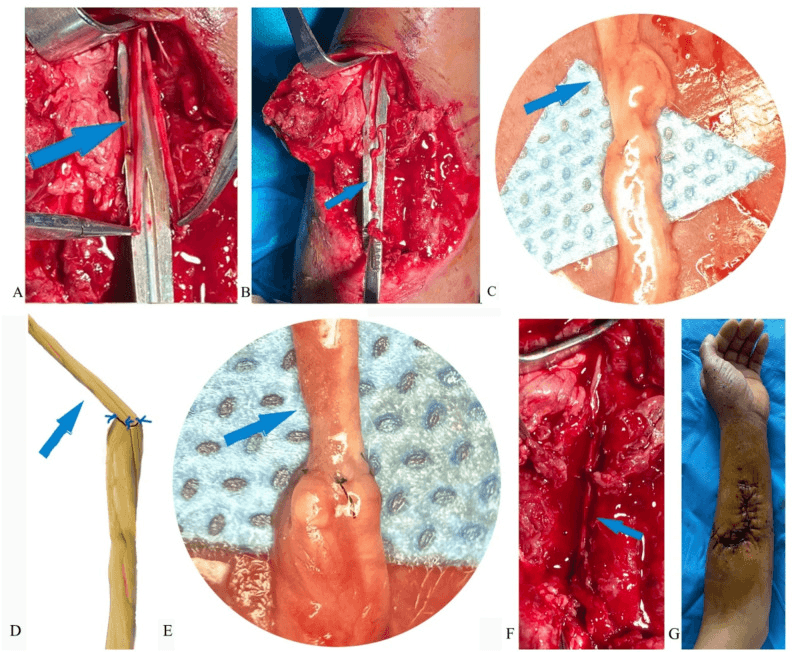
Complications
After surgery. Careful patient selection and meticulous technique are essential.
- Neuroma formation - the most feared complication; extremely painful and difficult to treat
- Incomplete release - failure to fully decompress the nerve leads to persistent symptoms
- Recurrence - scarring can reform the fascial band, causing re-entrapment
- Iatrogenic nerve injury - direct injury during dissection worsens prognosis
- Wound complications - infection, dehiscence or hypertrophic scarring
Untreated disease. Early intervention prevents progression to chronic pain syndromes. Left alone, the condition can bring:
- Chronic pain - persistent burning and dysaesthesia affecting quality of life
- Numbness - progressive sensory loss in the radial dorsal hand
- CRPS - complex regional pain syndrome (type II) may develop
- Hyperaesthesia - increased sensitivity that makes normal activities painful
- Psychological impact - chronic pain affects sleep, work and mental health
Rehabilitation
Weeks 0-2. Immediate gentle range of motion to prevent adhesions, with a splint for comfort only.
Weeks 2-6. Scar massage and texture handling (rice buckets) to prevent hypersensitivity. Fluidotherapy and mirror therapy may be useful for CRPS.
Weeks 6-12. A progressive return to normal activities, including work duties, watching for recurrence as activity escalates and continuing to avoid identified triggers such as watches and tight clothing. The goal is full functional recovery without recurrence, and work modifications may be needed for occupational cases.
Prognosis
Outcomes by treatment. Patient selection is the key determinant of surgical success. The decompression figure comes from the largest surgical series (Lanzetta and Foucher, 1993), where conservative treatment in the same, non-randomised cohort achieved 71% excellent or good results.
- Conservative management - 70-80% resolution with removal of the compressing agent alone
- Corticosteroid injection - 60-70% improvement; may need repeat injection
- Surgical decompression - 74% good or excellent outcomes in well-selected patients
- Neuroma surgery - unpredictable; 50-60% improvement at best
- Chronic cases - may require multidisciplinary pain management
Prognostic factors. Addressing the underlying cause prevents recurrence.
- Duration - shorter duration (under 3 months) is associated with better outcomes
- Aetiology - external compression (watch, handcuff) has the best prognosis
- Iatrogenic cause - post-surgical cases have poorer outcomes because of scarring
- CRPS - a poor prognostic sign requiring specialist input
- Age - younger patients generally recover better
- Psychological factors - anxiety and catastrophising predict poorer outcomes
Recovery time. Serial assessment helps identify patients failing to progress.
- Neurapraxia (external compression) - 6-12 weeks for full recovery
- Axonotmesis (moderate injury) - 3-6 months; may have residual symptoms
- Neurotmesis (severe injury) - permanent deficit unless the nerve is repaired
- After surgery - 3-6 months for the optimal surgical outcome
- Desensitisation programmes - may require 6-12 months of consistent therapy
Guidelines, Registries & Global Practice
Global Epidemiology
- Wartenberg's syndrome is uncommon and under-recognised; precise incidence figures are lacking and most evidence is from case series (largest is 52 cases, Lanzetta & Foucher 1993).
- Coexistent De Quervain's tenosynovitis is reported in roughly half of cases, making mixed presentations the dominant clinical pattern worldwide.
- High-risk groups are consistent across regions: people detained in over-tight handcuffs (the SRN is the most commonly injured nerve - Grant & Cook 2000), wearers of tight watches/bracelets/wristbands, manual workers with repetitive pronation-supination, and post-operative patients after first dorsal compartment release.
Society Guidance (side by side)
- Position on SRN entrapment / cutaneous nerve injury
- No condition-specific guideline; consensus is conservative-first management and meticulous SRN protection during De Quervain's release
- Position on SRN entrapment / cutaneous nerve injury
- Addresses De Quervain's, not Wartenberg's directly; emphasises differentiating tendon from nerve pathology before surgery
- Position on SRN entrapment / cutaneous nerve injury
- Peripheral nerve injury principles: confirm diagnosis clinically, image with ultrasound, escalate to specialist hand unit for surgery
- Position on SRN entrapment / cutaneous nerve injury
- Treat as a focal compression neuropathy: remove external cause, neurolysis only for confirmed refractory entrapment
There is no high-level (Level 1) guideline or RCT for this condition anywhere; management is consensus- and case-series-driven.
Registry Evidence
- This is a soft-tissue nerve condition with no implant, so it is NOT captured by arthroplasty registries (NJR, AJRR, AOANJRR, SHAR). Outcome data come from single-centre series and reviews, not national registries — a key reason the evidence base remains Level 3 to 5.
High- vs Limited-Resource Practice Variation
- High-resource settings: High-resolution ultrasound (and occasionally MR neurography) confirms the diagnosis and excludes ganglion/mass; nerve conduction studies and specialist hand therapy (desensitisation, custom splinting) are available; surgery reserved for confirmed refractory cases.
- Limited-resource settings: Diagnosis is clinical (Tinel's, false-positive Finkelstein, hyperpronation test); first-line management is removal of the offending compressor plus simple splinting and activity modification, which resolves the majority. Imaging and electrodiagnostics are used selectively. This pragmatic pathway is appropriate given that conservative care alone achieves around 70% good/excellent results.
Iatrogenic Prevention (universal principle)
- The SBRN crosses the first dorsal compartment and is at real risk during De Quervain's release. Identify and protect the nerve first, use loupe magnification, and include nerve injury/neuroma in informed consent. A "failed" or "worse after" De Quervain's release should be assumed to be an SRN problem until proven otherwise.
Related pages: Superficial Radial Nerve Anatomy holds the course this syndrome depends on - the emergence between brachioradialis and extensor carpi radialis longus that is compressed by the scissoring of those tendons in pronation - and Radial Nerve Anatomy places it in the parent trunk; De Quervain Tenosynovitis is the diagnosis this one hides behind, coexisting in half of Lanzetta's cases and sharing a positive Finkelstein test, which is why a first dorsal compartment release performed without excluding it is the classic failure and the classic claim; Intersection Syndrome completes the differential for dorsoradial forearm pain; Radial Tunnel Syndrome, Posterior Interosseous Syndrome and Posterior Interosseous Nerve Anatomy are the motor members of the same family, and separating sensory from motor is the first step in naming any radial neuropathy; Ulnar Nerve Anatomy and Cubital Tunnel Syndrome cover Wartenberg's SIGN, a different entity in a different nerve that shares only the eponym; Carpal Tunnel Syndrome is the commonest concurrent compression in the double-crush discussion; Distal Radius Fractures are a frequent iatrogenic and traumatic cause; and Neuroma Management with CRPS of the Hand cover the two outcomes that make failed surgery here so difficult to retrieve.
Controversies & Areas of Uncertainty
Surgical decompression reports around 74% good/excellent results in selected patients (Lanzetta & Foucher 1993), but there are no controlled trials. Failures are frequently due to an incorrect original diagnosis or recurrent perineural scarring. Many surgeons remain cautious because a painful neuroma is worse than the original complaint.
Recurrent perineural scarring after neurolysis has driven interest in adhesion barriers (e.g. amniotic membrane wrapping), but evidence is limited to very small series and the technique is not standard of care.
Some authorities regard Wartenberg's as a clinical diagnosis (Tinel's, false-positive Finkelstein, hyperpronation test); others advocate routine ultrasound. Normal nerve conduction studies do NOT exclude it, particularly in dynamic compression.
Peri-neural corticosteroid can relieve symptoms, but there is no robust trial evidence and intraneural injection risks worsening the nerve. Image guidance is increasingly recommended where available.
MCQ Practice Points
Q: Where does the SRN exit the deep fascia? A: Between the Brachioradialis and ECRL tendons, at the junction of middle/distal thirds of forearm.
Q: Which sign is most specific for Wartenberg's Syndrome vs De Quervain's? A: Tinel's sign over the nerve (and absence of mechanical tenderness over the compartment).
Q: What is the most common cause of 'Cheiralgia Paresthetica'? A: External compression (Watches, Handcuffs).
Q: Which nerve is involved in Wartenberg's SIGN? A: The Ulnar Nerve.
Q: What is the first line treatment? A: Removal of constricting items (Watch, Bracelet) and Splinting.
Q: What is the prognosis after handcuff neuropathy? A: Usually good (neurapraxia, recovery over weeks), but NOT guaranteed - the SRN is the commonest nerve injured by handcuffs and injury can be severe or permanent (Grant & Cook 2000). Observe, reassure cautiously, and document.
Viva Scenarios
Practise clinical reasoning and management decisions out loud
“A patient had surgery for De Quervain's Tenosynovitis 3 months ago. The pain is now WORSE, burning in nature, and shooting to the index finger. Scar is tender.”
“A 25-year-old male complains of numbness over the back of his hand after being arrested last night. Wrists are bruised.”
“A junior registrar tells you the patient has a 'Positive Wartenberg's'. What do they mean?”
“A 45-year-old woman presents with radial wrist pain, positive Finkelstein's test, AND numbness over the dorsal first webspace. Tinel's is positive over the first dorsal compartment.”
Anatomy
- Superficial Radial Nerve (SRN) = pure sensory
- Exits between BR and ECRL at middle/distal third forearm
- Dorsal thumb/1st webspace/index/middle sensation
- Parent = Radial Nerve (divides at radiocapitellar joint)
- Cheiralgia Paresthetica = eponymous name
Clinical
- Burning Pain & Paresthesia (dorsal radial hand)
- Tinel's Positive over SRN (differentiates from De Quervain's)
- Finkelstein can be positive (Mock De Quervain's)
- No motor weakness (pure sensory nerve)
- External compression (watch, cast, handcuffs) common cause
Treatment
- Remove offending item (watch, bracelet, tight cast)
- Splint in neutral to rest nerve
- Desensitization program
- Injection rarely needed
- Surgical release of fascia (last resort if fails 6 months)
Evidence Base
Dellon & Mackinnon: Radial Sensory Nerve Entrapment (Landmark)
- 51 patients with radial sensory nerve entrapment, usually after crush/twisting injury or repetitive pronation-supination
- Pain and burning over the dorsoradial wrist, worse with pinch and grip
- Positive Tinel's sign where the nerve exits the deep fascia, a FALSE-positive Finkelstein test, and a positive hyperpronation provocative test
- Both non-operative and surgical outcomes reported
Lanzetta & Foucher: Largest Surgical Series
- 52 cases of SRN entrapment (Wartenberg's syndrome) treated 1988 to 1992
- Conservative treatment achieved 71% excellent/good results
- Surgical decompression achieved 74% excellent/good results
- De Quervain's disease coexisted in 50% of cases; diagnose Wartenberg's BEFORE operating on the tenosynovitis to avoid complications and medicolegal problems
Grant & Cook: Prospective Handcuff Neuropathy Study
- Prospective 27-month study of 41 patients with hand symptoms attributed to over-tight handcuffs
- Of nerves tested electrodiagnostically: 22 superficial radial, 12 median, 9 ulnar neuropathies
- Clinical and electrodiagnostic correlation was best for superficial radial neuropathy
- Injury to the superficial radial nerve can be SEVERE and PERMANENT, not always self-limiting
Tryfonidis et al.: Anatomical Variation Predisposing to Entrapment
- 20 cadaveric upper limbs studying the SBRN relative to the brachioradialis tendon
- In 4 of 20 limbs the SBRN became subcutaneous by PIERCING the brachioradialis tendon, creating a tendinous band that compressed the nerve and blocked gliding in ulnar flexion
- Communication between SBRN and lateral cutaneous nerve of forearm in 2 limbs may explain the small sensory deficit
- Recommend looking for and releasing this anomaly at surgery
Chang et al.: High-Resolution Ultrasound of Cutaneous Nerves
- Review of high-resolution ultrasound for limb cutaneous nerves and their entrapment syndromes, including the SBRN
- Cutaneous nerves run superficially in the subcutaneous layer, making them vulnerable to entrapment and external compression
- Ultrasound enables direct visualisation and dynamic assessment despite few bony landmarks
- Supports US as a first-line imaging tool for superficial nerve entrapment
Dang & Rodner: Forearm Radial Compression Neuropathies (Review)
- Review of uncommon forearm radial nerve compression neuropathies
- Groups posterior interosseous nerve syndrome, radial tunnel syndrome and superficial radial nerve compression (Wartenberg's syndrome)
- Emphasises that a missed forearm compression diagnosis has far-reaching consequences
- Provides a structured diagnostic approach distinguishing motor (PIN) from sensory (SBRN) pathology
Wartenberg: Original Eponymous Description (Historical)
- Original description of 'cheiralgia paraesthetica' (isolated neuritis of the superficial radial nerve)
- Noted association with external pressure over the dorsoradial wrist
- Pre-PubMed historical reference; not independently indexed
- Established the eponym still in use today