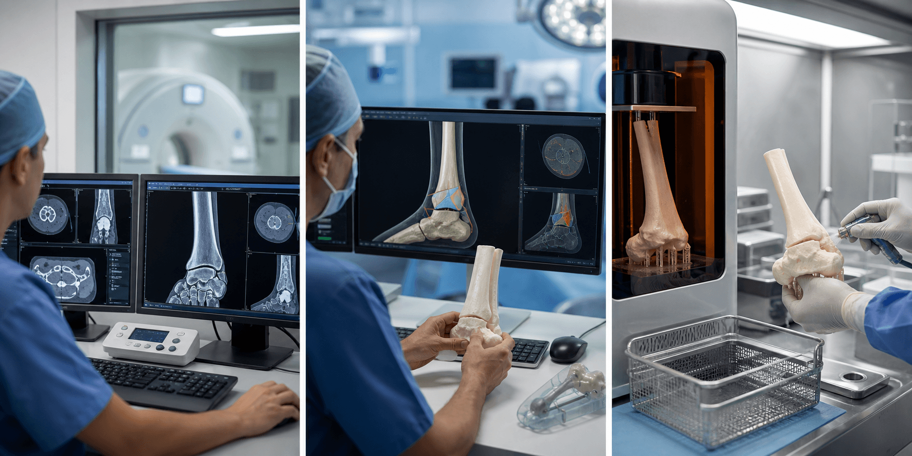
Additive Manufacturing in Orthopaedic Surgery
Anatomical Models: Pre-operative planning, education
Patient-Specific Instruments: Cutting guides, drill guides
Custom Implants: Tumour reconstruction, revision arthroplasty
Key: 3D printing transforms 2D imaging into tangible surgical tools
- CT provides best data for 3D printing (bone segmentation)
- Thin-slice (less than 1mm) CT required for accuracy
- Patient-specific instruments improve implant positioning
- Custom implants for complex reconstruction
- Regulatory requirements for implantable devices
- “DICOM to STL conversion is the key processing step
- “Segmentation quality determines print accuracy
- “Anatomical models reduce operative time in complex cases
- “PSI can improve component alignment in arthroplasty
3D printing is an emerging topic with increasing exam relevance. Understand the workflow from imaging to print, key applications (models, PSI, custom implants), and imaging requirements (thin-slice CT). Know the advantages and limitations of this technology.
PRINTPRINT Framework
Hook:Good orthopaedic printing starts with the imaging dataset, not the printer.
Overview

Three-dimensional printing is a downstream use of imaging, not a separate imaging modality. The quality of the printed model depends first on acquisition, then segmentation, then the clinical relevance of the model that is produced.
For orthopaedics, the strongest indications are complex articular trauma, deformity planning, implant templating, and patient-specific education where spatial understanding genuinely changes decision-making.
Clinical Imaging
Imaging Atlas


PRINTPRINT Workflow
Hook:Remember the 5 stages from image to operating theatre
STERILESTERILE Requirements
Hook:STERILE workflow ensures safe clinical application
Systematic Approach to 3D Printing
The Five-Stage Workflow
Stage 1: Imaging Acquisition
- Thin-slice CT (less than 1mm) provides optimal data
- MRI for soft tissue assessment in complex cases
- DICOM format required for processing
Stage 2: Segmentation and Modelling
- DICOM to STL conversion
- Threshold-based bone segmentation
- Manual refinement for accuracy
- Quality control checks
Stage 3: Design and Planning
- Virtual surgical planning
- Guide design (cutting planes, drill trajectories)
- Implant templating
- Sterilisation considerations
Stage 4: Manufacturing
- Material selection (PLA, nylon, titanium)
- Printer calibration and quality control
- Surface finishing for theatre use
- Sterilisation validation
Stage 5: Clinical Application
- Pre-operative team briefing with model
- Intra-operative guide placement
- Post-operative validation
- Sterilisation: Gamma irradiation or autoclave compatible materials
- Accuracy: Less than 1mm deviation for implantable devices
- Documentation: Full traceability from imaging to implant

Workflow Overview


| Step | Process | Key Considerations |
|---|---|---|
| 1. Image Acquisition | CT/MRI scan | Thin slices (less than 1mm), minimal artefact |
| 2. DICOM Transfer | Send to processing software | Anonymisation, secure transfer |
| 3. Segmentation | Separate structures of interest | Quality determines accuracy |
| 4. 3D Model Creation | Generate surface mesh | STL file format |
| 5. Design/Modification | Add guides, cutting slots | Engineering input for instruments |
| 6. Printing | Additive manufacturing | Material selection for purpose |
| 7. Post-Processing | Cleaning, sterilisation | Regulatory compliance for implants |
Imaging Requirements

| Parameter | Recommendation | Rationale |
|---|---|---|
| Modality | CT preferred for bone | Superior bone-soft tissue contrast |
| Slice thickness | Less than 1mm (ideally 0.5-0.625mm) | Reduces stair-stepping artefact |
| Field of view | Include relevant anatomy | Sufficient margins for planning |
| Metal artefact | MARS protocol if hardware present | Improves segmentation accuracy |
| Contrast | Usually unnecessary for bone | May help tumour delineation |
Clinical Applications

| Application | Benefit | Examples |
|---|---|---|
| Pre-operative planning | 3D visualisation of pathology | Complex fractures, tumour resection |
| Template/trialling | Pre-bend plates, trial implants | Pelvic fractures, spine deformity |
| Patient education | Tangible explanation of surgery | Joint replacement, deformity correction |
| Surgical training | Practice complex procedures | Resident education, rare procedures |
| Intraoperative reference | Anatomical guide in OR | Tumour margins, fracture reduction |


Printing Technologies

| Technology | Mechanism | Applications |
|---|---|---|
| FDM (Fused Deposition) | Extruded thermoplastic | Anatomical models, low-cost |
| SLA (Stereolithography) | UV-cured resin | High detail models, surgical guides |
| SLS (Selective Laser Sintering) | Laser-fused powder | Durable guides, nylon models |
| DMLS (Direct Metal Laser Sintering) | Metal powder laser fusion | Titanium implants |
| EBM (Electron Beam Melting) | Metal powder electron beam | Porous metal implants |
Quality Assurance

| Aspect | Requirement | Verification |
|---|---|---|
| Dimensional accuracy | Less than 1mm deviation | Calliper measurement, CT comparison |
| Anatomical fidelity | Matches patient anatomy | Overlay on source CT |
| Sterilisation | Appropriate for OR use | Validated sterilisation process |
| Material biocompatibility | Non-toxic, implant grade | Material certification |
| Structural integrity | Withstands intended use | Mechanical testing |
Limitations

| Limitation | Explanation | Mitigation |
|---|---|---|
| Time | Days to weeks for complex prints | Early planning, in-house printing |
| Cost | Equipment, materials, expertise | Case selection, shared services |
| Imaging artefact | Metal hardware degrades segmentation | MARS protocols, manual editing |
| Regulatory complexity | Especially for custom implants | Partner with approved manufacturers |
| Learning curve | Segmentation and design skills | Training, dedicated staff |
Choosing the Right Technology
Examiners often probe whether a candidate can select the appropriate technology rather than reflexively reaching for a 3D print. The table below contrasts 3D printing with the alternatives it competes with for the same clinical problems.
| Feature | 3D-printed model/guide | On-screen virtual planning | Intra-op CT navigation | Robotic assistance |
|---|---|---|---|---|
| Tactile model in hand | Yes | No | No | No |
| Pre-bend plates / trial reduction | Yes | No | No | No |
| Intra-operative adjustability | Low (fixed at print) | n/a (planning only) | High | High |
| Real-time accuracy feedback | No | No | Yes | Yes |
| Lead time before surgery | Days to weeks | Hours | None | None |
| Capital cost | Low to moderate | Low | High | Very high |
| Best-suited problem | Complex articular/pelvic trauma, deformity, tumour | Rapid templating, teaching | Pedicle screws, tumour margins | Arthroplasty alignment |
Controversies and Areas of Uncertainty
| Question | Current position | Uncertainty |
|---|---|---|
| Does PSI improve TKA outcomes? | No clinically relevant benefit in routine primary TKA (Level I meta-analysis) | May still help in extra-articular deformity or retained hardware where canals cannot be referenced |
| Do 3D models help complex trauma? | Reduce operative time, blood loss, fluoroscopy in acetabular fractures | Mostly small single-centre series; few multicentre RCTs; outcome (not process) benefit less proven |
| Point-of-care vs industry printing | Models/guides increasingly printed in-house | Implants almost always require certified manufacturers; in-hospital QMS burden is high |
| Custom tumour/revision implants | Allow reconstruction otherwise impossible | Long-term survivorship data immature; failure modes and registry capture incomplete |
| Accuracy standard | Sub-millimetre dimensional fidelity achievable | No universal validation/verification standard; accuracy degrades with metal artefact and poor segmentation |
The single highest-yield controversy: PSI does NOT improve outcomes in routine total knee arthroplasty. Quote the Level I meta-analysis and reserve PSI/3D guides for cases where conventional referencing fails (severe extra-articular deformity, retained hardware, tumour margins).
Guidelines, Registries & Global Practice
| Body / Region | Framework | Practical implication |
|---|---|---|
| FDA (US) | Guidance on technical considerations for additively manufactured devices; point-of-care framework | Anatomical models and guides have clear pathways; patient-matched implants need validated processes |
| EU MDR (Europe) | Custom-made device provisions under MDR 2017/745; notified-body oversight for higher-risk implants | Documentation and conformity assessment scale with device risk class |
| ISO 13485 | Quality management system for medical devices | Required for any site manufacturing implantable or patient-contacting printed devices |
| AO Foundation / EFORT | Education and consensus on computer-assisted and patient-specific planning | Endorse case selection over routine use; emphasise verification against source imaging |
| RSNA-ACR / radiology bodies | Appropriateness and quality criteria for clinical 3D printing in radiology | Standardise indications, segmentation QA, and reporting for point-of-care labs |
Evidence Base

Landmark and Supporting Studies
Key Points
- Trauma planning: Comparative evidence supports reduced operative time, blood loss, and fluoroscopy with pre-operative 3D models in complex acetabular fractures.
- Deformity guides: Meta-analytic Level I evidence shows 3D-printed drill guides improve pedicle screw accuracy and lower radiation exposure.
- PSI in routine TKA: High-quality Level I evidence shows no clinically relevant outcome benefit — value is confined to selected, atypical anatomy.
- Custom implants: Geometry and stiffness matching are real advantages, offset by cost, multi-week lead time, and loss of intraoperative flexibility.
Clinical Decision Scenarios
Practise clinical reasoning and management decisions out loud
“You are planning surgery for a complex acetabular fracture. How might 3D printing assist your pre-operative planning?”
“A patient requires resection of a pelvic chondrosarcoma with reconstruction. How can 3D printing technology assist?”
“You are considering patient-specific instruments (PSI) for a complex total knee replacement in a patient with severe extra-articular deformity from a malunited tibial fracture.”
Imaging Atlas

Workflow
- CT acquisition (less than 1mm slices)
- DICOM to segmentation software
- Generate STL file
- Print and post-process
- Sterilise if for OR use
Applications
- Anatomical models (planning, education)
- PSI (cutting guides, drill guides)
- Custom implants (tumour, revision)
- Pre-contoured plates
Imaging Requirements
- CT preferred for bone
- Slice thickness less than 1mm
- MARS protocol if metal present
- Include relevant anatomy margins
Limitations
- Time (days to weeks)
- Cost (equipment, materials)
- Regulatory for implants
- Requires expertise