Congenital variant in deep posterior compartment | Posteromedial mass that hardens on plantarflexion | MRI is diagnostic | Excise only if symptomatic and refractory
- Not a tumour: it is normal skeletal muscle on MRI (iso-intense to soleus on all sequences) with its own tendon - biopsy is almost never indicated and risks unnecessary morbidity
- Becomes prominent on contraction: the mass or fullness posteromedial to the Achilles appears or hardens when the patient performs a heel rise or resisted plantarflexion - this is pathognomonic on examination
- MRI first for the mass: any unexplained posteromedial ankle "swelling" in a young adult gets an MRI before any invasive step; it maps insertion, excludes sarcoma or ganglion, and shows whether the tibial nerve is compressed
- Most are incidental: the majority of accessory soleus muscles are asymptomatic and require only reassurance; surgery is reserved for clear mechanical pain, documented nerve entrapment, or failure of conservative care in active patients
- Simple excision is effective: when indicated, complete excision from origin to insertion via a posteromedial approach, protecting the tibial neurovascular bundle, gives reliable pain relief and return to sport with low recurrence
- “Always ask the patient to stand on tiptoes or perform resisted plantarflexion during examination - the accessory soleus will visibly and palpably contract and become firmer while a lipoma or cyst will not
- “Tarsal tunnel symptoms (medial heel/plantar paraesthesia worse at night or with activity) in a young athletic patient without trauma should prompt a search for space-occupying lesions including accessory soleus on MRI
- “If you see an incidental "mass" in the deep posterior compartment on an MRI done for other reasons, describe it fully (origin, insertion, relation to neurovascular structures) and reassure the patient and referrer - it is a variant, not a sarcoma
- “Document the presence of an accessory soleus in the operative note even if incidental during Achilles or ankle surgery - future surgeons will thank you and it explains any unusual anatomy
Overview
The accessory soleus is a congenital anatomic variant: an extra muscle belly in the deep posterior compartment of the leg. It is present in 0.7-5.5% of individuals depending on the series, imaging or cadaveric, and is bilateral in a minority of cases. Most people who have one live their entire lives without symptoms, and the finding is incidental on an MRI performed for unrelated foot or ankle pathology.
When it declares itself. The symptomatic muscle produces a posteromedial ankle mass or pain that is activity-related, often first noticed in adolescence or young adulthood during sport or military training. Because it follows normal skeletal muscle signal on every MRI sequence and has its own tendon, it is readily diagnosed without biopsy once the examiner and radiologist are familiar with the entity.
Why it is examined. It is common enough to appear in clinic and on imaging lists, yet unfamiliar enough that candidates who do not know it misdiagnose the mass as a tumour or recommend an unnecessary biopsy. It links surface anatomy, the deep posterior compartment and the contents of the tarsal tunnel, MRI interpretation, and a simple surgical principle: a symptomatic accessory muscle in a young active patient can be cured by complete excision when conservative measures have failed.
It turns up in three common scenarios:
- The young athlete or recruit with a "swelling" that hurts on running
- The patient with unexplained unilateral tarsal tunnel symptoms
- The incidental finding on an MRI already performed for Achilles or ankle pathology
In each, the examiner wants to hear the same answer: recognise the contraction sign, order MRI rather than biopsy, describe the insertion, and reserve excision for clear refractory cases after conservative treatment has been tried.
Anatomy and Pathophysiology
Origin and course. The accessory soleus arises from the anterior (deep) surface of the soleus muscle, the fibula, or the soleal line on the tibia, usually distal to the normal soleus origin. It lies anterior or anteromedial to the Achilles tendon within the deep posterior compartment and remains muscular well into the distal leg or retromalleolar region. There it tapers to a tendon that inserts separately from the Achilles, and where that tendon goes is the basis of the classification below.
Nerve and blood supply. Innervation is by a branch of the tibial nerve, separate from the branch to the soleus in most descriptions, and the blood supply comes from muscular branches of the posterior tibial artery. It is normal skeletal muscle in every respect, which is exactly why its MRI signal is identical to the soleus and why its histology is unremarkable.
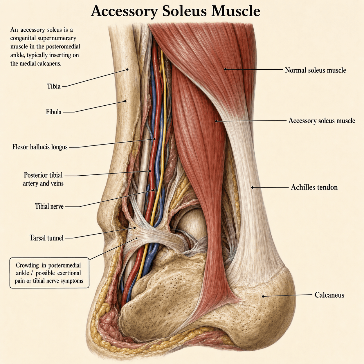
Present from birth, usually symptomatic only in the second or third decade. The extra belly does not form de novo in the athlete. The muscle is enclosed by the deep posterior compartment fascia and, distally, by the flexor retinaculum, so any increase in its volume, such as hypertrophy with training, or simply the presence of extra bulk in a tight space, produces pressure effects during activity. The variable distal insertion reflects where the aberrant slip's tendon condensed during development, and that in turn determines the later clinical pattern and the extent of any excision.
If asked "is this acquired or congenital?", answer congenital: the accessory soleus is a variant of soleus differentiation present from birth, and only the symptoms are acquired. These are the two things examiners test most: that the lesion is unequivocally benign (a differentiation variant, never a neoplasm) and that it is congenital yet characteristically late-presenting.
How it causes symptoms. During plantarflexion the accessory soleus contracts and increases in cross-sectional area, raising pressure against the Achilles, the neurovascular bundle, or the calcaneal insertions. Because the extra muscle is working inside a non-compliant fascial envelope, exertional compartment-like pain can occur. The visible or palpable mass is simply the contracting muscle belly itself; there is no true sheath or bursa in most cases.
Nerve compression. In the retromalleolar region or tarsal tunnel the muscle acts as a space-occupying lesion, producing local pain, a swelling sensation, or true compression neuropathy of the tibial nerve, with medial heel and plantar paraesthesia, Tinel's sign and night symptoms. It can compress the tibial nerve or its medial calcaneal branch against the flexor retinaculum or the medial calcaneus. The medial calcaneal insertion is the pattern most often responsible for focal pain and compression of the nerve or its branches within or just proximal to the tarsal tunnel.
Benign, always. The variant does not degenerate into sarcoma and does not represent a neoplastic process. Misinterpreting it as a soft-tissue tumour is the most common serious error, and recognising the normal muscle signal and the contraction sign avoids it.
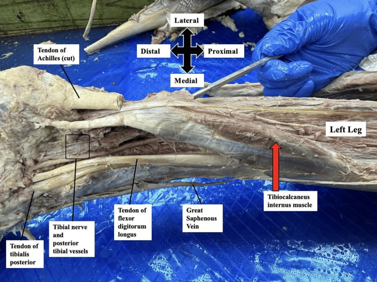
Classification
By insertion. There is no single universally used classification. The practical scheme for surgeons and examiners is based on the distal insertion of the accessory tendon, because the insertion determines both where the symptoms arise and how far the surgical dissection must go.
- Anatomic Location
- Accessory belly merges with or inserts onto the Achilles proper
- Typical Symptoms / Notes
- Visible bulk on contraction; less likely to cause focal nerve compression or bony pain
- Anatomic Location
- Inserts on upper posterior calcaneus, deep to Achilles insertion (Kager's fat pad)
- Typical Symptoms / Notes
- Posteromedial fullness and pain with running; occupies space that is normally fat only
- Anatomic Location
- Medial calcaneal tuberosity, sustentaculum tali region or flexor retinaculum area
- Typical Symptoms / Notes
- Posteromedial pain, tarsal tunnel syndrome, positive Tinel's; the pattern examiners associate with nerve compression
- Anatomic Location
- Extends to lower calcaneus or plantar aponeurosis
- Typical Symptoms / Notes
- Arch pain, heel pain, or a mass that can be mistaken for plantar fibromatosis
- Anatomic Location
- Ends in Kager's fat or on the flexor retinaculum
- Typical Symptoms / Notes
- Mobile, less fixed mass; can still produce pressure or be mistaken for a ganglion or lipoma
By presentation. Clinically there are four pictures:
- Asymptomatic, the majority. Incidental on MRI or discovered during surgery for other pathology, with no corresponding mass or activity-related pain. It needs only documentation and reassurance.
- Mass or pain. Visible or palpable posteromedial bulk that hardens on contraction, with activity-related aching or pressure pain. The most common presentation in athletes and military recruits.
- Nerve compression. Tarsal tunnel syndrome (medial and plantar paraesthesia, Tinel's, night pain) from the muscle occupying space in or proximal to the tarsal tunnel. EMG may confirm it.
- Exertional compartment picture. Deep posterior leg pain and tightness with exercise, relieved by rest, from an accessory soleus expanding within the fascial envelope.
When describing an accessory soleus on MRI or in the operating note, always state the origin, the course relative to the Achilles and neurovascular structures, and the precise insertion. This information guides both prognosis and the surgical plan if excision is required.
Clinical Presentation
Who. Patients are typically adolescents or young adults, 15-40 years, often involved in running, jumping sports or military training. There is no history of trauma in the classic case.
History. The usual story is a gradual-onset posteromedial ankle or distal leg "swelling" or pain, noticed during or after activity. The pain is aching, pressure or cramping, and worse with prolonged standing, running, or tight boots. Some patients report plantar tingling or burning that is worse at night or after exercise, a tarsal tunnel picture.
Look. With the patient standing, inspect from behind for asymmetric fullness or a visible mass posteromedial to the Achilles tendon, just above or at the level of the medial malleolus. The other side is usually flat in that location, and the skin is normal, with no erythema or sinus.
Feel. The mass is firm, non-fluctuant and distinct from the Achilles tendon, and a groove or plane may be felt between them. It is usually non-tender or only mildly tender.
The contraction sign. Palpate while the patient performs a single-leg heel rise or resisted plantarflexion: the mass hardens, rock-hard under the fingers, and sometimes becomes visibly larger. A lipoma, ganglion or sarcoma will not change with contraction. This single manoeuvre is the most useful physical finding, highly suggestive of the diagnosis, and should be documented.
Move. Ankle and subtalar range of motion are usually full. Pain may be reproduced at the extreme of passive dorsiflexion (stretch) or with resisted plantarflexion, and strength is normal unless chronic disuse or nerve compression has caused weakness.
Special tests. Tinel's percussion over the tarsal tunnel, posterior to the medial malleolus and along the course of the tibial nerve, may elicit plantar paraesthesia if the accessory soleus is compressing the nerve, and the dorsiflexion-eversion test for tarsal tunnel may be positive. Compare pulses and sensation; a vascular or neurological deficit is rare but should be documented if present.
Unilateral tarsal tunnel in a young patient. In a young athletic patient with unilateral tarsal tunnel symptoms and no trauma, image for space-occupying lesions, this variant included, before attributing the symptoms to idiopathic tarsal tunnel syndrome.
Differential diagnosis. Each of the alternatives has a discriminating feature:
- Lipoma - soft, does not contract
- Ganglion cyst - fluctuant, transilluminates, no muscle signal on MRI
- Achilles tendinopathy or paratendinitis - no separate mass
- Sarcoma or other neoplasm - progressive night pain, heterogeneous on MRI, rapid growth
- Chronic exertional compartment syndrome - bilateral, no mass
- Idiopathic tarsal tunnel syndrome - no space-occupying lesion on imaging
Investigations
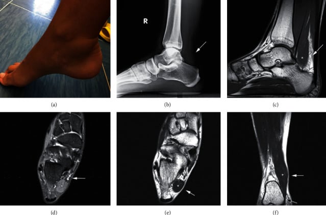
Radiographs. Usually normal. They may show a non-specific soft-tissue shadow anterior to the Achilles, with loss of Kager's fat triangle as in both cases illustrated on this page, or a small enthesophyte at the calcaneal insertion of the accessory tendon. They are not diagnostic, but they are useful to exclude a bony tumour or fracture in the work-up of ankle pain.
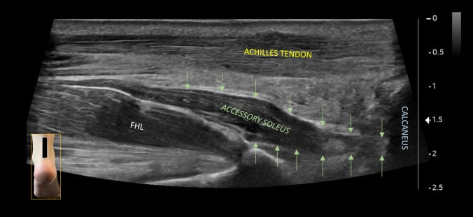
Ultrasound. A useful first-line or bedside tool where it is available. It shows a structure with normal skeletal muscle echotexture (hypoechoic with hyperechoic septa) that contracts dynamically on plantarflexion, and it can map the tendon insertion and assess compression of the tibial nerve. It is operator-dependent, and a negative scan does not exclude the diagnosis when clinical suspicion is high.
MRI, the gold standard. MRI is the investigation of choice. The accessory soleus appears as a fusiform or pennate muscle belly of identical signal to the soleus and gastrocnemius on T1-weighted, T2-weighted and proton-density sequences, with no fat-suppression dropout and no enhancement after gadolinium, unlike many tumours. A separate tendon runs from the belly to an insertion distal to the normal soleus, apart from the Achilles. Axial and sagittal planes best show the relationship to the neurovascular bundle and the exact insertion.
What MRI excludes. It rules out lipoma (high T1 fat signal), ganglion (fluid signal, thin wall) and sarcoma (heterogeneous, invasion, perilesional oedema), and it shows whether the tibial nerve is compressed.
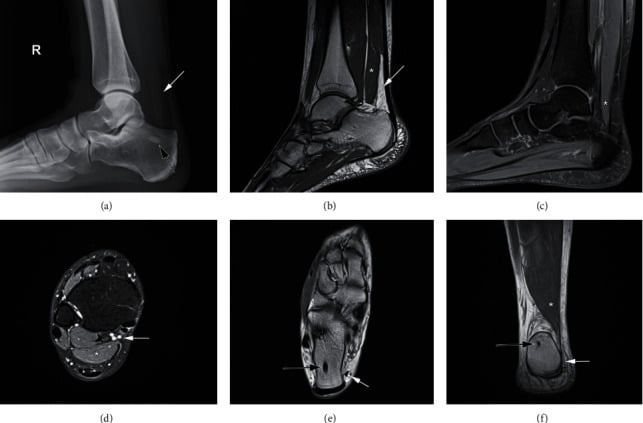
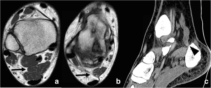
Other tests. CT is rarely helpful except when bony detail of the insertion is needed for surgical planning. Nerve conduction studies and EMG are indicated when there is clinical or imaging evidence of tibial nerve compression; they confirm the level and severity but are not required to diagnose the accessory muscle itself.
A mass that follows muscle signal exactly in a young patient is the accessory soleus until proven otherwise. Biopsy is almost never required, and is contraindicated in the typical imaging picture: it risks sampling error, unnecessary surgery and patient anxiety, and a good history with a clear MRI description avoids it.
Management
The incidental finding. Many accessory soleus muscles are found on MRI performed for Achilles tendinopathy, ankle instability or other reasons. If there is no corresponding clinical mass, pain on contraction, or nerve compression, reassurance and a description in the report are sufficient; no treatment or follow-up imaging is needed.
Conservative care. The majority of symptomatic cases settle with conservative treatment. Reasonable measures are:
- Activity modification: reduce running volume and avoid boots that compress the area
- A heel-raise insert or orthosis to off-load the posterior structures
- Physiotherapy focused on gastrocnemius-soleus flexibility and eccentric control
- A short course of NSAIDs
Many young athletes can be kept active with load management while the tissues adapt. A 3- to 6-month trial is appropriate before considering surgery in most cases.
Even when surgery is ultimately required, a documented trial of conservative care strengthens the indication and sets patient expectations.
Indications for surgery. Surgery is indicated for persistent, activity-limiting pain or documented nerve compression after an adequate conservative trial, in a patient who needs to return to sport or military duty. The presence of an accessory soleus on imaging alone, without correlating symptoms, is not an indication for excision.
Choosing the operation. Complete excision is the operation described below, but the pooled evidence shows it is only one of the surgical options actually used, and not even the commonest. In the quantitative systematic review of symptomatic cases, fasciotomy (fascial decompression) was the single most frequently performed procedure, in about a fifth to a quarter of treated cases, ahead of excision and isolated tibial-nerve decompression. All surgically treated patients were reported satisfied whichever route was chosen, but that finding comes from pooled published cases and is a publication artefact rather than evidence that the operations are equivalent (see the Evidence section). Be ready to justify the choice rather than reflexively "excise everything".
Fasciotomy for a pure exertional picture. When the dominant problem is an exertional, compartment-like syndrome, deep posterior leg pain and tightness with exercise that is relieved by rest, releasing the tight investing fascia of the deep posterior compartment relieves the pressure while preserving the muscle. This mirrors the principle used for chronic exertional compartment syndrome and is attractive when there is no discrete mass and no focal nerve entrapment. It is a smaller operation, with less dissection around the neurovascular bundle.
Excision for a mass or nerve compression. Complete excision of the muscle-tendon unit is favoured when the muscle is itself the space-occupying culprit: a symptomatic focal posteromedial mass, or documented tibial-nerve or tarsal-tunnel compression where the muscle is flattening the nerve. Removing the offending bulk addresses both the mass effect and the neuropathy in one step.
Excision with tarsal tunnel decompression. Where the accessory soleus compresses the tibial nerve within the tarsal tunnel, excision combined with tarsal tunnel decompression may be required, as in the reported case that presented with a neuropathic picture and no swelling. Excision alone can be enough: in the Badr case, removing the muscle without any tarsal tunnel release resolved the neuropathy.
Match the operation to the mechanism. Never perform an isolated tarsal tunnel release and leave the muscle in place: retinacular release that leaves the muscle behind is the classic trap, and is insufficient when the muscle is the compressor.
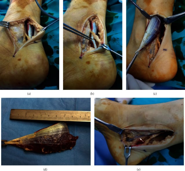
Excision through a posteromedial approach. The whole muscle-tendon unit comes out; partial excision risks recurrence of symptoms.
- Plan on the MRI. Check the insertion preoperatively; it decides how far distally the tendon must be followed.
- Position prone or lateral with the affected leg up. A tourniquet is optional but helpful for a bloodless field.
- Incise longitudinally over the posteromedial ankle, centred over the mass and extended proximally and distally as needed to reach the origin and insertion.
- Protect the nerve first. Identify and gently mobilise the tibial nerve and posterior tibial vessels before dissecting the muscle; in many cases they lie just anterior or deep to it. The sural nerve is posterior and lateral and usually not at risk in a medial approach.
- Excise by detaching the accessory muscle at its origin, dissecting it free from the surrounding tissues, and releasing its tendon at the calcaneal or other insertion.
- Close in layers, over a drain if needed.
- Send the specimen. Histology shows normal skeletal muscle, which confirms the diagnosis and excludes rare mimics.
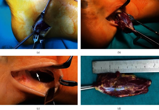
Aftercare. Avoid prolonged immobilisation. Begin range-of-motion exercises as pain allows, with progressive weight-bearing and strengthening over 6-12 weeks; most patients return to sport by 3-4 months.
Open or endoscopic. Endoscopic or minimally invasive excision has been described in small series for selected cases, but open excision remains the standard for reliable complete removal and nerve protection.
Counselling. Tell the patient before surgery that this is a variant, not a tumour. Preoperative counselling reduces anxiety and sets realistic expectations for recovery.
Complications
Of the condition. The variant itself, or the failure to recognise it, can lead to:
- Chronic exertional pain limiting sport or duty
- Missed or delayed diagnosis leading to inappropriate interventions: tarsal tunnel release alone, biopsy, or oncology referral
- Tarsal tunnel syndrome with permanent sensory change or weakness if long-standing compression is ignored
Of surgery. Incomplete excision, nerve injury and wound problems are set out with their prevention in the table, alongside misdiagnosis as a neoplasm. Beyond them, deep vein thrombosis and infection are met with standard prophylaxis and sterile technique, and perceived weakness is rare because the normal soleus and gastrocnemius compensate for loss of the small accessory belly.
- Mechanism
- Leaving proximal origin or distal tendon attached, with recurrent mass effect or pain
- Prevention / Management
- Preoperative MRI mapping of entire muscle-tendon unit; excise from origin to insertion
- Mechanism
- Failure to identify and protect the nerve, or its calcaneal branches, lying adjacent to the muscle
- Prevention / Management
- Wide exposure, nerve stimulation if available, gentle retraction only; protect the nerve throughout
- Mechanism
- Posteromedial skin is thin and under tension
- Prevention / Management
- Meticulous layered closure, avoid excessive undermining, early motion once healed
- Mechanism
- Operating without MRI or misreading normal muscle signal as tumour
- Prevention / Management
- MRI before any invasive step; describe normal muscle signal and separate tendon explicitly in the report
Guidelines, Registries & Global Practice
-
Epidemiology is consistent worldwide: cadaveric and imaging series from multiple continents report prevalence between 0.7% and 5.5%, with no strong evidence of major geographic or ethnic variation. Bilateral cases occur in approximately 10-15% of individuals with the variant.
-
No dedicated society guidelines: because the accessory soleus is a rare cause of symptoms rather than a primary disease, major organisations (AAOS, BOA, EFORT, AOFAS) have not issued specific clinical practice guidelines. Management is guided by level IV and V evidence (case series, small comparative studies, and two recent systematic/narrative reviews).
-
Diagnostic standard is MRI: across published series the shift from routine biopsy or exploration in the 1980s-1990s to MRI-first diagnosis has been universal where MRI is available. In resource-limited settings, ultrasound plus plain films plus a clear contraction sign on examination can be sufficient to avoid invasive steps.
-
Surgical outcomes are reported from athletic and military cohorts: good-to-excellent pain relief and return to pre-symptom activity levels are described in greater than 85-90% of operated cases when complete excision is performed. These results come from series in Europe, North America, the Middle East and Asia; the principles (protect the tibial nerve, remove the entire unit, confirm normal muscle on histology) are the same everywhere.
-
Registries: not applicable. The accessory soleus is not an implant or arthroplasty-related condition; no joint registries capture it.
-
Global practice variation: the main variation is access to MRI for definitive non-invasive diagnosis and the threshold for surgery in borderline symptomatic cases. In high-performance military or elite sport settings, earlier imaging and lower threshold for excision are common; in general orthopaedic practice a longer trial of conservative care is usual. The core teaching - recognise the variant, image before you cut, excise completely when operating - remains constant.
Exam Viva Scenarios
Practise clinical reasoning and management decisions out loud
“A 22-year-old military recruit presents with a 4-month history of posteromedial right ankle 'swelling' and aching pain when running in boots. On examination there is a firm mass posteromedial to the Achilles that becomes visibly larger and rock-hard when he performs a heel rise. How do you approach this?”
“A 19-year-old footballer has 6 months of burning pain and tingling in the medial heel and plantar foot that is worse after training and at night. There is a subtle posteromedial fullness that hardens on resisted plantarflexion. Tinel's sign is positive over the tarsal tunnel. MRI shows an accessory soleus inserting on the medial calcaneus with the tibial nerve flattened against it. How do you manage?”
“You are reviewing the MRI of a 35-year-old runner with mid-portion Achilles tendinopathy. The report mentions an 'accessory muscle in the deep posterior compartment consistent with accessory soleus'. The patient has no posteromedial mass or pain on the opposite side of the Achilles on examination. What do you tell the patient and the referrer?”
Key Facts
- Congenital variant, 0.7-5.5% population, often incidental
- Posteromedial mass that hardens on resisted plantarflexion or heel rise
- MRI: normal skeletal muscle signal + separate tendon = diagnostic
- Most need only reassurance; operate for refractory pain or nerve compression
Classification (Insertion)
- To Achilles tendon - bulk without focal compression
- Superior calcaneus (Kager's) - space occupation deep to Achilles
- Medial calcaneus - commonest symptomatic, tarsal tunnel risk
- Inferior/plantar or free-ending - less common patterns
Investigations
- X-ray: normal or non-specific soft-tissue shadow
- US: dynamic confirmation of muscle that contracts
- MRI gold standard - maps origin, course, insertion, nerve relation
- NCS/EMG only if clinical nerve compression suspected
Management
- Conservative first: activity mod, heel raise, physio, 3-6 months
- Surgery: complete excision (origin to insertion) via posteromedial approach
- Protect tibial nerve and branches throughout
- Send specimen (normal skeletal muscle); early motion postop
Red Flags / Traps
- Biopsy or oncology referral before MRI (the mass follows muscle signal)
- Partial excision (recurrence of symptoms)
- Isolated tarsal tunnel release when the muscle is the compressor
- Missing the contraction sign on examination
Evidence
Clinical Presentation of the Accessory Soleus Muscle: A Quantitative Systematic Review
- Quantitative systematic review of 41 articles reporting 134 symptomatic accessory soleus muscles in 122 patients
- Presenting features: exertional pain in 60% and posteromedial ankle swelling in 59%; a soft mass that becomes hard on plantarflexion in 44% (the contraction sign), a positive Tinel's sign in 11%, and foot numbness/dysaesthesia in 5%
- It is also a lump that need not hurt - a soft NON-TENDER mass was recorded in 30% and a tender mass in only 9%, which is why the painless posteromedial swelling is so readily mistaken for a soft-tissue tumour
- Diagnostic modalities used across cases: plain radiographs 62.7%, MRI 52.2%, CT 24.6%, ultrasound 12.7%, and EMG 13.4%
- Of 128 treated cases, conservative management was used in 37% and surgery in 50% - fasciotomy 22.6%, excision 20.3%, posterior tibial nerve decompression 7.8%, combined 1.7%; all surgically treated patients were satisfied
The Accessory Soleus Muscle: A Narrative Review of the Literature
- Narrative review: 148 articles screened, 59 studies (predominantly case reports) eligible for analysis of anatomy, presentation, diagnosis, associated pathologies and treatment
- Awareness of the variant and its clinical presentation allows diagnosis without surgical exploration; recognising the anomaly is key to correct diagnosis and treatment planning
- Insertion-based classification is emphasised, guiding both the location of symptoms and the surgical approach
- The authors note the literature remains inconclusive on why some patients are symptomatic and on optimal therapy protocols; conservative care suits mild cases and complete excision is reserved for refractory symptoms
Extrinsic Compression Neuropathy of the Tibial Nerve Secondary to Accessory Soleus Muscle in a Young Teenager
- Case report of a 13-year-old girl with posteromedial ankle pain and plantar paraesthesia caused by an accessory soleus compressing the tibial nerve
- Diagnosis was made on history, physical examination and imaging; MRI demonstrated the accessory muscle and its relationship to the neurovascular bundle
- Complete surgical excision of the accessory soleus - performed without any tarsal tunnel release - produced complete resolution of symptoms, sustained free of symptoms for eighteen months postoperatively with no recurrence
- The case illustrates that nerve compression can occur in adolescents and that removing the compressing muscle can be curative even without a formal tarsal tunnel decompression
Symptomatic Accessory Soleus Muscle: A Cause for Exertional Compartment Syndrome in a Young Soldier
- Case report of a young soldier with exertional posteromedial leg pain and swelling caused by an accessory soleus within the deep posterior compartment
- MRI demonstrated a bulky accessory soleus (bilateral) and ruled out tumour and other causes; because symptoms were severe and activity-limiting on the right, surgical resection was chosen
- Surgical excision of the accessory soleus produced complete resolution of pain, numbness and swelling with no recurrence at twelve months and return to full activity
- Highlights that accessory soleus can produce a compartment-syndrome-like picture in high-demand individuals even without classic tarsal tunnel findings