Tibial Diaphysis | Biphasic Histology | Soap-Bubble Appearance | Wide Excision
- Tibial diaphysis is the classic location (85% of cases)
- Biphasic histology - epithelial cells in osteofibrous stroma
- Soap-bubble appearance on X-ray - multilocular lytic lesion
- Wide en bloc excision required - curettage has high recurrence
- Cytokeratin positive - distinguishes from other bone tumours
- “Anterior tibial cortex involvement is characteristic
- “May arise from or coexist with osteofibrous dysplasia
- “Lymph node and lung metastases occur in 15-30%
- “Late recurrence possible - follow up for decades
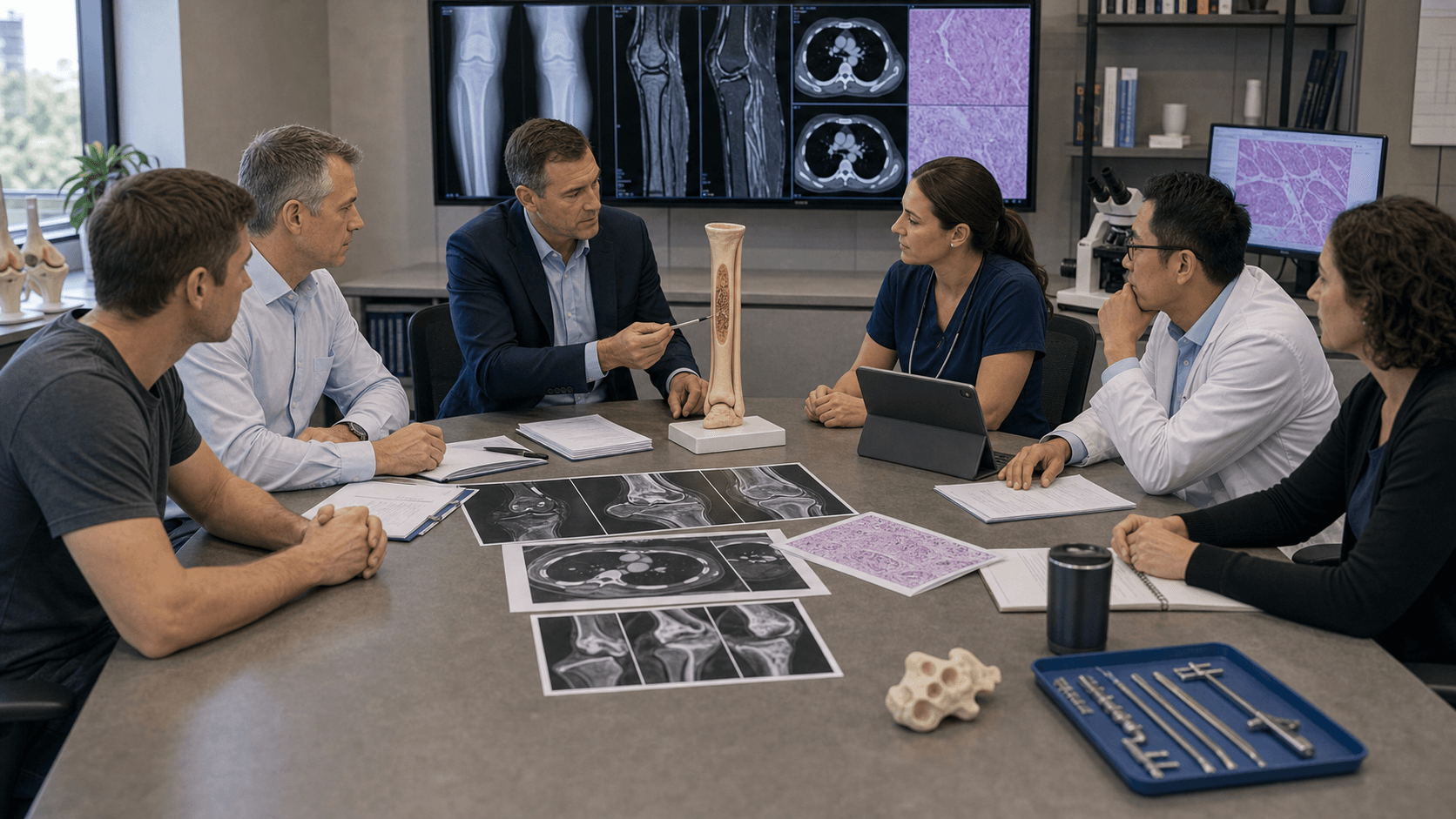
Clinical Imaging
Imaging Atlas


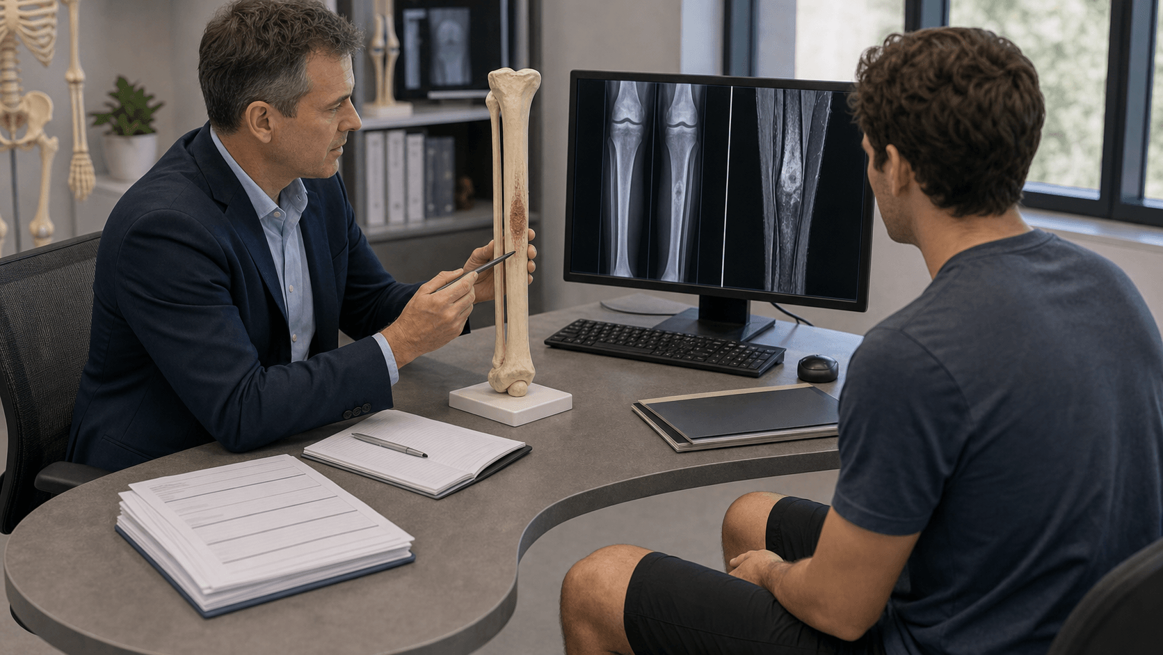
Tibial diaphysis is the hallmark location (85%). The lesion characteristically involves the anterior cortex. If you see a lytic tibial diaphyseal lesion in a young adult, consider adamantinoma in the differential.
The tumour contains epithelial nests within an osteofibrous stroma. Epithelial cells are cytokeratin 14 and 19 positive. This biphasic pattern distinguishes it from pure osteofibrous dysplasia.
Wide en bloc resection is mandatory. Curettage or marginal excision leads to unacceptably high local recurrence (up to 90%). Intralesional surgery is the strongest predictor of recurrence and metastasis.
Recurrence can occur up to 36 years after initial resection. Patients require lifelong surveillance. The 10-year survival is excellent at 92% but late metastases occur.
| Diagnosis | Age | Location | Key Features |
|---|---|---|---|
| Adamantinoma | 20-50 years | Tibial diaphysis (anterior cortex) | Soap-bubble, cytokeratin positive |
| Osteofibrous dysplasia | Under 10 years | Tibial diaphysis (anterior cortex) | Self-limiting, anterior bowing |
| Fibrous dysplasia | Any age | Any bone, often polyostotic | Ground-glass matrix, shepherd's crook |
| Ewing sarcoma | 5-25 years | Diaphysis any bone | Permeative, onion-skin periosteal reaction |
| Osteosarcoma | 10-25 years | Metaphysis long bones | Aggressive, Codman triangle, sunburst |
TIBIATIBIA - Key Features of Adamantinoma
Hook:TIBIA helps remember this is the classic tibial tumour with unique biphasic histology
WIDEWIDE - Surgical Principles
Hook:WIDE margins are essential - never curettage adamantinoma
SOAPSOAP - Imaging Features
Hook:The SOAP-bubble appearance on X-ray is characteristic of adamantinoma
Overview and Epidemiology
Adamantinoma is a rare low-grade malignant bone tumour with distinctive biphasic histology featuring epithelial cells within an osteofibrous stroma. It has a remarkable predilection for the tibial diaphysis and represents less than 0.5% of all primary bone tumours.
Key epidemiological features:
- Incidence: Accounts for less than 1% of primary malignant bone tumours
- Age distribution: Peak between 20-50 years (range 2-86 years)
- Gender: Slight male predominance (5:4 ratio)
- Location: 80-85% occur in the tibial diaphysis
Risk factors:
- No established genetic predisposition identified
- Possible association with osteofibrous dysplasia (OFD)
- History of prior trauma reported in 60% of patients (uncertain significance)
- No association with radiation or chemical exposure
The name "adamantinoma" derives from Greek "adamantinos" meaning "very hard", coined by Fischer in 1913. The term was originally used for odontogenic tumours with similar histology. Despite the name suggesting a relationship to dental tumours, skeletal adamantinoma is a distinct entity.
Pathophysiology
Histological Features
Adamantinoma is characterised by its distinctive biphasic histology with epithelial and osteofibrous components that may be intermingled in various proportions.
Epithelial Component:
- Nests, cords, or tubules of epithelial cells
- Positive for cytokeratins 14, 19 (but negative for CK8, CK18)
- Four histological patterns: basaloid, tubular, spindle-cell, squamous
- Peripheral palisading of cells may be seen
Osteofibrous Component:
- Fibrous stroma with woven bone trabeculae
- Similar to osteofibrous dysplasia
- Contains scattered osteoclast-like giant cells
- Positive for vimentin
Relationship to Osteofibrous Dysplasia
A controversial but clinically important relationship exists between adamantinoma and osteofibrous dysplasia (OFD):
- Osteofibrous Dysplasia
- Children (under 10)
- OFD-like Adamantinoma
- Adolescents
- Classic Adamantinoma
- Adults (20-50)
- Osteofibrous Dysplasia
- Rare or absent
- OFD-like Adamantinoma
- Scattered, small
- Classic Adamantinoma
- Prominent nests
- Osteofibrous Dysplasia
- Negative or focal
- OFD-like Adamantinoma
- Focal positive
- Classic Adamantinoma
- Strongly positive
- Osteofibrous Dysplasia
- Benign, self-limiting
- OFD-like Adamantinoma
- Low malignant potential
- Classic Adamantinoma
- Malignant
- Osteofibrous Dysplasia
- Observation (most)
- OFD-like Adamantinoma
- Wide excision
- Classic Adamantinoma
- Wide excision
Proposed pathogenic relationship (a genuine controversy):
- The Leiden group demonstrated keratin-positive epithelial cells emerging individually from the osteofibrous stroma, with progressively organised basement membrane around cohesive epithelial islands - evidence that OFD-like adamantinoma may be a precursor to classic adamantinoma via fibrous-to-epithelial transformation (Hazelbag, Hum Pathol 1997).
- The Mayo Clinic counterargument: in 80 OFD cases followed for up to 31 years, no lesion progressed to adamantinoma and two matured into fibrous dysplasia, suggesting OFD is a variant of fibrous dysplasia rather than an obligate precursor (Park & Unni, Hum Pathol 1993).
- Reconciled view: OFD, differentiated (OFD-like) adamantinoma and classic adamantinoma form a histological continuum, but pure OFD does not inevitably become malignant - hence cytokeratin immunohistochemistry is essential to detect the epithelial component.
Molecular Features
- Aneuploidy is confined to the epithelial cells. DNA cytometry shows aneuploid nuclei only in cells of epithelial phenotype, while the fibrous cells remain diploid - direct evidence that the malignant clone is epithelial and the osteofibrous tissue is reactive (Hazelbag, Am J Pathol 1995).
- p53 abnormalities (immunoreactivity and loss of heterozygosity at the p53 locus) are detectable in roughly half of tumours and again localise to the epithelial component (Hazelbag, Am J Pathol 1995).
- No specific recurrent fusion gene has been identified (unlike the EWSR1 rearrangement of Ewing sarcoma), which is a useful diagnostic discriminator.
- GNAS mutations characteristic of fibrous dysplasia are not a feature, helping separate adamantinoma/OFD from fibrous dysplasia.
- Lung metastases contain only keratin-positive epithelial cells, with no osteofibrous element, confirming the epithelial cells as the metastasising population (Hazelbag, Am J Pathol 1995).
Classification Systems
Histological Classification
| Pattern | Features | Frequency | Prognosis |
|---|---|---|---|
| Basaloid | Nests of basaloid cells with peripheral palisading | Common | Standard |
| Tubular | Epithelial cells forming tubular/glandular structures | Common | Standard |
| Spindle-cell | Elongated spindle-shaped epithelial cells | Less common | May be confused with sarcoma |
| Squamous | Squamous differentiation with keratinisation | Less common | May have better prognosis |
| Osteofibrous-like (Differentiated) | Predominantly fibrous with scattered epithelial cells | 10-20% | Better prognosis |
Most tumours show mixed patterns. The osteofibrous-like (differentiated) subtype has a notably better prognosis and occurs in younger patients.
Clinical Assessment
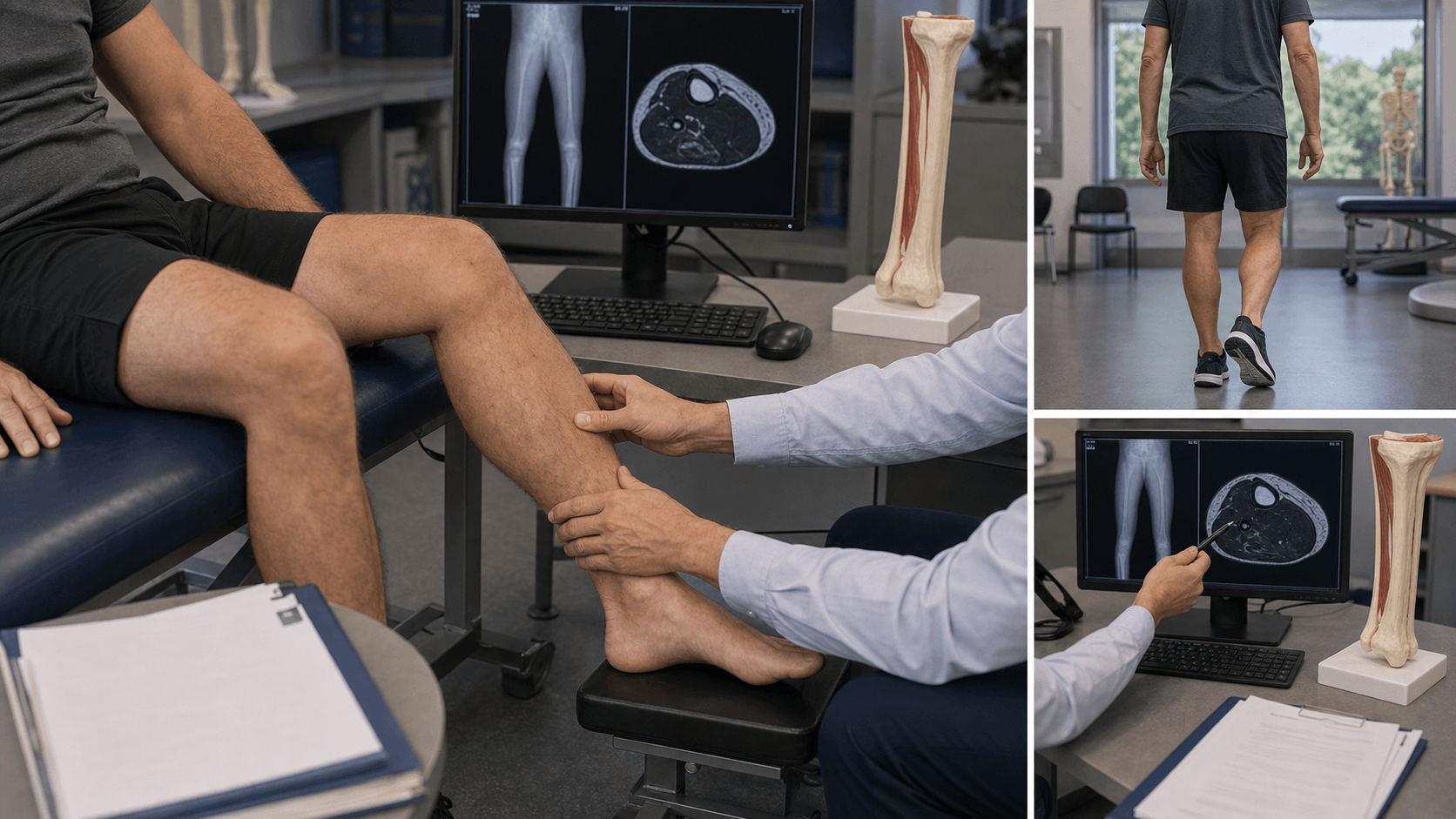
History
Classic Presentation:
- Slowly progressive swelling over the anterior shin
- Dull, aching pain - often present for months to years
- Approximately 30% have symptoms for more than 5 years before diagnosis
- May have history of prior trauma (reported in 60%)
Duration of symptoms is an important prognostic factor - shorter symptom duration (less than 5 years) is associated with higher recurrence risk.
Red Flag Symptoms:
- Rapid increase in swelling
- New or worsening pain
- Constitutional symptoms (weight loss, fever) - rare but concerning
- Pathological fracture (occurs in 16-23% of cases)
Physical Examination
Standard Findings:
- Palpable firm mass over anterior tibial shaft
- Overlying skin may be normal or tense
- Possible anterior tibial bowing
- Tenderness on palpation
- Usually no warmth or erythema (unless fracture)
Examination Sequence:
- Assessment
- Inspection
- Findings
- Anterior tibial swelling, possible bowing
- Assessment
- Palpation
- Findings
- Firm, fixed mass arising from bone
- Assessment
- Range of motion
- Findings
- Usually preserved at knee and ankle
- Assessment
- Neurovascular
- Findings
- Usually normal, check dorsalis pedis
- Assessment
- Lymph nodes
- Findings
- Palpate popliteal and inguinal nodes
Differential Diagnosis
| Diagnosis | Key Differentiating Features |
|---|---|
| Osteofibrous dysplasia | Age under 10, anterior bowing, self-limiting, cytokeratin negative |
| Fibrous dysplasia | Ground-glass matrix, polyostotic form common, GNAS mutation |
| Ewing sarcoma | Age 5-25, permeative, systemic symptoms, EWSR1 translocation |
| Osteomyelitis | Fever, elevated WCC/CRP, sequestrum on CT |
| Metastatic carcinoma | Known primary, older age, multiple lesions common |
Investigations
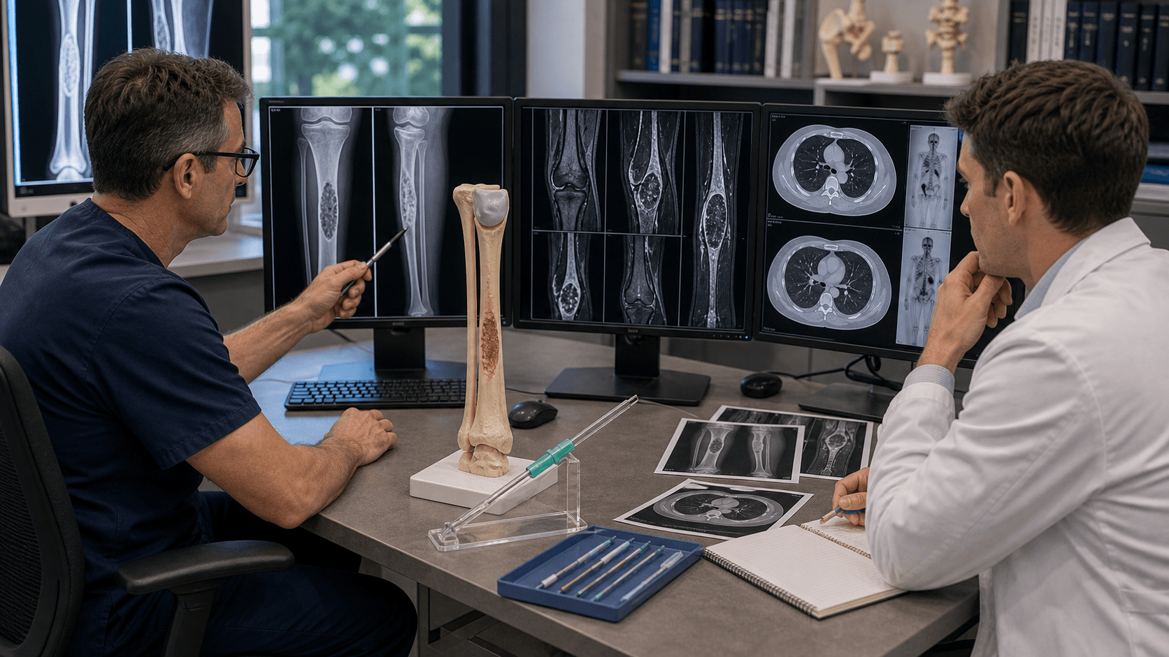
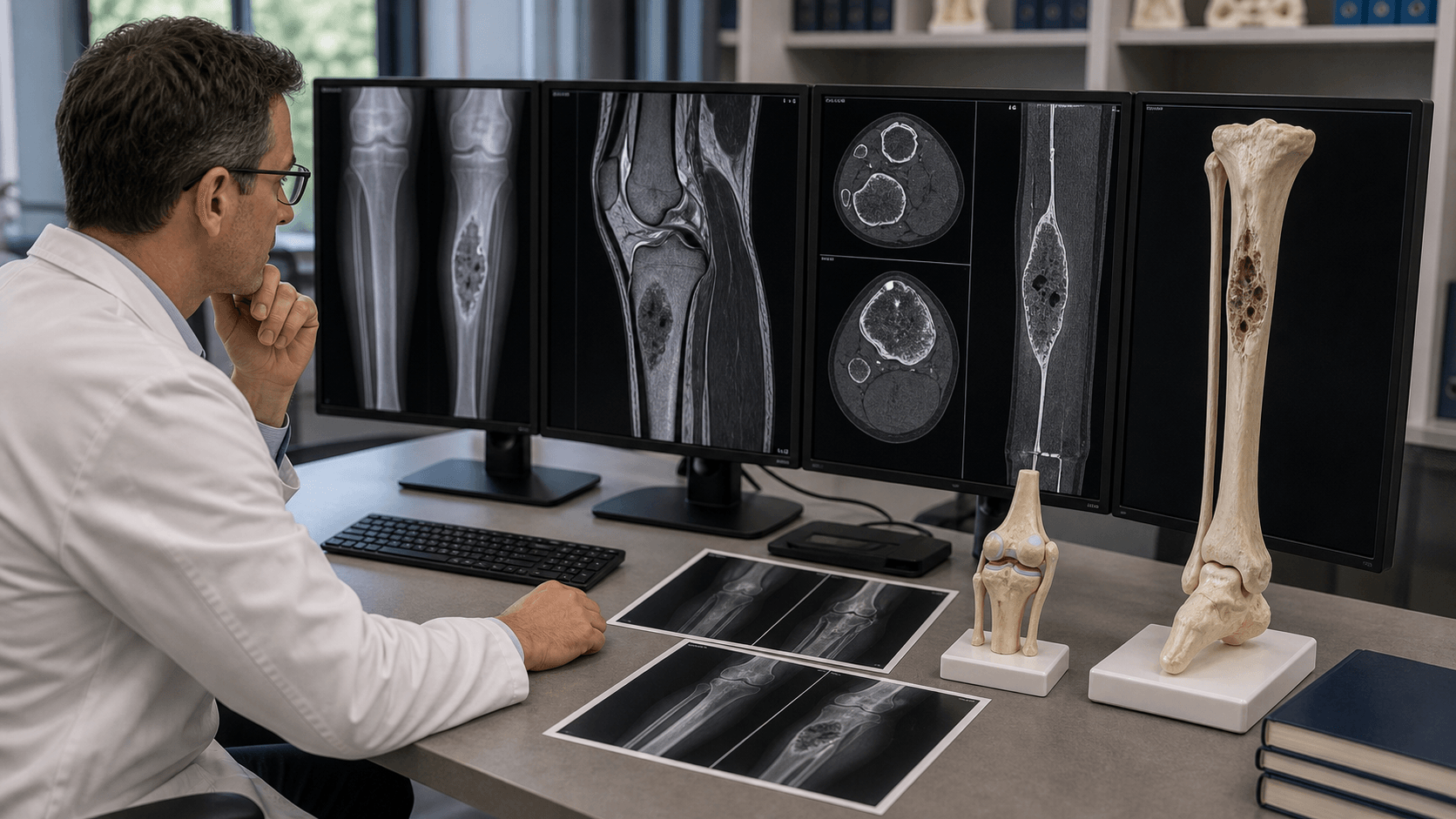
Imaging Algorithm
Step 1: Plain Radiographs
- First-line investigation for tibial swelling
- Characteristic "soap-bubble" or multilocular lytic appearance
- Eccentric location in anterior cortex
- Sclerotic margins with cortical expansion
- May show multifocality (skip lesions)
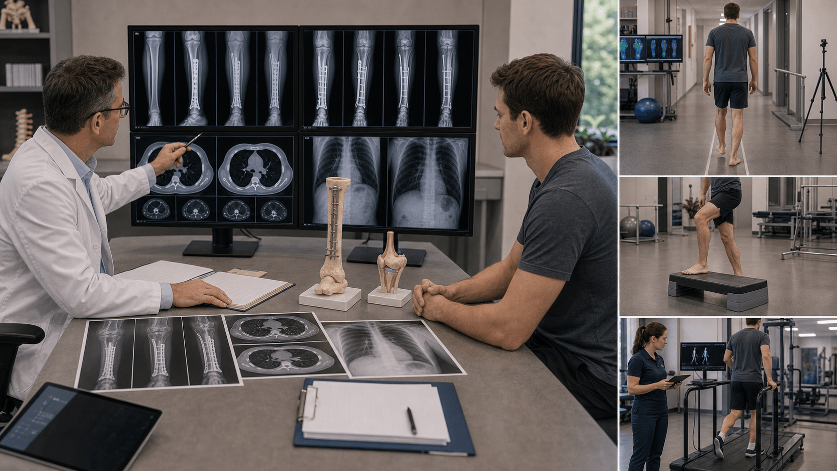
Step 2: CT Scan
- Better cortical definition than X-ray
- Defines extent of bone destruction
- Identifies pathological fracture
- CT chest for pulmonary metastasis staging
Step 3: MRI
- Gold standard for local staging
- Defines intramedullary extent
- Identifies soft tissue extension
- Detects skip lesions (multifocality)
Imaging Characteristics
| Modality | Key Findings | Role |
|---|---|---|
| Plain radiograph | Soap-bubble appearance, eccentric, sclerotic margins | Initial assessment |
| CT scan | Cortical destruction, extent of bone involvement | Surgical planning, staging (chest) |
| MRI | T1 low, T2 high signal, contrast enhancement, skip lesions | Local staging, soft tissue assessment |
| Bone scan | Increased uptake, identifies multifocal disease | Screening for metastases |
| PET-CT | FDG avid, useful for staging and recurrence | Staging, surveillance |
Biopsy
Principles:
- Core needle biopsy preferred (CT or ultrasound-guided)
- Open biopsy if core inconclusive
- Biopsy tract must be excised with definitive resection
- Place biopsy tract in line with planned surgical incision
Always discuss biopsy approach with the operating surgeon before performing. The biopsy tract becomes contaminated and must be excised en bloc with the tumour. Poorly placed biopsies can compromise limb salvage.
Laboratory Studies
- FBC, ESR, CRP - usually normal
- LDH, ALP - usually normal or mildly elevated
- Calcium - rarely hypercalcaemia (paraneoplastic)
- Immunohistochemistry: Cytokeratin 14, 19 positive; CK8, 18 negative
Management
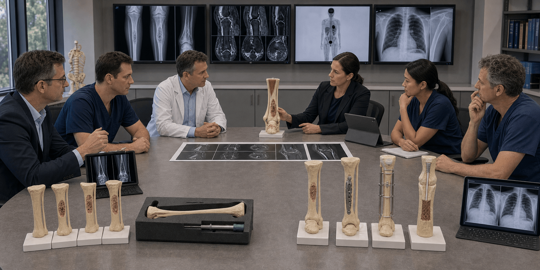
Surgical Principles
Wide En Bloc Resection is the treatment of choice. The goal is complete tumour removal with a cuff of normal tissue.
Margin Types:
- Description
- Through the tumour
- Local Recurrence
- Up to 90%
- Description
- Through reactive zone
- Local Recurrence
- 30-50%
- Description
- Through normal tissue
- Local Recurrence
- Under 10%
- Description
- Entire compartment
- Local Recurrence
- Under 5%
Curettage or intralesional excision is contraindicated. This is the strongest predictor of local recurrence and subsequent metastasis. All adamantinomas require wide surgical margins.
Surgical Goals:
- Complete tumour removal with wide margins
- Excision of biopsy tract
- Preservation of limb function when possible
- Durable reconstruction
Limb Salvage Reconstruction Options:
| Method | Advantages | Disadvantages |
|---|---|---|
| Intercalary allograft | Anatomical reconstruction, joint preservation | Nonunion, fracture, infection risk |
| Vascularised fibula autograft | Living bone, good incorporation | Donor morbidity, hypertrophy takes time |
| Allograft-prosthesis composite | Immediate stability, joint replacement | Loosening, wear, infection |
| Distraction osteogenesis | Autologous bone, no donor site | Prolonged treatment, pin site issues |
| Endoprosthesis | Immediate function | Loosening, wear, revision |
The choice of reconstruction depends on resection length, patient age, expected activity level, and institutional expertise.
Complications
Tumour-Related Complications
| Complication | Incidence | Management |
|---|---|---|
| Pathological fracture | 16-23% | Stabilisation then definitive resection |
| Local recurrence | 10-35% (margin dependent) | Re-resection with wider margins, amputation |
| Pulmonary metastases | 15-30% | Metastasectomy if possible, systemic therapy |
| Lymph node metastases | 5-10% | Lymphadenectomy |
Surgical Complications
- Incidence
- 5-15%
- Risk Factors
- Large resection, diabetes
- Incidence
- 10-30% (allograft)
- Risk Factors
- Long segment, chemotherapy
- Incidence
- 15-25%
- Risk Factors
- Stress risers, activity
- Incidence
- 5-10%
- Risk Factors
- Prolonged surgery, immobility
- Incidence
- Under 5%
- Risk Factors
- Tumour proximity
Managing Recurrence
Local recurrence is the most significant complication. Assessment includes:
- Imaging: MRI of local site, CT chest, bone scan/PET
- Biopsy: Confirm recurrence histologically
- Surgical planning: Review margins from primary surgery
Treatment Options:
- Wide re-excision if margins achievable
- Amputation if limb salvage not possible
- Palliative care for unresectable/metastatic disease
Local recurrence significantly increases the risk of subsequent metastatic disease. Achieving adequate margins at initial surgery is critical to prevent this cascade of complications.
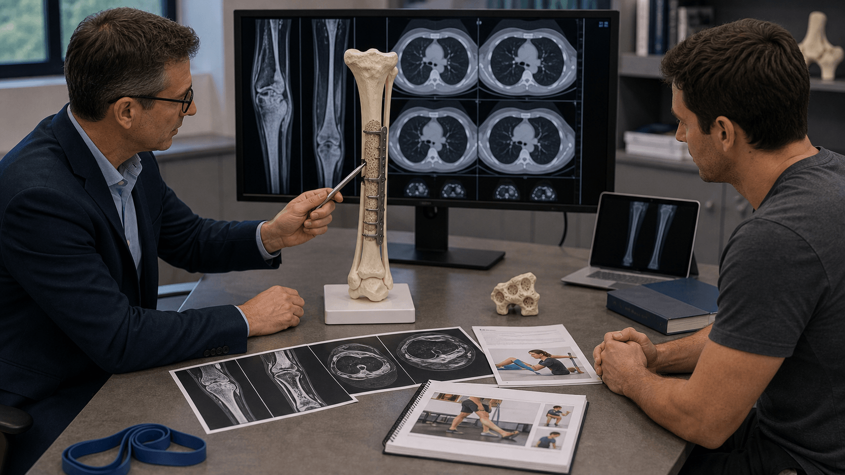
Postoperative Care
Immediate Postoperative Care
Day 0-3:
- DVT prophylaxis (mechanical and pharmacological)
- Drain management - remove when output under 50ml per 24 hours
- Pain management - PCA then oral analgesia
- Non-weight bearing on affected limb
Week 1-6:
- Wound surveillance - watch for infection
- Protected weight bearing (depends on reconstruction)
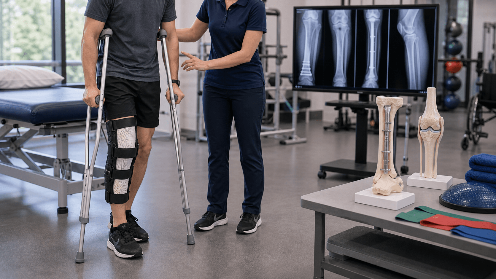
- Physiotherapy for ROM and muscle strengthening
- Monitor for DVT/PE
Rehabilitation Protocol
- Timeframe
- Weeks 0-6
- Goals
- Protected weight bearing, ROM, wound healing
- Timeframe
- Weeks 6-12
- Goals
- Progressive weight bearing, strength
- Timeframe
- Months 3-6
- Goals
- Full weight bearing, functional activities
- Timeframe
- After 6 months
- Goals
- Return to recreational activities
Reconstruction-Specific Considerations:
- Allograft: Non-weight bearing 6-12 weeks until union
- Vascularised fibula: Progressive weight bearing as fibula hypertrophies
- Endoprosthesis: Earlier weight bearing (2-4 weeks)
Return to Activity
- Desk work: 4-6 weeks (depends on pain control)
- Manual work: 3-6 months
- Sports: Generally low-impact activities recommended
- Driving: When off opioids and can weight bear for emergency stop
Intercalary allograft reconstruction requires careful monitoring for union. Radiographic union at the allograft-host junction may take 12-18 months. Protect the construct during this period.
Outcomes and Prognosis
Survival Outcomes
Overall Survival:
- 5-year survival: 95-98%
- 10-year survival: 87-92%
- Metastatic disease significantly worsens prognosis
Disease-Free Survival:
- Recurrence-free survival at 10 years: approximately 72%
- Late recurrence is a significant concern
Prognostic Factors
Factors Associated with Worse Outcome:
| Factor | Better Prognosis | Worse Prognosis |
|---|---|---|
| Histological subtype | Osteofibrous-like (differentiated) | Classic with high epithelial component |
| Surgical margins | Wide or radical | Intralesional or marginal |
| Symptom duration | Greater than 5 years | Less than 1 year |
| Age | Older (over 20) | Younger (under 20) |
| Pain at presentation | Minimal or absent | Significant pain |
| Squamous differentiation | Present | Absent |
Functional Outcomes
Limb Salvage:
- Achievable in 80-90% of cases at specialised centres
- MSTS functional score typically 70-85% of normal
Quality of Life:
- Generally good with successful limb salvage
- May require walking aids initially
- Long-term surveillance can cause anxiety
Evidence and Guidelines
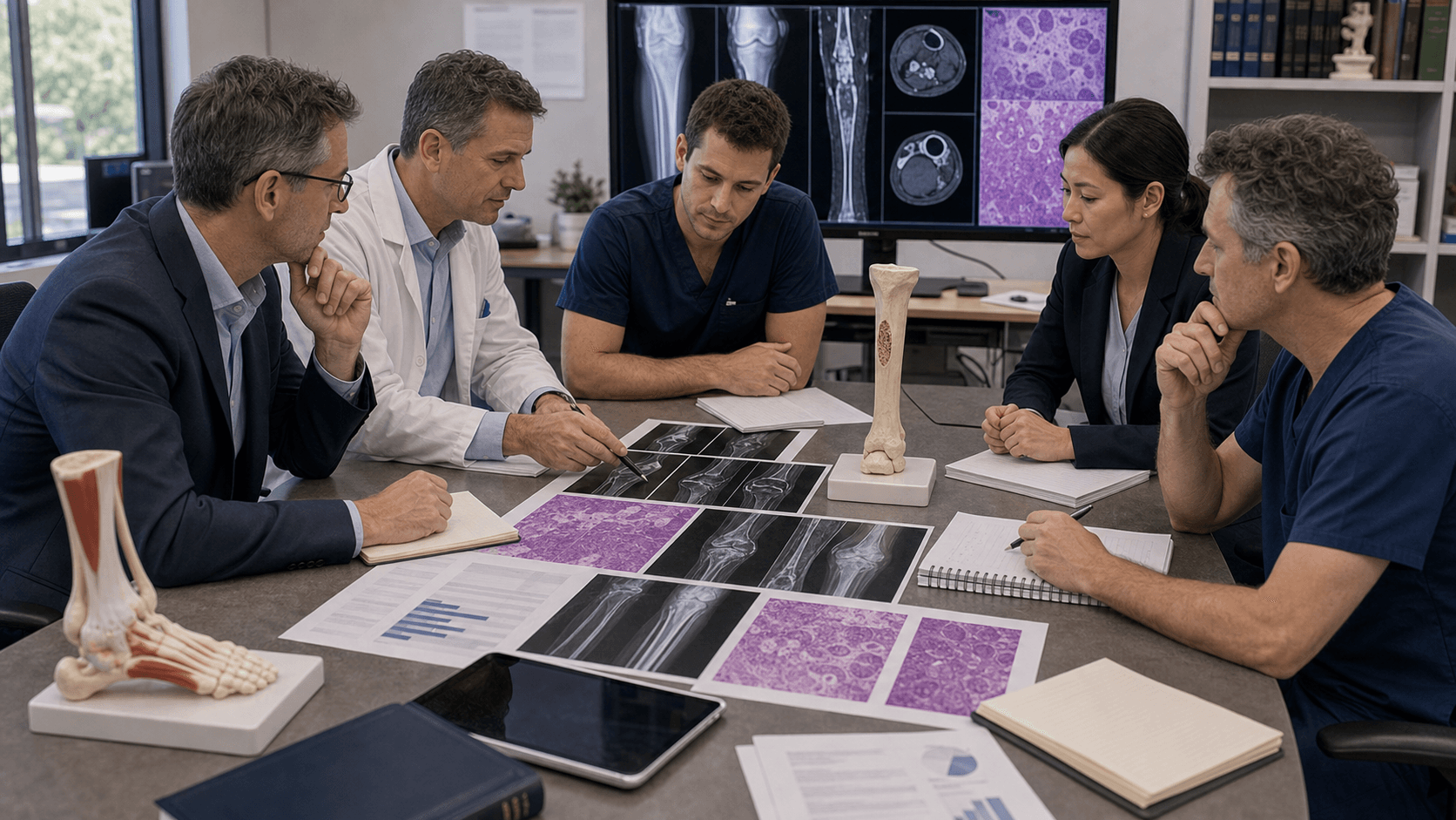
Defining Series: 85 Adamantinomas of Long Bones
- 70 of 85 tumours arose in the tibia (11 also involving the fibula); peak age 10-30 years
- 26 of 85 (31%) developed local recurrence; 13 (15%) lung metastasis; 6 (7%) lymph-node metastasis
- 9 of the patients with lung metastasis had preceding local recurrence
- Risk factors for recurrence/metastasis: male sex, pain, symptoms under 5 years, and inadequate index surgery (biopsy, curettage, simple excision); lack of squamous differentiation was the only adverse histological feature
- 11 patients died of disease; 41 remained alive disease-free 1 month-47 years after treatment
Long-term Outcomes of Adamantinoma and OFD
- 10 adamantinomas and 5 OFD lesions reviewed; mean follow-up 16 years (range 2-47)
- Local recurrence in 40% of adamantinomas and 40% of OFD, all following surgical removal
- Every patient treated by curettage (both entities) developed local recurrence
- In the en bloc resection subgroup, local recurrence fell to 29% (2 of 7)
- Overall prognosis good, but recurrence is frequent irrespective of strategy - authors advocate a more radical approach for adamantinoma
Osteofibrous Dysplasia Does Not Progress to Adamantinoma
- 80 cases of long-bone OFD reviewed; cytokeratin positivity seen in 2 of 6 OFD lesions stained, but all fibrous dysplasia controls were negative
- No OFD lesion progressed to adamantinoma over follow-up (mean 5.4 years, range 1 month-31 years)
- Two OFD lesions in young boys matured histologically into fibrous dysplasia
- Authors conclude OFD is probably a variant of fibrous dysplasia, not an obligate adamantinoma precursor
- Surgery reserved for extensive lesions, pseudoarthrosis or marked tibial bowing; overall prognosis good
Fibrous-to-Epithelial Transformation Hypothesis
- Extracellular-matrix mapping in 21 adamantinomas: basement-membrane proteins (collagen IV, laminin) surrounded cohesive epithelial islands but were interrupted around isolated keratin-positive cells
- OFD-like tumours showed scattered keratin-positive cells emerging from osteofibrous tissue
- Pattern mirrors the glandular component of biphasic synovial sarcoma
- Supports OFD being a potential precursor with individual epithelial cells transforming from the osteofibrous stroma
Malignant Clone Resides in the Epithelial Cells
- DNA flow/image cytometry: aneuploidy detected in 6 of 15 tumours (40%), confined to cells with an epithelial phenotype; all fibrous cells were diploid
- p53 immunoreactivity in 12 of 25 tumours (48%), restricted to epithelial cells, with confirmed loss of heterozygosity at the p53 locus
- Lung metastases contained only keratin-positive epithelial cells - the osteofibrous component was absent
- Indicates a malignant epithelial component with a reactive (benign) osteofibrous stroma
Dedifferentiated Adamantinoma
- Three patients with sarcomatoid transformation of the epithelial component (one primary, two recurrences)
- Dedifferentiated areas showed high mitotic count, pleomorphism and osteoid/chondroid matrix while retaining pankeratin positivity
- One patient died of metastatic disease
- A keratin-positive sarcomatoid cortical tibial tumour should still raise adamantinoma in the differential
WHO Classification of Bone Tumours
- Adamantinoma classified as a locally aggressive, rarely metastasising malignant bone tumour of uncertain histogenesis
- Two recognised entities: classic adamantinoma and differentiated (OFD-like) adamantinoma
- Diagnosis rests on demonstrating keratin-positive epithelial cells within an osteofibrous background
- Differentiated/OFD-like adamantinoma occurs in younger patients and behaves less aggressively than classic disease
Clinical Decision Scenarios
Practise clinical reasoning and management decisions out loud
“A 28-year-old man presents with a 2-year history of anterior shin swelling. Radiographs show a multilocular lytic lesion in the mid-tibial diaphysis with a soap-bubble appearance and sclerotic margins. There is cortical expansion but no periosteal reaction.”
“A 45-year-old woman had curettage of a tibial lesion 5 years ago at an outside institution. She was told it was 'benign'. She now presents with increasing pain and swelling. MRI shows a large tibial lesion with soft tissue extension. Review of original pathology confirms adamantinoma.”
“A 7-year-old child is referred with anterior tibial bowing and an incidental finding of a lytic lesion in the tibial diaphysis on X-ray. The lesion appears intracortical with slight expansion. The child is asymptomatic.”
MCQ Practice Points
Q: What is the most common location for adamantinoma?
A: Tibial diaphysis - 85% of adamantinomas occur in the mid-tibial shaft, characteristically involving the anterior cortex. This location is virtually pathognomonic. Other sites (fibula, humerus, femur) are rare.
Q: What is the characteristic histological feature of adamantinoma?
A: Biphasic pattern with epithelial nests or cords within an osteofibrous stroma. The epithelial cells are positive for cytokeratins 14 and 19 (but negative for CK8, CK18). This cytokeratin profile distinguishes it from metastatic carcinoma.
Q: What is the surgical treatment of choice for adamantinoma?
A: Wide en bloc resection with adequate margins. Curettage is contraindicated and associated with up to 90% local recurrence. Even marginal excision has 30-50% recurrence. The minimum acceptable margin is wide (through normal tissue).
Q: What is the prognosis and metastasis rate for adamantinoma?
A: 10-year survival is approximately 92% with adequate surgical margins. Metastases occur in 15-30% of cases, primarily to lungs and lymph nodes. Late recurrence (up to 36 years post-surgery) mandates lifelong follow-up.
Q: What is the characteristic radiographic appearance of adamantinoma?
A: "Soap-bubble" appearance - multilocular lytic lesion with sclerotic margins, eccentric in the anterior tibial cortex. The lesion may show cortical expansion without periosteal reaction. Skip lesions (multifocality) can occur in the same bone.
Guidelines, Registries & Global Practice
Global Epidemiology
Adamantinoma accounts for well under 1% of all primary bone tumours, making it one of the rarest malignant bone neoplasms encountered worldwide. The disease is remarkably consistent across populations: the tibia is the dominant site (around 70-85% of cases), the peak incidence is in the second to fifth decades, and there is no described geographic, ethnic or environmental predisposition. The tumour has not been linked to radiation or to a hereditary cancer syndrome. Because the absolute numbers are so small, no national cancer registry generates adamantinoma-specific incidence figures of high precision, and the evidence base is built almost entirely from single-institution and tertiary-referral case series rather than trials.
Side-by-Side Guideline and Society Guidance
There is no disease-specific randomised evidence; all major bodies converge on the same principles, derived from retrospective series (evidence level IV) and expert consensus. Differences are largely organisational rather than oncological.
| Body / Region | Core Recommendation | Evidence Basis |
|---|---|---|
| WHO Classification (IARC, 5th ed 2020) | Defines classic vs differentiated (OFD-like) adamantinoma; diagnosis requires keratin-positive epithelial cells in osteofibrous background | Consensus / expert panel |
| ESMO-EURACAN-GENTURIS (Europe) | All suspected primary bone tumours referred to a reference sarcoma centre BEFORE biopsy; biopsy by the treating surgical team; wide en bloc resection for adamantinoma | Expert consensus, level IV-V |
| NICE / BSG (United Kingdom) | Suspected bone sarcoma referred to a recognised bone sarcoma centre or diagnostic clinic; MDT-directed surgery; chemotherapy/radiotherapy not standard for adamantinoma | Service guidance, level IV-V |
| NCCN (United States) | Bone cancer pathway: biopsy and definitive surgery at a centre with sarcoma expertise; wide resection for adamantinoma; systemic therapy reserved for unresectable/metastatic disease | Category 2A consensus |
| BOA / BOOS (UK orthopaedics) | Centralisation of bone tumour surgery; do not biopsy or operate on suspicious bone lesions outside a tumour unit | Standards of practice |
Registry and Centralisation Evidence
Because adamantinoma is so uncommon, the strongest registry-level lesson is centralisation: outcomes for primary bone tumours improve when biopsy and surgery are performed at high-volume sarcoma units, and a poorly planned biopsy outside such a unit can compromise limb salvage. National and supranational sarcoma networks (for example the European EURACAN reference network and equivalent national bone tumour services) exist precisely to pool these rare cases. There is no arthroplasty-style implant registry relevant to adamantinoma, but limb-salvage reconstruction outcomes (allograft, vascularised fibula, endoprosthesis) are tracked within institutional and bone tumour databases.
Practice Variation
The principal real-world variation is in reconstruction technique and resource availability rather than oncological strategy. Wide resection is universal, but the choice between intercalary allograft, vascularised fibular autograft, allograft-prosthesis composite and endoprosthesis depends on resection length, patient age and - importantly in lower-resource settings - access to bone banks and custom implants. Where advanced prostheses and allografts are unavailable, free vascularised fibular grafting is a well-described and effective alternative (Farooque, J Orthop Case Rep 2024). Across all systems, lifelong surveillance for late local recurrence and pulmonary metastasis is the shared standard, reflecting documented recurrences decades after index surgery.
Key Diagnosis
- Tibial diaphysis location (85%) - anterior cortex
- Soap-bubble appearance on X-ray
- Biphasic histology - epithelial + osteofibrous
- Cytokeratin 14, 19 positive
Treatment Principles
- Wide en bloc resection mandatory
- NEVER curettage (90% recurrence)
- Excise biopsy tract with specimen
- Reconstruction: allograft, vascularised fibula, prosthesis
Prognosis
- 10-year survival approximately 92%
- Metastases in 15-30% (lung, lymph nodes)
- Late recurrence up to 36 years
- Lifelong follow-up required
Exam Triggers
- Young adult with tibial diaphyseal lytic lesion
- Anterior cortex soap-bubble lesion
- Multilocular lytic with sclerotic margins
- Positive cytokeratin staining on biopsy
Common Mistakes
- Performing curettage or marginal excision
- Not staging with CT chest before surgery
- Forgetting to excise biopsy tract
- Stopping follow-up after 5 or 10 years