AO Spine Thoracolumbar Injury Classification System
Examiners expect you to classify thoracolumbar injuries using the AO Spine system (not the old Denis three-column model). For any given fracture image, state the morphologic type and subgroup (e.g. A3 burst fracture), the neurologic status (NL grade 0–4), and any modifiers. Then give a clear operative versus non-operative recommendation with justification. Type C injuries and any incomplete neurologic deficit (NL2 or NL3) almost always mandate operative management. Complete cord injuries (NL4) at the thoracolumbar level still warrant decompression and stabilisation to facilitate rehabilitation.
The AO Spine TL Classification — Morphologic Types
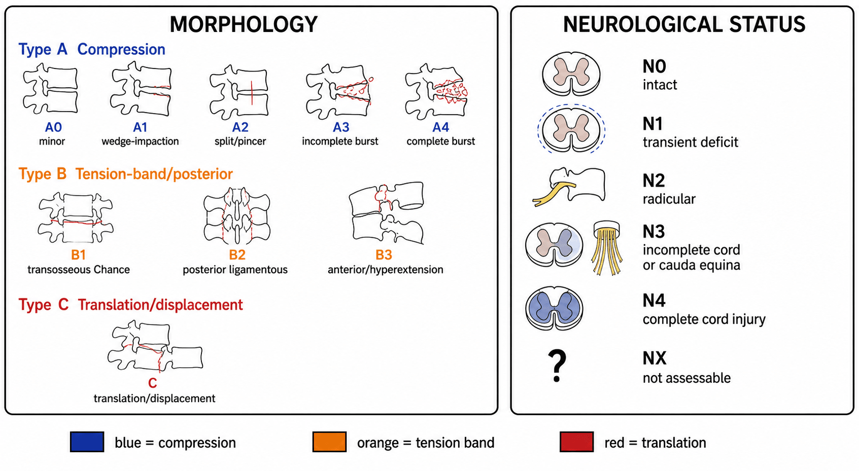
The system has three main morphologic types, each with subgroups. Severity increases from A through C.
- Mechanism
- Axial loading with or without flexion
- Key Feature
- Vertebral body failure; posterior tension band intact
- Subgroups
- A0: minor, A1: wedge, A2: pincer/coronal split, A3: incomplete burst, A4: complete burst
- Mechanism
- Distraction with flexion or extension
- Key Feature
- Failure of posterior or anterior tension band; some body fracture may coexist
- Subgroups
- B1: transosseous (lamina/spine process), B2: posterior soft-tissue (PLC), B3: anterior (hyperextension through disc)
- Mechanism
- Translation or rotation across any axis
- Key Feature
- Displacement of one vertebral segment relative to another; highly unstable
- Subgroups
- Any A or B injury with additional translation or dislocation
Axial · Band · Carry-awayThe three types: A, B, C
Hook:Axial → Band → Carry-away: severity rises from A through C, and a translation always makes it C.
A Type C injury is defined by displacement between adjacent vertebrae in any plane. Even a seemingly modest listhesis on lateral imaging represents a complete ligamentous and/or bony disruption of all columns. The label 'C' always overrides — a burst fracture with translation is classified as C, not A.
Type A Subgroups in Detail
Type A injuries are the most common thoracolumbar fractures and involve the vertebral body under axial loading. The posterior ligamentous complex remains intact.
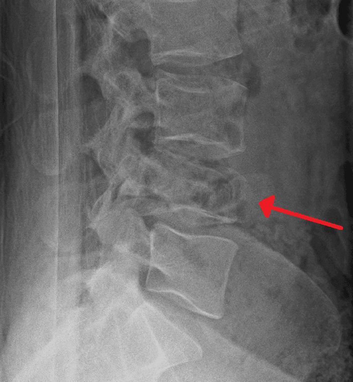
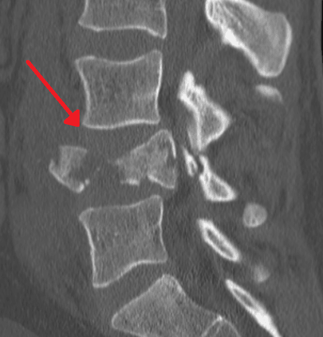
- Description
- Minor, non-structural
- Radiographic Hallmark
- Spinous or transverse process fracture, endplate avulsion
- Typical Management
- Non-operative
- Description
- Wedge compression
- Radiographic Hallmark
- Single endplate fracture, less than 18 degrees kyphosis, less than 50 percent height loss
- Typical Management
- Non-operative (brace) unless significant deformity
- Description
- Pincer / coronal split
- Radiographic Hallmark
- Sagittal split of the vertebral body, both endplates may be involved
- Typical Management
- Usually non-operative; monitor for displacement
- Description
- Incomplete burst
- Radiographic Hallmark
- Fracture of one endplate with retropulsion into the canal; posterior wall fractured but posterior arch intact
- Typical Management
- Non-operative if NL0 and acceptable kyphosis; operative if NL greater than 0 or progressive deformity
- Description
- Complete burst
- Radiographic Hallmark
- Both endplates and posterior wall fractured; greater canal compromise than A3
- Typical Management
- Operative if NL greater than 0 or significant kyphosis; non-operative possible in NL0 with acceptable alignment
Minor · Wedge · Split · Half-burst · Full-burstType A subgroups: 0-1-2-3-4
Hook:The posterior wall is the line in the sand: A1/A2 spare it, A3/A4 (burst) break it.
The key distinction between A1 (wedge) and A3 (burst) is involvement of the posterior vertebral body wall. A wedge fracture spares the posterior wall. A burst fracture disrupts the posterior wall and may retropulse fragments into the spinal canal. This is why CT is mandatory — plain films cannot reliably assess the posterior wall.
Type B and C Injuries
Type B injuries involve failure of the tension band (posterior or anterior) with distraction. Type C injuries have translation or rotation in any plane.
- Disruption Type
- Transosseous (posterior bony)
- Mechanism
- Flexion-distraction (Chance-type through bone)
- Key Imaging Finding
- Horizontal fracture through the pedicles and/or lamina; 'bony Chance'
- Disruption Type
- Posterior soft-tissue (PLC)
- Mechanism
- Flexion-distraction through ligaments
- Key Imaging Finding
- Interspinous widening, facet subluxation/dislocation, positive STIR in PLC on MRI
- Disruption Type
- Anterior tension-band (hyperextension)
- Mechanism
- Extension through the anterior disc/ALL
- Key Imaging Finding
- Anterior disc space widening, possible retrolisthesis; seen in ankylosing conditions
A Type B2 injury (soft-tissue PLC disruption) can be subtle on plain radiographs. If there is any interspinous widening, facet perch, or kyphotic angulation out of proportion to the visible bony injury, obtain an MRI with STIR sequence to assess the PLC. Missing an unstable B2 injury and treating it as a Type A can lead to progressive deformity and neurologic deterioration.
- Detail
- Any translation or dislocation of one vertebral segment relative to the adjacent segment, in any plane
- Detail
- Obvious listhesis, rotation, or facet dislocation on CT/sagittal reformats; may have bilateral facet dislocation
- Detail
- By definition grossly unstable — complete disruption of all stabilising structures
- Detail
- Almost always operative — reduction and instrumented stabilisation; urgent if neurologically incomplete
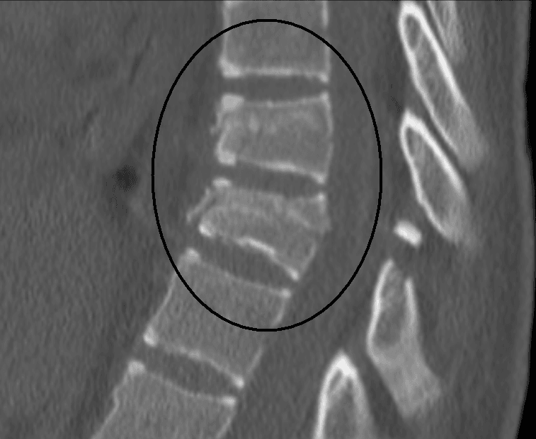
A Chance fracture is a B1 injury (bony tension-band failure). If the same mechanism disrupts the soft tissues (PLC, disc) instead of bone, it is a B2. Both are unstable. The classic mechanism in a Chance fracture is a lap-belt injury in a motor vehicle collision — flexion over the lap belt with the posterior elements pulled apart.
A fracture in a fused, rigid spine (ankylosing spondylitis or DISH) is a different and far more dangerous entity — exactly what the M2 modifier flags:
- It behaves like a long-bone fracture. The brittle, fused column has long lever arms, so even a low-energy injury (a ground-level fall) produces a transverse fracture through all three columns, typically an extension (B3) pattern through the ossified disc or across bone — and it is highly unstable.
- It is easily MISSED and prone to deterioration. The fracture is hard to see against the ankylosed bone and the pain is often blamed on the underlying disease; there is a high rate of epidural haematoma and delayed/secondary neurological deterioration. Mortality is high.
- Image the WHOLE spine. Plain films are unreliable → CT of the entire spine (look for non-contiguous fractures) plus MRI (epidural haematoma, occult fracture, PLC). Have a very low threshold in any ankylosed patient with new back pain after even trivial trauma.
- Management: usually long-segment posterior instrumented fixation (several levels above and below — short constructs fail in this stiff bone). Positioning is critical — transfer and position the patient maintaining their pre-existing fixed kyphotic deformity; forcing them supine/flat or into extension can displace the fracture and injure the cord. Avoid over-reduction.
Neurologic Status and Modifiers
Neurologic status is graded separately and is the most important modifier for treatment decisions.
- Status
- Neurologically intact
- Description
- No motor, sensory, or bowel/bladder deficit
- Operative Implication
- Morphology alone guides decision
- Status
- Transient deficit, now resolved
- Description
- Deficit was present but has fully recovered
- Operative Implication
- Consider morphology; less urgent than active deficit
- Status
- Nerve root injury (radiculopathy)
- Description
- Radicular symptoms or signs
- Operative Implication
- Strong operative indication: decompress and stabilise
- Status
- Incomplete cord OR cauda equina injury
- Description
- ASIA B/C/D, or any degree of cauda equina injury
- Operative Implication
- Urgent operative decompression and stabilisation
- Status
- Complete spinal cord injury
- Description
- ASIA A; no motor or sensory function below the level
- Operative Implication
- Operative for stabilisation and rehabilitation, decompress if within window
Intact · Transient · Root · Incomplete · CompleteNeuro grades and modifiers
Hook:N rises with severity (NX = not assessable). Modifiers: M1 = indeterminate PLC on MRI; M2 = patient/comorbidity factor (e.g. ankylosing spinal disorder, polytrauma).
Decision Framework: Operative versus Non-operative
- Neuro Status
- NL0
- Recommendation
- Non-operative (brace, mobilise)
- Key Rationale
- Stable fracture pattern, intact PLC, no canal compromise
- Neuro Status
- NL0, acceptable alignment
- Recommendation
- Trial of non-operative treatment
- Key Rationale
- Monitor for progressive kyphosis or late neuro deficit; brace 8–12 weeks
- Neuro Status
- NL1–NL4
- Recommendation
- Operative: decompression and stabilisation
- Key Rationale
- Neuro deficit drives operative management; decompress the canal
- Neuro Status
- Any
- Recommendation
- Operative stabilisation
- Key Rationale
- Tension-band disruption = unstable; non-operative treatment risks failure
- Neuro Status
- Any
- Recommendation
- Operative: reduction and instrumented fixation
- Key Rationale
- Gross instability; nearly universal operative indication
The TL AOSIS score integrates morphology, neurology, and modifiers into a single number. Per the AO Spine surgical algorithm (Vaccaro 2016): a score of 3 or less favours a trial of conservative treatment, a score greater than 5 (i.e. 6 or more) favours surgery, and a score of 4 or 5 is the grey zone where operative or non-operative treatment is both acceptable. Examiners may ask you to calculate the score, but the principle — neurology and tension-band/translation injuries drive surgery — matters more than the exact point values.
The thresholds are useless without the inputs. Add the points from the three axes:
- Morphology: A0 = 0, A1 = 1, A2 = 2, A3 = 3, A4 = 5; B1 = 5, B2 = 6, B3 = 7; C = 8.
- Neurology: N0 = 0, N1 = 1, N2 = 2, N3 = 4, N4 = 4, NX = 3 (not assessable scores like an incomplete deficit, so it errs toward surgery).
- Modifiers: M1 (indeterminate PLC) = 1; M2 (comorbidity) = 0.
Sum, then map: total 3 or less → trial of conservative care, 4 or 5 → grey zone (either acceptable), 6 or more → surgery.
Worked examples: an A1 wedge, neurology intact = 1 + 0 = 1 → brace; an A3 incomplete burst, intact = 3 → conservative, but add an indeterminate PLC (M1, +1) = 4 → grey zone; an A4 complete burst with an incomplete cord injury (N3) = 5 + 4 = 9 → surgery; any Type C starts at 8, so it is always surgical. Notice that any single tension-band (B) or translation (C) morphology, or an N3/N4 deficit, already pushes the total to 5 or more — which is why "B/C or a deficit means operate" is the shortcut.
Guidelines, Registries and Global Practice
- Key Recommendation
- Classify all TL injuries using AO Spine TL system; use TL AOS score to guide operative decision
- Evidence Level
- Expert consensus
- Key Recommendation
- MRI recommended when PLC integrity uncertain; operative treatment for unstable injuries and neuro deficit
- Evidence Level
- Grade B/C
- Key Recommendation
- Class III evidence supports operative decompression for incomplete cord injury within 24 hours where feasible
- Evidence Level
- Class III
- Key Recommendation
- Adopts AO Spine classification; recommends CT for all suspected TL fractures and MRI for PLC assessment
- Evidence Level
- Level 2b/3
- Epidemiology: Thoracolumbar fractures account for approximately 90 percent of all spinal fractures, with the thoracolumbar junction (T11–L2) being the most common site due to the transition from the rigid thoracic kyphosis to the mobile lumbar lordosis. High-energy mechanisms (falls from height, motor vehicle collisions) predominate in younger patients; low-energy mechanisms (ground-level falls) are more common in the elderly with osteoporosis.
- Global practice variation: Operative rates for thoracolumbar burst fractures without neurologic deficit vary from approximately 30 percent in some European centres to over 70 percent in parts of North America and Asia, reflecting genuine equipoise and cultural/systemic factors rather than evidence-based consensus. The AO Spine classification is used worldwide but the TL AOS treatment score threshold for operative intervention is applied variably.
- Registry evidence: National trauma registries (e.g. the UK TARN, US NTDB, German TraumaRegister DGU) report thoracolumbar fracture incidence and treatment patterns. The Spine Tango registry (Eurospine) collects AO Spine classification data prospectively and has demonstrated correlation between classification type and operative rates across European centres.
Exam Viva
Practise clinical reasoning and management decisions out loud
“A 32-year-old man falls from 4 metres onto his feet. He presents with severe thoracolumbar pain. CT shows a L1 burst fracture with retropulsion of the posterior wall into the canal and both endplates fractured. He has an incomplete paraplegia with some preserved motor function in the lower limbs (ASIA C). How do you classify this injury and what is your management plan?”
“A 45-year-old woman was an unrestrained passenger in a high-speed head-on collision. She has severe back pain and is ASIA A (complete paraplegia) at the T12 level. CT shows a fracture-dislocation at T12–L1 with approximately 50 percent anterolisthesis of T12 on L1 and bilateral facet dislocation. MRI confirms complete canal occlusion and extensive soft-tissue disruption. How do you classify and manage this injury?”
Three morphologic types
- Type A — Compression: body fracture under axial load; PLC intact (A0 minor, A1 wedge, A2 pincer, A3 incomplete burst, A4 complete burst)
- Type B — Tension-band: posterior or anterior tension-band disruption with distraction (B1 bony Chance, B2 PLC soft-tissue, B3 anterior hyperextension)
- Type C — Translation: displacement between adjacent vertebrae in any plane; always unstable; any A or B injury with added translation becomes C
Neurologic status (NL 0–4)
- NL0: intact. NL1: resolved transient deficit. NL2: nerve root (radiculopathy). NL3: incomplete cord (ASIA B/C/D) OR any cauda equina injury. NL4: complete cord (ASIA A)
- Neurologic status is the strongest driver of operative decision-making
- NL2 or greater is a strong operative indication regardless of morphologic type
Treatment principles
- A0–A2, NL0: non-operative (brace, mobilise)
- A3–A4, NL0: non-operative if acceptable alignment; operate if progressive kyphosis or late neuro deficit
- Any type with NL2–NL4: operative — decompression and stabilisation
- Type B or C, any neuro status: operative stabilisation
- TL AOSIS score: 3 or less non-operative; over 5 (6 or more) surgical; 4 or 5 is the grey zone (either acceptable)
Key exam traps
- PLC assessment is unreliable on plain films — always use MRI STIR sequence when PLC integrity is uncertain
- A Chance fracture through bone is B1; through ligaments is B2 — both are unstable
- Do not classify using the old Denis three-column system in an exam setting — use AO Spine
- Complete cord injury (NL4) still requires operative stabilisation for rehabilitation
Evidence Base
Every citation below has been checked against its source record in PubMed. The lineage runs from TLICS (Vaccaro 2005) → the consensus AO Spine TL system (Vaccaro 2013) → the surgical algorithm and TL AOSIS thresholds (Vaccaro 2016) → an independent reliability check (Urrutia 2015), with the Cochrane review (Abudou 2013) framing the operative-vs-conservative debate for neurologically intact burst fractures.
A new classification of thoracolumbar injuries: the importance of injury morphology, the integrity of the posterior ligamentous complex, and neurologic status (TLICS)
- Proposed the Thoracolumbar Injury Classification and Severity Score (TLICS) built on three characteristics: injury morphology, posterior ligamentous complex (PLC) integrity, and neurologic status
- A composite severity score stratified patients into surgical versus non-surgical groups and guided the operative approach
- Introduced PLC integrity as a key independent variable in thoracolumbar treatment decision-making
AOSpine thoracolumbar spine injury classification system: fracture description, neurological status, and key modifiers
- Developed the AO Spine TL classification by structured international consensus to achieve universal adoption where Magerl and TLICS had not
- Three morphologic types — A (compression), B (tension-band), C (displacement/translation) — with subgroups, plus separate neurologic grading and patient-specific modifiers
- Reliability for identifying the morphologic type was substantial (kappa = 0.72) across the international development group
The surgical algorithm for the AOSpine thoracolumbar spine injury classification system
- Surveyed AO Spine members across the six AO world regions to build a globally derived treatment algorithm for thoracolumbar injuries
- Thresholds: a thoracolumbar AO Spine injury score (TL AOSIS) of 3 or less should have a trial of conservative care; a score of more than 5 should have surgery
- A TL AOSIS of 4 or 5 is the grey zone — operative or non-operative treatment is both acceptable