Avascular Necrosis of the Humeral Head
Progressive ischemic bone death leading to structural collapse and secondary arthritis
Cruess Classification
Critical Must-Knows
- MRI double-line sign is pathognomonic (T2: inner low + outer high signal)
- Crescent sign = point of no return (mechanical failure, joint preservation unlikely)
- ALWAYS screen opposite shoulder - 30-60% bilateral (78% if steroid-induced)
- Core decompression ONLY effective pre-collapse (Stage I-II): 50-70% success
- TSA superior to hemiarthroplasty when glenoid involved (Stage V-VI)
- Young patients (under 50): 24% revision rate at 10 years - counsel lifetime burden
Clinical Pearls
- "AVN ≠ primary OA: younger age, bilateral, intact cuff, systemic etiology
- "Arcuate artery (AHCA branch) provides 80% humeral head blood supply
- "Steroid risk: over 2000mg cumulative, over 20mg/day, over 3 months duration
- "Resurfacing requires intact cuff (ABSOLUTE) - cuff deficiency = failure
- "Bilateral simultaneous surgery CONTRAINDICATED - need one functional arm

Clinical Warning
AVN ≠ Osteoarthritis
Crucial Distinction: Patients are younger, often bilateral, and have intact rotator cuffs.
Management Dilemma
Young Age: High revision risk with TSA. Intact Cuff: Reverse TSA often contraindicated/suboptimal (wastes a good cuff). Result: Complex decision-making (Resurfacing vs Stemless TSA vs Hemi).
Bilateral Trap
30-60% Bilateral: Always screen the other shoulder. Steroid AVN = 78% bilateral.
At a Glance
| Category | Details |
|---|---|
| Definition | Progressive ischemic necrosis of humeral head bone and marrow leading to structural collapse and secondary glenohumeral arthritis |
| Incidence | Humeral head is the 2nd most common osteonecrosis site after the femoral head (around 10% of cases); AVN accounts for roughly 3-5% of shoulder arthroplasties in joint registries |
| Age | 30-50 years (two decades younger than primary OA) |
| Sex | Male predominance (2-3:1) |
| Bilateral | 30-60% at presentation or during follow-up (highest in steroid-induced: 78%) |
| Etiology | Non-traumatic (80%): Steroids (35-40%), Alcohol (20-25%), Idiopathic (20-25%), Sickle cell, SLE, Gaucher Traumatic (20%): Proximal humerus fractures (4-part: 75% AVN risk), shoulder dislocation |
| Pathophysiology | Vascular insult → Osteocyte death → Repair attempt → Bone weakening → Subchondral fracture → Collapse → Secondary arthritis |
| Key Anatomy | Arcuate artery (AHCA branch) = 80% supply, enters posterolateral to bicipital groove; Watershed zone = superomedial head |
| Classification | Cruess (6 stages): I-II Pre-collapse, III Crescent, IV-VI Collapse/arthritis Ficat-Arlet (0-V): Correlates radiographic progression Modified Ficat: Adds MRI volumetric assessment |
| Diagnosis | MRI: Double-line sign (pathognomonic), band sign, lesion volume X-ray: Normal (Stage I) → Sclerosis (II) → Crescent (III) → Collapse (IV-VI) |
| Treatment | Stage I-II: Observation, core decompression ± biologics Stage III: Controversial (observation vs decompression vs arthroplasty) Stage IV: Hemiarthroplasty, resurfacing (young + intact cuff), TSA (older) Stage V-VI: TSA (cuff intact) or reverse TSA (cuff deficient + age over 65) |
| Outcomes | Core decompression: 50-70% success (no progression) at 5 years in Stage I-II Hemiarthroplasty: 20% revision at 10 years TSA: 12% revision at 10 years Young (under 50): 24% revision at 10 years (all implant types) |
| Complications | Infection (1-2%, higher if immunosuppressed), Instability (2-4%), Neurovascular injury (1-2%), Glenoid loosening (10-15% at 10 years), Contralateral progression (30-60%) |
| Exam Focus | Cruess staging, MRI double-line sign, Crescent sign significance, Core decompression indications, Hemi vs TSA vs reverse decision-making, Young patient arthroplasty challenges, Bilateral screening |
Common Mnemonics
A-S-E-P-T-I-CASEPTIC Causes of AVN
| A | Alcohol Chronic consumption over 400mL/week - fat emboli and direct toxicity |
| S | Steroids over 2000mg cumulative prednisone, over 20mg/day, over 3 months - most common non-traumatic cause |
| E | Ethanol (see Alcohol above) - emphasizes alcohol as major contributor |
| P | Pancreatitis/Pregnancy Fat emboli from pancreatic necrosis; pregnancy-related hypercoagulability |
| T | Trauma/Thrombophilia Proximal humerus fractures (4-part 75% risk); inherited clotting disorders |
| I | Idiopathic 20-25% of cases - no identifiable cause despite investigation |
| C | Connective tissue/Caisson SLE, RA (often steroid-related); decompression sickness in divers |
| A | Alcohol Chronic consumption over 400mL/week - fat emboli and direct toxicity | P | Pancreatitis/Pregnancy Fat emboli from pancreatic necrosis; pregnancy-related hypercoagulability | C | Connective tissue/Caisson SLE, RA (often steroid-related); decompression sickness in divers |
| S | Steroids over 2000mg cumulative prednisone, over 20mg/day, over 3 months - most common non-traumatic cause | T | Trauma/Thrombophilia Proximal humerus fractures (4-part 75% risk); inherited clotting disorders | ||
| E | Ethanol (see Alcohol above) - emphasizes alcohol as major contributor | I | Idiopathic 20-25% of cases - no identifiable cause despite investigation |
Hook:Remember non-traumatic causes are 'ASEPTIC' conditions - helps differentiate from post-traumatic AVN which has different prognosis and management. The double-E reminds you that alcohol (ethanol) is a major player alongside steroids.
Overview and Epidemiology
Definition
Avascular necrosis (AVN, also termed osteonecrosis) of the humeral head represents a spectrum of disease from reversible bone marrow ischemia to irreversible structural collapse and secondary glenohumeral arthritis. Unlike primary osteoarthritis, AVN typically affects younger patients with intact rotator cuffs but compromised subchondral bone vascularity.
The pathophysiological cascade progresses from initial vascular insult through attempted bone repair to mechanical failure and ultimately degenerative arthritis. The critical feature distinguishing AVN from other shoulder pathology is the underlying ischemic bone death, which creates unique challenges in surgical management.
Epidemiology
Incidence and Prevalence:
- The humeral head is the second most common site of osteonecrosis after the femoral head (each shoulder and knee account for roughly 10% of all osteonecrosis cases)
- AVN accounts for approximately 3-5% of shoulder arthroplasties across major joint registries
- Humeral head involvement is uncommon among osteonecrosis patients overall (~7% in early cohort data), but is frequently bilateral and often coexists with hip osteonecrosis (~80%)
- Increasing recognition due to improved MRI access and awareness; true incidence likely underestimated (asymptomatic cases not diagnosed)
Demographics:
- Peak age: 30-50 years (two decades younger than primary osteoarthritis)
- Sex distribution: Male predominance 2-3:1 in many series (reflects higher alcohol consumption and steroid exposure); steroid-driven cohorts may show female predominance
- Bilateral involvement: 30-60% at presentation or develop during follow-up (up to ~74-78% in steroid-induced disease)
- Mean time to bilateral disease: 1-2 years from unilateral diagnosis
Registry Perspective (Global): Across national arthroplasty registries, patients undergoing shoulder replacement for AVN are typically younger than those with primary OA, creating challenges in prosthesis selection, activity counselling and lifetime revision burden. A consistent registry signal worldwide is a higher revision rate in AVN patients under 50 years compared with older cohorts.
Geographic and Population Variation:
- Higher rates in populations with increased systemic steroid use (inflammatory and autoimmune disease prevalence, transplant programmes)
- Alcohol-related AVN correlates with regional drinking patterns
- Sickle cell disease is a leading cause in populations of African, Mediterranean, Middle Eastern and South Asian ancestry
- Socioeconomic factors and MRI availability influence access to early diagnosis (early disease is frequently missed in limited-resource settings reliant on plain radiographs)
Etiology and Risk Factors
Non-Traumatic Causes (80% of cases):
Corticosteroids (35-40%):
- Dose threshold: Greater than 2000mg cumulative prednisone equivalent
- Daily dose risk: Greater than 20mg/day
- Duration risk: Greater than 3 months continuous use
- Route effect: IV pulse therapy higher risk than oral equivalent
- Mechanism: Direct osteocyte toxicity, fat embolism, intravascular coagulation
- Common indications: Organ transplant immunosuppression, SLE, inflammatory bowel disease, severe asthma, hematologic malignancy
Alcohol (20-25%):
- Threshold: Greater than 400mL/week chronic consumption
- Mechanism: Direct cellular toxicity, fat metabolism disruption, intravascular fat emboli
- Pattern: Chronic heavy use greater risk than intermittent binge (though both contribute)
- Dose-response: Risk increases with quantity and duration
Idiopathic (20-25%):
- No identifiable risk factor despite thorough investigation
- May represent unrecognized genetic susceptibility or environmental factors
- Bilateral rate similar to other etiologies
- Prognosis and treatment response similar to known-cause AVN
Hemoglobinopathies (5-10%):
- Sickle cell disease: Most common - vaso-occlusive crises cause bone infarction
- Thalassemia (less common)
- Mechanism: Intravascular sickling causes mechanical vascular occlusion
Connective Tissue Diseases (5%):
- Systemic lupus erythematosus (SLE): Multifactorial (disease + steroids + vasculitis)
- Rheumatoid arthritis (usually steroid-related)
- Mixed connective tissue disease
Other Causes (Less than 5% each):
- Gaucher disease: Lysosomal storage disorder causing marrow infiltration
- Caisson disease (decompression sickness): Nitrogen bubble emboli in divers, tunnel workers
- Pancreatitis: Fat emboli from necrotic pancreatic tissue
- Pregnancy: Unclear mechanism, may relate to hypercoagulability
- HIV: Multifactorial (disease, medications, coagulopathy)
- Radiation therapy: Direct vascular injury
- Chemotherapy: Endothelial toxicity
Traumatic Causes (20% of cases):

Proximal Humerus Fractures:
- Overall AVN risk: 3-5% at 2 years post-fracture
- 4-part fractures: 75% AVN rate (disruption of arcuate artery)
- Valgus-impacted 4-part: 25-30% (some vascular preservation)
- Head-splitting fractures: 40-50% (direct vascular injury)
- Time to AVN development: 6 months to 3 years (mean 18 months)
Shoulder Dislocation:
- Incidence: 1-2% overall (higher with recurrent dislocations)
- Mechanism: Circumflex vessel injury during dislocation or reduction
- Hill-Sachs lesions: Large defects may have associated AVN component
Iatrogenic:
- Surgical fixation: Overly aggressive dissection disrupting arcuate artery
- Multiple surgeries: Cumulative vascular insult
- Screws/plates: Direct vascular injury from hardware
These etiological factors have important implications for management, including need for medical optimization, bilateral screening, and counseling about ongoing risk with continued exposure.
Anatomy and Biomechanics
Vascular Anatomy of the Humeral Head
-
Anterior Humeral Circumflex Artery (AHCA) - Primary supply (80%):
- Origin: Axillary artery (lateral to pectoralis minor)
- Course: Runs along inferior border of subscapularis
- Arcuate artery branch: CRITICAL vessel
- Enters humeral head just posterolateral to bicipital groove
- Ascends within bone to supply anterolateral 2/3 of head
- Most vulnerable to injury in displaced fractures and surgical dissection
- Supplies: Greater tuberosity, anterolateral humeral head, long head biceps
-
Posterior Humeral Circumflex Artery (PHCA) - Secondary supply (20%):
- Origin: Axillary artery (travels with axillary nerve through quadrangular space)
- Course: Posterior to surgical neck
- Supplies: Posterior 1/3 of humeral head via multiple capsular vessels
- Less clinically significant than AHCA (smaller contribution)
-
Terminal Intraosseous Distribution:
- Vessels arborize in subchondral bone creating terminal vascular network
- Watershed zone: Superomedial humeral head (junction of AHCA and PHCA territories)
- Limited intraosseous anastomoses (poor collateral circulation)
- End-artery pattern makes head vulnerable to single-vessel injury
Anatomical Vulnerabilities:
- Single dominant vessel reliance: AHCA (arcuate artery) disruption causes extensive necrosis affecting 80% of head
- Epiphyseal-metaphyseal junction: Watershed zone most susceptible to ischemia
- Capsular reflection: Creates anatomical separation limiting collateral flow between metaphysis and epiphysis
- Intracapsular location: Limits external soft tissue collateral development
- Terminal vessel architecture: End-artery pattern with minimal anastomoses
Surgical Relevance:
- Fracture fixation: Overzealous dissection anterolateral to bicipital groove risks arcuate artery injury
- Deltopectoral approach: Staying medial to biceps groove protects AHCA
- Inferior capsular release: Avoid extending dissection too far posteriorly (PHCA in quadrangular space)
- Screw placement: Avoid long screws in anterolateral quadrant (may disrupt arcuate artery)
Classification Systems
Cruess Classification (Most Commonly Used)
The Cruess classification is the most clinically practical system, based primarily on plain radiographic findings and correlating well with treatment algorithms. It is the gold standard for communication among surgeons and in clinical decision-making.
Cruess Classification of Humeral Head AVN
| Stage | Radiographic Findings | MRI Findings | Symptoms | Treatment Options |
|---|---|---|---|---|
| **I: Early** | Normal radiographs, increased bone density may be subtle | Diffuse marrow edema, band sign, no clear demarcation | Minimal to moderate pain, often activity-related | Observation + risk factor modification, Core decompression ± biologics |
| **II: Sclerosis** | Patchy sclerosis and cyst formation, no collapse, preserved contour | Band sign or double-line sign, demarcated lesion, volume assessment | Moderate pain with activity, some night pain | Core decompression + bone grafting, Biological augmentation, Close surveillance |
| **III: Crescent Sign** | Subchondral fracture visible as crescent-shaped lucency (point of no return) | Subchondral fracture line clearly evident, beginning structural disruption | Significant pain with activity and at rest, limited ROM | Controversial: Observation vs Core decompression (low success) vs Arthroplasty |
| **IV: Collapse** | Humeral head flattening and contour loss, joint space maintained | Structural collapse visible, glenoid cartilage still normal | Severe pain, stiffness, grinding sensation | Hemiarthroplasty, Humeral head resurfacing (young + intact cuff), TSA (older) |
| **V: Early Arthritis** | Joint space narrowing begins, early glenoid sclerosis and changes | Glenoid edema, subchondral sclerosis, cartilage thinning | Severe pain, marked stiffness, functional limitation | TSA (if glenoid suitable for component), Hemiarthroplasty (preserve glenoid if young) |
| **VI: Advanced Arthritis** | Advanced degenerative changes both sides, osteophytes, severe narrowing | Severe glenoid involvement, possible bone loss, cuff may be compromised | Disabling pain and dysfunction, severe ROM loss | TSA (if glenoid bone stock adequate), Reverse TSA (if cuff deficient) |
Clinical Warning
The Crescent Sign
Stage III Point of No Return: Represents distinct subchondral fracture.
Clinical Implication
Mechanical Failure: Joint preservation (core decompression) is no longer effective. Arthroplasty usually required.
Clinical Assessment
History Taking - Red Flags for AVN
Presenting Complaint:
- Pain onset: Insidious (non-traumatic) vs acute (post-traumatic, post-fracture)
- Pain character: Deep, aching, boring quality (bone pain), worse with loading activities
- Pain location: Anterior and lateral shoulder most common, may radiate to deltoid insertion
- Night pain: Common in progressive stages (distinguishes from simple impingement)
- Bilateral symptoms: CRITICAL to ask - 30-60% have or will develop contralateral disease
Risk Factor Assessment (ESSENTIAL):
Steroid Exposure (Most Important):
- Indication: Why on steroids (transplant, SLE, IBD, severe asthma, malignancy, other)
- Route: IV pulse therapy vs oral maintenance (pulse higher risk)
- Dose: Current daily dose, cumulative dose estimation (greater than 2000mg prednisone threshold)
- Duration: How long on treatment (greater than 3 months threshold)
- Temporal relationship: Symptom onset typically 6-18 months post-initiation or dose escalation
- Ongoing exposure: Still on steroids (affects progression risk and surgical planning)
Alcohol History:
- Quantity: Drinks per week (greater than 400mL spirits/week threshold, roughly greater than 400g pure alcohol)
- Pattern: Chronic daily vs binge weekend drinking (both increase risk)
- Duration: Years of heavy consumption
- Current status: Still drinking vs abstinent (critical for surgical planning)
Medical Conditions:
- Sickle cell disease: Ask about crises, other sites of AVN (hip most common)
- SLE and connective tissue disorders: Disease activity, other organ involvement
- HIV: Treatment status, CD4 count, medication history
- Gaucher disease: Enzyme replacement therapy status
- Pancreatitis: Acute necrotizing episodes
- Diving/decompression illness: Professional or recreational diving, depth, decompression protocols
Trauma History:
- Proximal humerus fracture: When, what type, how treated (ORIF vs conservative)
- Shoulder dislocation: How many, directions, associated injuries
- Prior shoulder surgery: What procedures, complications
- Time interval: AVN develops 6 months to 3 years post-trauma (peak 18 months)
Functional Impact Assessment:
- Activities of daily living: Dressing (reaching behind back), grooming (hair), feeding (hand to mouth)
- Occupation: Manual labor vs sedentary, overhead work, lifting requirements, disability implications
- Sleep: Which positions possible, night wakening frequency
- Recreation: Sports, hobbies affected
- Previous treatments: Physiotherapy, injections, medications, effectiveness
Contralateral Screening (MANDATORY):
- "Do you have any pain or symptoms in your other shoulder?"
- "Have you had imaging of your other shoulder?"
- Document need for bilateral screening imaging in management plan
Clinical Pearl
Always ask about contralateral shoulder symptoms and document plan for bilateral imaging - 30-60% of AVN patients have bilateral disease, which may be asymptomatic radiographically. In steroid-induced AVN, bilateral rate is 78%. Examiners expect this in your assessment and will view omission as a significant oversight.
Physical Examination
Examination should be systematic, bilateral, and focused on rotator cuff integrity (critical for arthroplasty planning).
Inspection:
- Muscle wasting: Deltoid, supraspinatus, infraspinatus fossae (chronic disease indicator)
- Asymmetry: Compare shoulder heights, contours with opposite side
- Scars: Previous surgery or trauma
- Posture: Shoulder held in protective position (internal rotation, adduction)
- Skin: Cushing's stigmata if steroid-related (striae, bruising, thin skin)
Palpation:
- Bony landmarks: Acromion, clavicle, coracoid, humeral head
- Tenderness: Typically anterior and lateral humeral head (deep palpation)
- AC joint: Exclude concurrent pathology (test cross-body adduction)
- Biceps groove: Long head biceps often attritional in AVN
- Cervical spine: Rule out referred pain (palpate spinous processes, test ROM)
Range of Motion (Compare to Opposite Side):
Active ROM (Patient-Generated):
- Forward elevation: Normal 160-180 degrees (reduced in advanced AVN, typically 90-120 degrees)
- Abduction: Normal 160-180 degrees (similar pattern to elevation)
- External rotation at side: Normal 60-80 degrees (early loss suggests capsular involvement)
- Internal rotation: Assess by posterior reach (T7-T12 normal, buttock/lumbar in restricted)
Passive ROM (Examiner-Generated):
- Compare to active (if passive greater than active, suggests rotator cuff or pain issues)
- End-feel: Firm (capsular) vs hard (bony block from osteophytes) vs empty (pain)
- Pattern: Global restriction suggests capsular contracture vs selective loss
Strength Testing (CRITICAL for Arthroplasty Planning):
Rotator Cuff Integrity:
-
Supraspinatus:
- Empty can test (Jobe test): Resist abduction at 90 degrees in scapular plane, thumb down
- Grading: 0-5 MRC scale (document actual grade, not just "weak")
-
Infraspinatus:
- Resisted external rotation at side: Elbow 90 degrees flexed, resist external rotation
- Hornblower's sign (if positive, severe cuff deficiency)
-
Subscapularis (Multiple tests - use all):
- Lift-off test: Hand behind back, lift off from lumbar spine (most specific)
- Belly-press test: Press hand into abdomen (positive if elbow drops posterior to trunk)
- Bear-hug test: Hand on opposite shoulder, resist attempt to pull away
-
Teres minor:
- Hornblower's sign: Externally rotate at 90 degrees abduction (inability suggests teres minor weakness)
Deltoid:
- Resisted abduction 90 degrees (anterior, middle, posterior heads)
- Important for reverse TSA outcomes
Special Tests:
- Impingement signs (Neer, Hawkins-Kennedy): Usually negative in isolated AVN (not impingement pathology)
- Instability testing: Load-and-shift, apprehension, relocation (typically stable unless prior dislocation)
- Neurovascular: Axillary nerve sensation (lateral shoulder patch), radial pulse, motor function
Bilateral Examination (ESSENTIAL):
- ALWAYS examine opposite shoulder completely
- Document ROM and strength comparisons
- Look for early signs of bilateral disease (pain on deep palpation, subtle ROM loss)
- Aids in surgical planning if bilateral procedures anticipated
Rotator cuff assessment is CRITICAL before arthroplasty planning - a deficient cuff in a young AVN patient (who may not be suitable for reverse TSA due to age) creates a major management dilemma. Document strength testing clearly using MRC grading, and consider ultrasound or MRI if any weakness detected on examination.
Investigations
Investigations
Plain Radiographs (First-Line)
Plain radiographs remain the initial imaging modality despite limited sensitivity in early disease.
Views Required:
-
True AP (Grashey view):
- Patient rotated 40 degrees posterior oblique
- Best shows glenohumeral joint space
- Identifies glenoid changes in late disease
- Beam perpendicular to scapula
-
Scapular Y (lateral):
- Shows anterior-posterior humeral head position
- Crescent sign best seen on this view (profile of subchondral fracture)
- Assesses head sphericity and collapse
-
Axillary lateral:
- Essential for complete assessment (never omit)
- Shows posterior glenoid wear
- Detects subluxation
- Arm abducted 45 degrees, beam through axilla
Radiographic Findings by Cruess Stage:
- Stage I: Normal (60% of early AVN missed on X-ray alone)
- Stage II: Patchy sclerosis, cyst formation (1-5mm lucencies), increased bone density, mottled appearance
- Stage III: Crescent sign - subchondral lucency (1-3mm thick curvilinear lucency beneath articular surface)
- Stage IV: Flattening of humeral head, loss of sphericity, step-off deformity
- Stage V-VI: Joint space narrowing, glenoid sclerosis, osteophytes, subchondral cysts both sides
Sensitivity by Stage:
- Early AVN (Stage I-II): 40-60% (many false negatives)
- Late AVN (Stage III-VI): 90-100% (reliable for collapsed disease)
Limitations:
- Poor early detection (need MRI for Stage I disease)
- Cannot assess lesion volume or location (prognostic factors)
- Cannot evaluate rotator cuff
- 2-3 month delay from MRI detection to X-ray changes
Despite limitations, X-rays are cost-effective first step and sufficient for late-stage disease monitoring.
Magnetic Resonance Imaging (Gold Standard)
MRI is the definitive diagnostic test for AVN, detecting disease 6 months before radiographic changes.
Indications:
- Suspected AVN with normal radiographs (high index of suspicion)
- Contralateral screening in confirmed unilateral AVN (mandatory in steroid/alcohol cases)
- Pre-operative planning: Lesion volume, location, rotator cuff assessment, glenoid status
- Differentiation from other causes of shoulder pain (impingement, labral tears, cuff pathology)
- Monitoring: Serial MRI in non-operative management to detect progression
MRI Protocol:
- T1-weighted: Low signal in necrotic bone (dead marrow fat appears dark)
- T2-weighted: Classic double-line sign, high signal reactive zone
- STIR (fat-suppressed): Marrow edema, inflammation, reactive bone
- Sequences needed: Coronal, sagittal, axial (coronal best for AVN assessment)
- Contrast: Not routinely needed (unenhanced sequences sufficient)
Classic MRI Signs:

1. Band Sign (Single-Line):
- Appearance: Low signal T1 and T2
- Represents: Necrotic-viable bone interface
- Stage: Seen in early AVN (Stage I-II)
- Specificity: Moderate (can see in other marrow processes)
2. Double-Line Sign (Pathognomonic):
- Appearance:
- Inner line: Low signal on T1 and T2 (necrotic bone)
- Outer line: High signal on T2 (granulation tissue, revascularization zone)
- Represents: Repair interface (body attempting to revascularize)
- Stage: Typically Stage II (established AVN with repair response)
- Specificity: 80% for AVN (not seen in other conditions)
- This is the finding examiners want you to identify
3. Geographic Pattern:
- Well-demarcated wedge-shaped lesion
- Apex toward center of head, base toward articular surface
- Follows vascular territory distribution
Volumetric Assessment (Prognostic):
- Less than 30% head involvement: Low progression risk (15-20%)
- 30-50%: Moderate risk (50-60% progress to collapse)
- Greater than 50%: High risk (greater than 80% progress to collapse)
- Measured on coronal sequences
- Important for counseling and treatment selection
Additional Information from MRI:
- Rotator cuff integrity: Essential for arthroplasty planning (full-thickness tears, retraction, atrophy)
- Glenoid status: Cartilage thickness, subchondral bone quality, version
- Soft tissues: Long head biceps (often attritional), labrum, joint effusion
- Opposite shoulder: If bilateral MRI obtained, compare stages
Sensitivity and Specificity:
- Sensitivity: 95-100% (detects AVN 6 months before X-ray changes)
- Specificity: 95-98%
- Negative predictive value: Greater than 99% (negative MRI essentially rules out AVN)
- Gold standard for early diagnosis
Clinical Warning
Pathognomonic Sign
Double-Line Sign (MRI T2): Seen in 80% of cases. Specific for AVN.
Anatomical Correlation
Inner Line (Low Signal): Necrotic bone. Outer Line (High Signal): Hypervascular granulation tissue (healing attempt).
Other Imaging Modalities
Computed Tomography (CT):
Indications:
- Pre-operative templating for arthroplasty (standard practice)
- Assessment of glenoid bone stock, version, inclination
- Quantification of humeral head collapse extent
- Planning for bone grafting procedures (assess defect size)
- Alternative if MRI contraindicated (pacemaker, severe claustrophobia)
CT Findings:
- Subchondral fracture: Better defined than X-ray (crescent sign clearly visible)
- Trabecular architecture: Disruption pattern, sclerosis distribution
- Humeral head deformity: Precise quantification of collapse, flattening
- Glenoid version: Normal 5-10 degrees retroversion (increased in OA, measure for correction)
- Glenoid inclination: Superior vs inferior tilt
- Subluxation: Posterior humeral head subluxation quantification
3D Reconstruction:
- Surgical planning for resurfacing arthroplasty
- Assessment of bone loss patterns
- Virtual templating for custom implants
- Patient education (visual demonstration)
Limitations:
- Radiation exposure
- Less sensitive than MRI for early AVN
- Cannot assess soft tissues (cuff, labrum, cartilage quality)
Bone Scintigraphy (Rarely Used Currently):
- Replaced by MRI in nearly all centers
- May show increased uptake before radiographic changes (but 2-4 weeks after MRI detection)
- Less specific than MRI (many false positives: infection, tumor, fracture, arthritis)
- Useful for whole-body screening if multiple joint involvement suspected (sickle cell, Gaucher)
- Three-phase bone scan: Increased uptake all phases in AVN (vs infection different pattern)
Ultrasound:
- No role in AVN diagnosis (cannot visualize intraosseous pathology)
- Can assess rotator cuff integrity (alternative to MRI if focused question)
- May show joint effusion (non-specific)
- Useful for procedural guidance (aspiration, injection)
Laboratory Investigations
Blood tests identify underlying etiology and optimize patient for surgery but do not diagnose AVN (imaging-based diagnosis).
Hematology:
- Full blood count: Anemia (chronic disease, sickle cell), thrombocytopenia (alcohol), macrocytosis (alcohol), sickling screen
- ESR/CRP: Typically normal in AVN (helps exclude infection, inflammatory arthritis)
- Sickle cell screen: If relevant ethnicity (African, Mediterranean, Middle Eastern) or family history
- Hemoglobin electrophoresis: Confirm sickle cell disease subtype if positive screen
Biochemistry:
- Lipid profile: Hyperlipidemia association with AVN (unclear causation)
- Liver function tests: Alcohol-related liver disease, baseline before surgery
- Renal function: Transplant patients, contrast studies, medication dosing
- Glucose/HbA1c: Diabetes (common in steroid users, affects surgical outcomes)
- Bone profile: Calcium, phosphate, ALP, vitamin D (general bone health)
Immunology (If Etiology Unclear):
- ANA (antinuclear antibody): Screening for SLE, connective tissue disease
- ENA (extractable nuclear antigens): If ANA positive, subtype SLE
- Anti-dsDNA: Specific for SLE
- Rheumatoid factor, anti-CCP: Exclude rheumatoid arthritis (usually steroid-related if RA + AVN)
- HIV serology: If risk factors present or unexplained immunosuppression
Thrombophilia Screen (Selected Patients):
- Indications: Bilateral AVN in young patient, family history thrombosis, recurrent thrombosis
- Tests: Protein C, Protein S, Antithrombin III, Factor V Leiden, Prothrombin G20210A mutation, Anticardiolipin antibodies, Lupus anticoagulant
- Yield: Low in general AVN population, higher if bilateral and no steroid/alcohol exposure
Other:
- Vitamin D, PTH: Baseline bone health, correct deficiency pre-operatively
- Cortisol: If Cushing's suspected (endogenous steroid excess)
Interpretation:
- Normal inflammatory markers (ESR, CRP) typical - distinguishes from septic arthritis, inflammatory arthritis
- Abnormal tests guide underlying condition management but don't alter AVN treatment directly
- Pre-operative optimization: Correct vitamin D, manage diabetes, screen for infection
Differential Diagnosis
AVN must be distinguished from other causes of a painful, stiff or collapsing shoulder, particularly in younger patients with an intact cuff. The combination of a younger patient, systemic risk factors, an intact cuff and an MRI double-line sign is the key discriminator.
Differential Diagnosis of Humeral Head AVN
| Condition | Key distinguishing features | Imaging clue | Inflammatory markers |
|---|---|---|---|
| **Avascular necrosis** | Younger patient, systemic risk factors (steroid/alcohol/sickle cell), intact cuff, often bilateral | MRI double-line sign / band sign; crescent sign then collapse with preserved glenoid early | Normal ESR/CRP |
| **Primary glenohumeral OA** | Older patient (60s-70s), no AVN risk factors, gradual stiffness | Inferior humeral osteophyte (goat's beard), posterior glenoid wear (Walch B), joint space loss | Normal |
| **Rotator cuff arthropathy** | Cuff-deficient, pseudoparalysis, anterosuperior escape | Superior migration, acetabularisation of acromion, femoralisation of head | Normal |
| **Inflammatory (rheumatoid) arthritis** | Polyarticular, morning stiffness, systemic symptoms; may coexist with steroid-induced AVN | Symmetric joint space loss, periarticular erosions, osteopenia, central glenoid wear | Raised ESR/CRP, RF/anti-CCP positive |
| **Septic arthritis** | Acute, hot, very painful shoulder, systemic upset, often immunocompromised | Effusion, rapid joint destruction; aspiration is diagnostic | Markedly raised; aspirate WCC over 50,000 with neutrophilia |
| **Transient osteoporosis / bone marrow oedema syndrome** | Self-limiting pain, no risk factors, resolves over months | MRI diffuse marrow oedema WITHOUT a demarcating band or double-line sign | Normal |
| **Subchondral insufficiency fracture** | Older osteoporotic patient, acute onset, no AVN risk factors | Subchondral low-signal line with surrounding oedema, no serpentine reactive interface | Normal |
| **Post-traumatic head collapse (malunion)** | Clear fracture history; vascular insult overlaps with post-traumatic AVN | Deformity following prior fracture/fixation, hardware | Normal |
Clinical Pearl
The single most useful discriminator on MRI is the double-line / serpentine band sign of AVN versus the non-demarcated diffuse oedema of transient bone marrow oedema syndrome - the latter is self-limiting and does NOT require surgery, so do not mistake it for early AVN.
Management Algorithm
C-O-R-ECORE: Decompression Candidate Selection
| C | Collapse Absent Must be pre-collapse (Cruess I-II). Once crescent sign (Stage III) appears, mechanical stability is lost. |
| O | Osteonecrosis Size Ideal candidate has small/medium lesion (under 40% volume). Success drops dramatically if over 50% head involvement. |
| R | Risk Modification Must be able to cease alcohol or minimize steroids. Continued exposure leads to failure and complications. |
| E | Early Intervention Time is bone. Outcome best if treated within 3 months of MRI diagnosis before progression. |
| C | Collapse Absent Must be pre-collapse (Cruess I-II). Once crescent sign (Stage III) appears, mechanical stability is lost. | R | Risk Modification Must be able to cease alcohol or minimize steroids. Continued exposure leads to failure and complications. |
| O | Osteonecrosis Size Ideal candidate has small/medium lesion (under 40% volume). Success drops dramatically if over 50% head involvement. | E | Early Intervention Time is bone. Outcome best if treated within 3 months of MRI diagnosis before progression. |
Hook:Use the CORE criteria to decide who gets joint preservation (Core Decompression). If they don't meet CORE (e.g., they have Collapse), they need Arthroplasty.
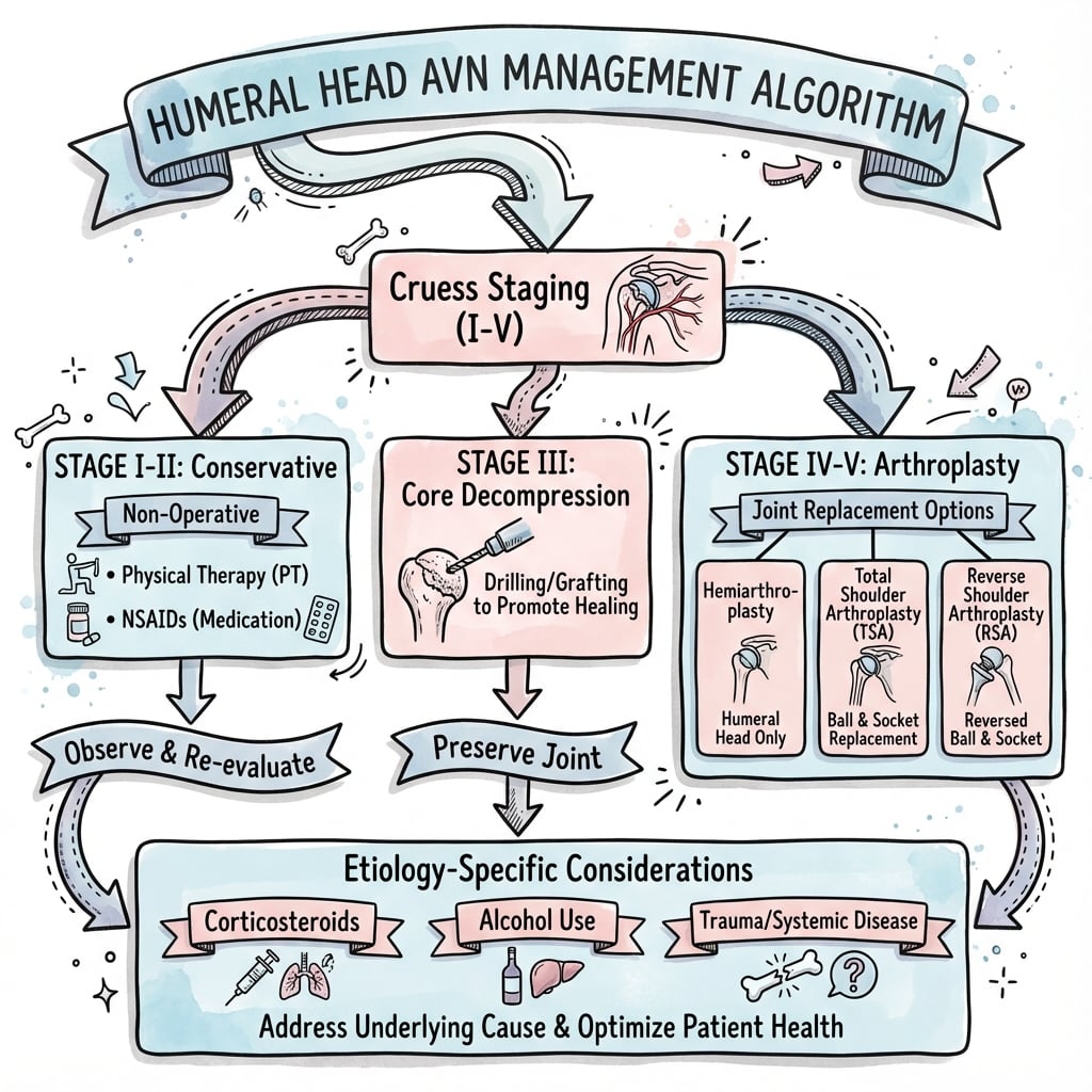
Surgical Technique
Core Decompression
Indications:
- Cruess Stage I-II (pre-collapse)
- Ficat Stage I-II
- MRI lesion less than 40% of head volume (greater than 40% has poor outcomes)
- Motivated patient willing to comply with 3-month activity restriction
- Age under 40 years (relative indication - younger patients better compliance, longer to arthroplasty)
Contraindications:
- Crescent sign present (Stage III) - mechanical failure already occurred, low success rate
- Collapse (Stage IV and beyond) - irreversible structural damage
- Greater than 50% head involvement - exceeds mechanical tolerance, very low success rate
- Infected shoulder - absolute contraindication
- Unable to comply with post-operative restrictions - premature loading causes fracture
- Non-modifiable risk factors (continued heavy steroid/alcohol) - relative contraindication
Pre-Operative Planning:
- Review MRI: Lesion location, volume, identify target for decompression
- Review CT if available: Assess bone quality, metaphyseal involvement
- Counsel realistic expectations: 50-70% halt progression (NOT reverse damage)
- Discuss augmentation options: Bone graft, BMAC, PRP (if available in research protocol)
- Optimize medical conditions: Cease smoking, vitamin D repletion, diabetes control
- Coordinate with treating physician: Minimize steroids if possible
Consent Discussion:
- Success rate: 70% Stage I, 50% Stage II (define success as no progression to collapse)
- Failure: 30-50% will progress to arthroplasty within 5 years
- Complications: Fracture (2-5%), infection (less than 1%), neurovascular injury (rare), persistent pain (30-40%)
- Post-operative requirements: Sling 4-6 weeks, no lifting 3 months (strict)
- Timeline: 6-12 months to know if successful (serial imaging)
This procedure "buys time" rather than providing definitive cure - important to set appropriate expectations.
Total Shoulder Arthroplasty for AVN
Pre-Operative Planning:
Imaging Review:
- CT scan: Assess glenoid version (normal 5-10 degrees retroversion), bone stock, humeral deformity from collapse
- Templating: Size humeral and glenoid components, plan version correction if needed
- MRI review: Confirm rotator cuff integrity (critical for anatomic TSA success)
- Assessment of bone quality: AVN often has mixed sclerosis and cystic areas (may need cement even in young patients)
Risk Stratification:
- Bleeding risk: Anticoagulation (common in AVN patients), immunosuppression affecting platelets
- Infection risk: Current immunosuppression (steroids, transplant medications), diabetes, malnutrition
- Bone quality: Osteoporosis from steroids/alcohol (fracture risk, component fixation concerns)
- Medical optimization: Vitamin D repletion, diabetes control (HbA1c less than 7.5%), nutrition (albumin greater than 35)
Implant Selection:
- Stemmed vs stemless humeral: Stemless preferred in AVN if metaphyseal bone quality adequate (bone preservation, easier revision)
- Cemented vs uncemented: Consider cement if osteoporotic bone (alcohol, steroids) even in younger patients
- Glenoid component: Cemented all-polyethylene standard (metal-backed higher failure in AVN population)
- Size: Avoid over-sizing (increases stiffness and nerve injury risk)
Patient Positioning:
Beach Chair (Most Common):
- Head secured in horseshoe headrest or specialized device
- Torso 30-45 degrees upright
- Operative arm free to extend and rotate
- Non-operative arm tucked or on arm board
- C-arm access from opposite side for intraoperative imaging
- Advantages: Familiar anatomy, easier assistant positioning, lower neuro risk
Lateral Decubitus (Alternative):
- Patient on beanbag or lateral positioning device
- Axillary roll under down-side axilla
- Arm suspended in traction device or supported
- Advantages: Easier humeral preparation, better visualization with gravity, more stable glenoid exposure
- Disadvantages: Unfamiliar anatomy orientation for some surgeons
Deltopectoral Approach:
-
Skin Incision:
- Start 2cm lateral and inferior to coracoid tip
- Extend 10-12cm distally along deltopectoral groove (palpable)
- Can extend proximally toward clavicle or distally toward deltoid insertion if needed
-
Superficial Dissection:
- Identify cephalic vein in deltopectoral groove
- Ligate crossing tributaries with electrocautery
- Retract vein laterally with deltoid (most common) or medially with pectoralis (alternative)
- Develop plane between deltoid (lateral) and pectoralis major (medial)
-
Deep Exposure:
- Incise clavipectoral fascia lateral to conjoined tendon
- Identify and protect conjoined tendon (retract medially)
- Coracoid process is medial landmark
- Tag muscle edges with stay sutures for later closure
-
Rotator Interval Exposure:
- Identify interval between subscapularis (inferior) and supraspinatus (superior)
- Long head biceps tendon lies in interval
- Tenotomy or tenodesis LHB: Routine in AVN (tendon often attritional, simplifies exposure)
- Open rotator interval capsule
Complications
Natural History (Untreated)
Stage-Specific Progression:
Stage I (MRI-Positive, X-Ray Negative):
- Spontaneous resolution: 10-15% (small lesions less than 15% volume, peripheral location)
- Stable disease: 15-20% (remain asymptomatic for years, lesion present but non-progressive)
- Progression to Stage II: 65-75% within 2 years (majority progress)
Stage II (Sclerosis, No Collapse):
- Stabilization: 20-30% (lesion persists but no collapse)
- Progression to collapse (Stage III-IV): 70-80% within 3-5 years
- Factors increasing progression: Central location, volume greater than 40%, continued steroid/alcohol exposure, younger age (faster bone turnover)
Stage III (Crescent Sign - Subchondral Fracture):
- Progression to collapse (Stage IV): Greater than 90% within 2 years (mechanical failure occurred)
- Rare stabilization: Less than 10% (small peripheral lesions only)
- Point of no return: Joint preservation unlikely to succeed
Stage IV-VI (Collapse and Arthritis):
- Inevitable progression: All progress to end-stage arthritis eventually
- Timeline to severe symptoms: 5-10 years from collapse (variable)
- Functional disability: Increases progressively with worsening arthritis
Predictors of Progression:
- Lesion volume greater than 40% of head (80% progress vs 20% if less than 30%)
- Central/medial location (3x higher collapse rate than peripheral)
- Bilateral disease (suggests systemic factors, higher progression risk)
- Ongoing steroid or alcohol exposure (accelerates all stages)
- Younger age at onset (longer disease duration, more cycles of loading)
Bilateral Disease Progression:
- 30-60% develop contralateral AVN (often within 1-2 years of index diagnosis)
- Steroid-induced highest bilateral rate (78%)
- Second shoulder may present at different stage than first
Surgical Complications
Core Decompression Complications:
Intraoperative (covered in detail in Surgical Technique section):
- Fracture during procedure: 1-2%
- Neurovascular injury: Less than 1%
- Failure to target lesion: 5%
Post-Operative:
- Infection: Less than 1%
- Persistent pain: 30-40% (often progression)
- Progression to collapse: 30-50% (Stage II)
- Fracture through decompression site: 2-5%
Arthroplasty Complications:
Major Complications (potentially requiring reoperation):
-
Infection (1-2%, higher in AVN due to immunosuppression):
- Risk factors: Steroids, transplant immunosuppression, diabetes, malnutrition, obesity
- Presentation: Wound drainage, fever, pain out of proportion, elevated CRP/ESR
- Workup: Joint aspiration (cell count greater than 3000 with greater than 80% PMNs suggests infection), culture
- Management:
- Acute (less than 3 weeks): Debridement, liner exchange, component retention, IV antibiotics 6 weeks
- Chronic (greater than 3 weeks): Two-stage revision (explant, antibiotic spacer 6-8 weeks, delayed reimplantation)
- Outcomes: Cure rate 85-90% (two-stage) vs 60-70% (DAIR - debridement and implant retention)
-
Instability (2-4%):
- Anterior (most common): Subscapularis failure (10-15% subscapularis repair failure), over-resection head, component malposition (excessive retroversion)
- Posterior: Component malposition (insufficient retroversion), posterior capsular laxity
- Inferior: Over-sizing components, deltoid dysfunction
- Prevention: Meticulous subscapularis repair, accurate component positioning, appropriate soft tissue balancing
- Management:
- Early (less than 6 weeks): Closed reduction, immobilization in IR (anterior) or ER (posterior), consider revision if recurrent
- Late (greater than 6 weeks): Revision arthroplasty (address soft tissue deficiency and/or component malposition)
-
Neurovascular Injury (1-2%):
- Axillary nerve (most at risk):
- Mechanism: Inferior capsular release (nerve 5-7mm from inferior glenoid rim), retractor placement, stretch from over-stuffing
- Presentation: Deltoid paralysis, numbness lateral shoulder
- Management: Observation (90% neurapraxia recover 6-12 months), consider exploration if sharp injury suspected
- Musculocutaneous nerve: Conjoined tendon retraction injury
- Brachial plexus: Positioning injury (stretch), over-stuffing
- Prevention: Gentle tissue handling, appropriate retractor placement, avoid over-sizing implants
- Axillary nerve (most at risk):
-
Periprosthetic Fracture (2-5%, higher in AVN due to bone quality):
- Intraoperative: Humeral shaft (aggressive reaming, osteoporotic bone), glenoid (over-reaming)
- Post-operative: Fall, trauma, stress fracture around stem
- Classification: Vancouver system adapted for humerus (AG periprosthetic greater tuberosity, AH shaft around stem, B stem loose, C distal to stem)
- Management:
- Non-displaced + stable stem: Conservative (sling, protected ROM)
- Displaced or unstable stem: ORIF with plate and cerclage wires, or revision to longer stem
Minor Complications:
-
Stiffness (10-15%):
- Risk factors: Pre-operative stiffness, poor compliance with PT, capsular contracture, over-stuffing
- Prevention: Appropriate sizing, early passive ROM, patient education
- Management: Intensive physiotherapy, manipulation under anesthesia (6-12 weeks post-op window), capsular release if refractory
-
Residual Pain (5-10%):
- Causes: Glenoid loosening, component malposition, rotator cuff issues, infection (rule out), nerve injury
- Workup: X-rays (loosening, malposition), CRP/ESR (infection), consider aspiration
- Management: Identify and address cause, may require revision
-
Heterotopic Ossification (2-3%):
- Risk factors: Post-traumatic AVN, male sex, HO history
- Prevention: Indomethacin 25mg TDS for 6 weeks post-op (if high risk)
- Significance: Rarely clinically significant (most asymptomatic), may limit ROM if severe
- Management: Observation (most), excision rarely needed
Late Complications (Greater than 1 year):
-
Aseptic Loosening:
- Glenoid (5-10% at 10 years): Most common late complication
- Radiolucent lines (greater than 2mm), component migration, screw breakage
- Revision to new glenoid component (if bone stock adequate) or reverse TSA
- Humeral (2-3% at 10 years): Less common
- Pain, subsidence, radiolucent lines
- Revision with longer stem, consider cement if uncemented originally
- Glenoid (5-10% at 10 years): Most common late complication
-
Glenoid Wear (Hemiarthroplasty specific, 30-50% at 10 years):
- Mechanism: Metal humeral head eroding polyethylene-like effect on glenoid cartilage
- Presentation: Recurrent pain (after initial good result), grating, crepitus
- Management: Conversion to TSA (requires adequate glenoid bone stock)
-
Rotator Cuff Failure (5% develop new cuff tear):
- Risk: Increases over time, supraspinatus most common
- Presentation: Pseudoparalysis, anterosuperior escape, pain
- Management: Revision to reverse TSA (if TSA fails due to cuff tear)
AVN-Specific Complication Considerations:
- Continued AVN progression: Opposite shoulder (30-60% bilateral rate requires surveillance)
- Bone quality issues: Higher fracture risk intra-operatively and post-operatively
- Immunosuppression: Higher infection risk (steroids, transplant medications)
- Young patient revisions: High lifetime revision burden (multiple surgeries expected)
Postoperative Care
(Covered comprehensively in Surgical Technique tabs above - Rehabilitation Protocol section)
Key points for exam purposes:
Immediate Post-Op (0-24 hours):
- Neurovascular checks (axillary nerve critical)
- Pain control (multimodal: block + oral/IV)
- Radiographs confirm position
- DVT prophylaxis initiated
Early Phase (0-6 weeks):
- Sling protection continuously
- Passive ROM only (protect subscapularis)
- Pendulums day 1, therapist-assisted passive ROM
- NO active subscapularis, lifting, or reaching
Intermediate Phase (6-12 weeks):
- Wean sling (week 6-8)
- Progress to active-assisted then active ROM
- Light isometric strengthening
- Goals: 120 degrees elevation, 40 degrees ER
Late Phase (3-6 months):
- Progressive resistance strengthening
- Return to ADLs and light work
- Goals: Near-normal ROM, 70% strength
Long-Term (6+ months):
- Full function within activity restrictions
- Permanent limits: No overhead heavy lifting (greater than 20kg), no contact sports
- Annual X-rays first 2 years
Compliance Critical: Subscapularis repair failure (10-15%) often due to non-compliance with restrictions in first 6 weeks.
Outcomes and Prognosis
Non-Operative Management:
- Symptom control: 40-60% achieve acceptable pain levels with analgesia, activity modification
- Progression to surgery: 50-70% within 5 years (Stage II patients)
- Quality of life: Moderate impairment, worse with bilateral disease
- Best outcomes: Small peripheral lesions (less than 30%), successful risk factor modification
Core Decompression (covered in detail earlier):
- Success (no progression): 50-70% at 5 years (Stage I-II)
- Conversion to arthroplasty: 30-50% by 5 years
- Pain relief: 60-70% report improvement even if radiographic progression (debulking effect)
- Younger patients: Better compliance, similar biological outcomes to older
Humeral Head Resurfacing:
- Survival (no revision): 80-85% at 5 years, 70-75% at 10 years
- Pain relief: Good to excellent in 75-80% (VAS improvement 5-6 points)
- Function: Oxford Shoulder Score improvement 15-20 points (moderate improvement)
- ROM: Forward elevation 120-140 degrees, ER 40-50 degrees (good but not full)
- Revision indications: Loosening (10%), glenoid erosion (8%), persistent pain (5%)
- Predictors of success: Age under 50, intact cuff, good bone quality, appropriate patient selection
Hemiarthroplasty:
- Pain relief: Good in 70-80% (inferior to TSA if glenoid involved)
- Function: Moderate improvement (limited by glenoid-sided pain if arthritis present)
- ROM: Variable (90-130 degrees elevation typical)
- Glenoid erosion: 30-50% at 10 years (progressive, predictable)
- Revision to TSA: 15-20% by 10 years (for glenoid erosion pain)
- Registry data: approximately 20% cumulative revision rate at 10 years (all causes)
Total Shoulder Arthroplasty (Anatomic):
- Pain relief: Excellent in 85-95% (VAS improvement 6-7 points, superior to hemi)
- Function: Good to excellent in 80-90% (ASES score 70-80 typical)
- ROM: Forward elevation 120-140 degrees, ER 40-50 degrees, IR to L1-L3
- Survival: approximately 88% at 10 years (pooled registry data)
- Revision rate: ~12% at 10 years (loosening 6%, instability 3%, infection 2%, other 1%)
- Younger patients (under 50): Higher revision rate (20% at 10 years - glenoid loosening main cause)
- Patient satisfaction: 85-90% satisfied or very satisfied
Reverse Total Shoulder Arthroplasty:
- Pain relief: Excellent in 90-95% (even better than anatomic TSA)
- Function: Good (limited by reduced rotation compared to anatomic)
- ROM: Forward elevation 100-130 degrees, ER 10-20 degrees (limited), IR to buttock/lumbar
- Survival: approximately 92% at 10 years (pooled registry data)
- Complications: Instability (4%), infection (2%), scapular notching (50-70% radiographic, mostly asymptomatic), acromial fracture (2%)
- Note: Typically used in older AVN patients with cuff deficiency (not young patients with intact cuffs)
Return to Work and Activities:
- Sedentary work: 3-6 months post-arthroplasty (typing, desk work)
- Light manual labor: 6-9 months (limit 10kg lifting)
- Heavy manual labor: Often require job modification or disability (20kg limit incompatible)
- Overhead work: Generally restricted long-term (limit 10kg overhead)
- Overhead sports: Generally discouraged (implant longevity concerns, instability risk)
- Golf, swimming, cycling: Generally permitted after 6 months
- Activities of daily living: 90% achieve independence by 6 months
Factors Influencing Outcomes:
Patient Factors:
- Age: Younger patients higher revision risk (24% under 50 vs 12% over 60 at 10 years) but better function
- Expectations: Higher expectations associated with lower satisfaction (counsel realistic goals)
- Compliance: Adhering to PT and activity restrictions improves outcomes
- Comorbidities: Diabetes, immunosuppression, smoking worsen outcomes
Disease Factors:
- Bilaterality: Second shoulder surgery typically done 6-12 months after first (staged)
- Etiology: Steroid-induced slightly worse outcomes than idiopathic (bone quality issues)
- Stage at presentation: Earlier intervention (before collapse) better long-term outcomes
Surgical Factors:
- Surgeon volume: High-volume surgeons better outcomes (especially glenoid component positioning)
- Component choice: TSA superior to hemi when glenoid involved
- Subscapularis repair: Integrity critical (failure rate 10-15%, leads to instability, poor function)
Bilateral AVN Outcomes:
- Staged procedures: 6-12 months apart (need one functional arm during recovery)
- Second side outcomes: Similar to first side (no significant difference)
- Patient satisfaction: Lower than unilateral (two recovery periods, bilateral restrictions)
- Economic impact: Significant (two surgeries, prolonged disability, young working-age patients)
Evidence Base
Core Decompression Superior to Nonoperative Management (Systematic Review)
- Core decompression success 76.6% vs nonoperative 13% (p less than 0.001)
- Stage III shoulders avoided arthroplasty in 63% after decompression
- Radiographic progression 24.2% (CD) vs 52.3% (nonop), p less than 0.001
- Clinical scores improved in 7 of 9 CD studies vs 1 of 6 nonop studies
- 291 shoulders (CD) and 359 shoulders (nonop) at mean 8.1 years
Foundational Cohort: Atraumatic Osteonecrosis of the Humeral Head
- 127 shoulders / 73 patients, mean age 41 years
- Corticosteroid association 82%; bilateral disease 74%
- Hip co-involvement 81% (advocates hip screening in shoulder AVN)
- Core decompression good-to-excellent in 78% at mean 6 years
- Severity did not correlate with steroid dose or duration
Natural History: Stage and Cause Predict Need for Arthroplasty
- 200 shoulders / 151 patients; corticosteroid cause in 112
- 3-year replacement rate: Stage 2 42%, Stage 4 55%, Stage 5 79%
- Traumatic cause progressed fastest (77.8% replaced by 3 years)
- Higher stage and greater head involvement predicted surgery
- Non-operative cohort: mean ASES 64.8 at 8.6 years
Surface Replacement Arthroplasty for Humeral Head Osteonecrosis
- 17 shoulders, mean head necrosis 18.6% (up to 30.9%)
- Constant score 31 to 62 points (p less than 0.0001)
- Flexion 87 to 139 degrees; abduction 64 to 120 degrees
- No implant loosening or revision at mean 3 years
- Non-traumatic osteonecrosis outperformed post-traumatic
Surgical Management of Humeral Head Osteonecrosis (PRISMA Systematic Review)
- 12 studies, 309 patients, 382 shoulders
- Core decompression effective for low-grade (pre-collapse) disease
- Arthroplasty (hemi/TSA) reserved for high-grade disease
- No randomised trials; overall evidence level IV
- Stage-stratified treatment supported across the literature
Steroid Dose and Osteonecrosis Risk (Across-Study Analysis)
- 22 studies pooled across-study; AVN of bone as outcome
- Daily total dose correlated with AVN rate (r 0.61-0.80)
- Oral dose strongly correlated (r 0.70-0.86)
- Bolus/pulse dose NOT associated with AVN risk in this analysis
- Cumulative/daily dose is the key driver of risk
Controversies and Areas of Uncertainty
The evidence base in humeral head AVN is weak (no randomised trials; mostly Level III-IV case series), so several decisions remain genuinely contested. Demonstrating awareness of these debates marks out the consultant-level candidate.
Core decompression at Stage III
Traditional teaching treats the crescent sign as the "point of no return." Yet a 2023 systematic review found 63% of Stage III shoulders avoided arthroplasty after decompression. Whether to offer decompression after early subchondral fracture - rather than proceeding to arthroplasty - is unresolved.
Role of biologics (BMAC / PRP)
Bone marrow aspirate concentrate, PRP and cell-based augmentation are biologically attractive but supported only by small uncontrolled series. There are no RCTs in the shoulder, and routine use cannot yet be recommended.
Stemless / resurfacing vs anatomic TSA in the young
Bone-preserving implants protect future revision options but a retained native glenoid predictably erodes. The trade-off between preserving bone now and accepting later glenoid-sided pain drives marked international practice variation.
Does bolus steroid dosing truly add risk?
Clinical teaching emphasises IV pulse therapy as especially dangerous, but the classic across-study analysis (Felson, Lancet 1987) found bolus dose NOT independently associated with AVN once daily/cumulative dose was accounted for. Cumulative and daily dose are the proven drivers.
Optimal contralateral screening
Given 30-78% bilateral disease, MRI screening of the asymptomatic shoulder seems logical, but evidence that early detection of an asymptomatic lesion changes outcome is limited, and cost-effectiveness is unproven outside high-risk (steroid) groups.
Reverse TSA in the cuff-deficient young patient
There is no good option for a young patient with collapse, glenoid arthritis and a deficient cuff. Reverse TSA works but carries a high lifetime revision burden; cuff repair plus hemiarthroplasty, or delaying surgery, are all defensible. This is a true area of equipoise.
Viva Scenarios
Clinical Decision Scenarios
Use these scenarios to practise clinical reasoning and management decisions
"A 38-year-old male renal transplant recipient presents with bilateral shoulder pain for 6 months. He is on maintenance prednisone 15mg daily and tacrolimus. Radiographs show bilateral humeral head sclerosis without collapse (Cruess Stage II). MRI confirms AVN with lesions involving approximately 40% of each humeral head (central location). He works as a builder and is very concerned about his ability to continue working. How would you manage this patient?"
Structured Viva Answer:
1. Problem Summary:
"This 38-year-old builder has bilateral Cruess Stage II AVN (pre-collapse), likely steroid-induced from transplant immunosuppression. Key concerns include: bilateral disease in working-age patient, high-demand occupation, ongoing steroid exposure, and 40% lesion volume indicating moderate progression risk."
2. Initial Assessment:
- "Complete assessment with bilateral MRI (lesion volume/location, cuff status)"
- "Baseline bloods: renal function, immunosuppression levels, Vitamin D"
- "Document occupational demands and functional baseline"
3. Risk Factor Modification (Critical):
- "Liaise with nephrologist: Minimise steroid dose if safe (steroid-sparing agents)"
- "Optimise bone health (Vitamin D, bisphosphonates)"
- "Smoking cessation if applicable"
4. Management Strategy:
- "Stage II pre-collapse represents window for joint preservation"
- "Primary Option: Bilateral staged **Core Decompression** ± bone graft/BMAC"
- "Given 40% volume: 50-60% chance of halting progression (conversely 40-50% failure rate)"
- "Observation: Reasonable if asymptomatic, but given young age and progression risk, intervention preferred"
5. Occupational Counseling (Difficult Discussion):
- "Building work involves heavy overhead loading - accelerates collapse"
- "Post-op restrictions: Strict NO lifting 3 months"
- "Long-term: If effective, may return to light duties. If arthroplasty needed, permanent 20kg restriction incompatible with heavy building"
- "Early vocational rehabilitation referral essential"
6. Surgical Plan (Core Decompression):
- "Staged procedures (6-12 months apart) to maintain one functional arm"
- "Technique: Percutaneous drilling (8-10mm) targeting lesion center under fluoro"
- "Augmentation: Iliac crest bone graft or BMAC to improve osteogenesis"
7. Long-Term Prognosis:
- "Buys time: Success = delaying arthroplasty by 5-10 years"
- "Failure: Conversion to arthroplasty (TSA or hemi) will likely be required eventually"
- "High lifetime revision burden for 38-year-old (24% revision at 10y)"
"A 55-year-old female with chronic alcohol use disorder presents with severe left shoulder pain. She reports drinking approximately 500mL spirits daily for 20 years. Radiographs show Cruess Stage V AVN with humeral head collapse and early glenoid involvement. MRI confirms intact rotator cuff. She continues to drink heavily and is not currently engaged with addiction services. How would you approach this patient?"
Structured Viva Answer:
1. Problem Summary:
"55F with Stage V AVN requiring TSA, but severe active alcohol use presents prohibitive surgical risk. Immediate priority is medical and addiction management, not surgery."
2. Risk Assessment:
- "Surgical risks: Infection (immunosuppression), Poor healing (malnutrition), Bleeding (coagulopathy), Fixation failure (osteoporosis)"
- "Medical risks: Withdrawal seizures/DTs, Cardiomyopathy, Liver disease"
3. Management Plan:
- "**NO elective surgery** while drinking 500mL spirits daily"
- "Referral to Addiction Medicine: Detox, rehabilitation, pharmacotherapy"
- "Goal: Minimum 6-8 weeks abstinence + medical optimization (Thiamine, Nutrition, LFTs, Coags)"
4. Counseling:
- "Frank discussion: 'Surgery now is dangerous. Infection risk is high. We need to get you safe first.'"
- "Non-operative interim: Analgesia, PT, injection (buy time)"
5. Definitive Surgical Plan (Once Optimized):
- "Anatomic TSA (Stage V + Intact Cuff)"
- "Cemented components (likely osteoporotic bone)"
- "Inpatient admission for withdrawal monitoring (CIWA protocol)"
"A 32-year-old woman with SLE on long-term steroids (prednisone 20mg daily for 5 years) has bilateral shoulder AVN. Right shoulder is Cruess Stage I (MRI positive, X-ray normal, minimal symptoms), left is Cruess Stage IV (collapsed head, glenoid intact, severe pain). MRI confirms intact rotator cuffs bilaterally. She has difficulty with ADLs and requests bilateral surgery 'to get it all over with at once.' What is your management approach?"
Structured Viva Answer:
1. Dilemma Breakdown:
- "Right Shoulder: Early Stage I (Preservation candidate)"
- "Left Shoulder: Late Stage IV (Arthroplasty candidate)"
- "Patient Factor: 32yo, SLE, Unrealistic expectations (Simultaneous surgery)"
2. Refusal of Simultaneous Surgery:
- "Explain SAFETY: Need one functional arm for hygiene/feeding"
- "Explain RISK: Higher VTE, Infection, Rehab failure"
- "Plan: Staged procedure (Left first) to restore function before addressing Right"
3. Surgical Plan (Left):
- "Stage IV (Collapsed head, Intact Glenoid)"
- "Preferred: Humeral Head Resurfacing or Stemless Hemi (Bone preservation)"
- "Avoid TSA if possible (Glenoid will fail in young patient)"
4. Surgical Plan (Right):
- "Stage I (Pre-collapse)"
- "Core Decompression ± Graft (Try to save joint)"
5. SLE Specifics:
- "Steroid stress dose (Hydrocortisone)"
- "Antibiotic prophylaxis (Immunosuppressed)"
- "Bone quality (Gentle handling)"
MCQ Practice Points
Cruess Phase 3 Feature
Q: Which of the following radiographic findings represents the transition from reversible disease to mechanical failure in humeral head avascular necrosis?
- A) Patchy sclerosis
- B) Cystic changes
- C) Crescent sign
- D) Joint space narrowing
- E) Osteophyte formation
A: C) Crescent sign
Explanation: The crescent sign (Cruess Stage III) represents a subchondral fracture, indicating that the structural integrity of the subchondral bone plate has failed. This is the "point of no return" where joint preservation procedures like core decompression have significantly lower success rates (20-30% vs 50-70%). Sclerosis/Cysts (Stage II) are pre-collapse. Joint space narrowing (Stage V) is secondary arthritis.
MRI Findings
Q: A 45-year-old male with a history of alcohol use presents with shoulder pain. Radiographs are normal. Which MRI finding is consistently pathognomonic for avascular necrosis?
- A) Diffuse marrow edema
- B) Double-line sign on T2
- C) Joint effusion
- D) Subchondral cyst
- E) Labral tear
A: B) Double-line sign on T2
Explanation: The double-line sign is pathognomonic for AVN. It consists of an inner low-signal line (dead bone) and an outer high-signal line (vascular granulation tissue/repair interface) seen on T2-weighted images. Diffuse edema (A) is seen in transient osteoporosis or infection. Cysts (D) and effusion (C) are non-specific.
Risk Factors
Q: Which of the following steroid regimens carries the HIGHEST risk for developing avascular necrosis?
- A) Prednisone 5mg daily for 10 years (RA)
- B) Inhaled corticosteroids for asthma
- C) Recent high-dose pulse IV methylprednisolone
- D) Prednisone 10mg daily tapered over 2 weeks
- E) Intra-articular steroid injection
A: C) Recent high-dose pulse IV methylprednisolone
Explanation: High-dose pulse therapy and high daily doses (over 20mg/day) are stronger risk factors than cumulative low-dose duration. The risk threshold is generally considered over 2000mg cumulative dose or over 20mg/day for over 3 months, but pulse therapy carries a particularly high risk of osteocyte death.
Management Principles
Q: A 32-year-old female with SLE has Cruess Stage II AVN of the humeral head (pre-collapse) involving 40% of the head volume. She has moderate pain. What is the most appropriate management?
- A) Hemiarthroplasty
- B) Total Shoulder Arthroplasty
- C) Core Decompression
- D) Reverse Total Shoulder Arthroplasty
- E) Observation only
A: C) Core Decompression
Explanation: In a young patient (32) with pre-collapse disease (Stage II), joint preservation is the goal. Core decompression is indicated to relieve intraosseous pressure and potentially halt progression. Arthroplasty (A/B/D) is reserved for post-collapse disease (Stage III-IV) or failure of preservation. Observation (E) is appropriate for asymptomatic or very small lesions, but she is symptomatic and at risk of progression.
Bilateral Disease
Q: A 28-year-old patient presents with symptomatic right shoulder AVN (Stage III). What is the approximate likelihood of finding AVN in the asymptomatic left shoulder on MRI screening?
- A) Less than 10%
- B) 20%
- C) 60%
- D) 90%
- E) 100%
A: C) 60%
Explanation: Bilateral involvement is very common in atraumatic AVN, typically quoted as 30-60%. In steroid-induced cases, it can be as high as 78%. Routine MRI screening of the asymptomatic contralateral shoulder is recommended because 45% of these asymptomatic lesions will progress to symptoms within 2 years, and early detection (Stage I) allows for more effective joint preservation treatment.
Arthroplasty in Young Patients
Q: For a patient under 50 years undergoing shoulder arthroplasty for AVN, what is the approximate cumulative revision rate at 10 years reported by national joint registries?
- A) 5%
- B) 12%
- C) 24%
- D) 35%
- E) 50%
A: C) 24%
Explanation: National joint replacement registries consistently report a high cumulative revision rate of roughly 24% at 10 years for patients under 50 years with AVN, compared to about 12% for those over 65. This highlights the "lifetime revision burden" young patients face and emphasizes exhausting joint preservation options and careful implant selection (e.g. bone-preserving stems) to facilitate future revision.
Guidelines, Registries & Global Practice
Side-by-Side Society Guidance
There is no high-level (RCT) evidence and no shoulder-specific society guideline dedicated to humeral head AVN; recommendations are extrapolated from hip osteonecrosis guidance and shoulder arthroplasty appropriate-use criteria. Where bodies do comment, the principles converge.
Guideline / Society Positions Relevant to Humeral Head AVN
| Body | Region | Position relevant to AVN | Evidence level |
|---|---|---|---|
| AAOS (Appropriate Use Criteria, glenohumeral OA/arthroplasty) | USA | Anatomic TSA favoured over hemiarthroplasty once glenoid is involved with an intact cuff; reverse TSA for cuff-deficient or elderly | Consensus / limited |
| BOA / BESS (British Elbow & Shoulder Society) | UK | Joint preservation for pre-collapse disease; arthroplasty for collapse; emphasise young-patient counselling and bone preservation | Consensus |
| AO Foundation / ARCO | International | Staging frameworks (ARCO/Ficat) and emphasis on early detection and risk-factor control extrapolated from femoral head osteonecrosis | Consensus |
| EFORT / European consensus | Europe | Stage-based algorithm; core decompression for early disease, stemless/resurfacing to preserve bone in the young | Consensus |
| IOC / sports medicine bodies | International | Recognise dysbaric (Caisson) osteonecrosis in divers; advocate decompression-protocol adherence and early imaging | Consensus |
Where recommendations genuinely differ: practice variation is greatest at the young patient with collapse and intact cuff decision. North American practice leans toward anatomic TSA/stemless once the glenoid is involved; many European centres favour bone-preserving surface replacement or hemiarthroplasty to protect future revision options. All bodies agree that reverse TSA is appropriate for cuff-deficient or elderly patients and is best avoided in the young patient with an intact cuff.
Exam Day Cheat Sheet
Clinical summary
Classifications (Must-Know)
- •**Cruess (6 stages)**: I (Normal) → II (Sclerosis) → III (Crescent/Fracture) → IV (Collapse) → V (Glenoid Arthritis)
- •**Ficat**: Radiographic progression (0-IV, similar to Hip)
- •**Modified Ficat**: Adds MRI volumetric assessment to Ficat stages
- •**Key Point**: Crescent sign (Stage III) is the 'point of no return' for joint preservation
Etiology (ASEPTIC Mnemonic)
- •**A**lcohol (over 400mL/week)
- •**S**teroids (over 20mg/day, over 2000mg cumulative)
- •**E**thanol (Alcohol)
- •**P**ancreatitis/Pregnancy
- •**T**rauma (4-part fracture, dislocation)
- •**I**diopathic (20-25%)
- •**C**onnective Tissue Disease (SLE)
Management Algorithm
- •**Pre-Collapse (I-II)**: Core Decompression (50-70% success)
- •**Crescent Sign (III)**: Arthroplasty usually needed (Decompression fails)
- •**Collapse (IV)**: Hemi or TSA (depends on glenoid)
- •**Arthritis (V-VI)**: TSA (if cuff intact) or Reverse (if cuff deficient/elderly)
Key Exam Phrases
- •The crescent sign indicates subchondral fracture and mechanical failure.
- •Double-line sign on T2 MRI is pathognomonic.
- •In young patients, revision rate is 24% at 10 years - counseling is critical.
- •Bilateral screening is mandatory (30-60% prevalence).
Key Takeaways
Diagnosis and Staging
- MRI gold standard: Double-line sign pathognomonic (T2: inner low + outer high signal)
- Cruess classification: Guides treatment (I-II preservation, III controversial, IV-VI replacement)
- Crescent sign: Point of no return (subchondral fracture = mechanical failure)
- ALWAYS screen opposite shoulder: 30-60% bilateral (78% steroid-induced)
Etiology and Prevention
- Steroids: Greater than 2000mg cumulative, greater than 20mg/day, greater than 3 months (35-40% cases)
- Alcohol: Greater than 400mL/week threshold (20-25% cases)
- Risk factor modification NON-NEGOTIABLE: Cease alcohol, minimize steroids with treating physician
- ASEPTIC mnemonic: Systematic approach to non-traumatic causes
Joint Preservation
- Core decompression: 50-70% success PRE-collapse (Stage I-II only)
- ONLY effective before crescent sign: Post-crescent low success (20-30%)
- 3-month activity restriction CRITICAL: Premature loading causes fracture
- Biologics investigational: BMAC, PRP promising but not standard care (research protocols)
Arthroplasty Outcomes
- TSA superior to hemiarthroplasty: When glenoid involved (Stage V-VI) - Level 2 evidence
- Young age (under 50): ~24% revision at 10 years (joint registries) - lifetime burden
- Reverse TSA: Cuff deficiency OR age over 70 (NOT young intact cuff patients)
- Resurfacing: Requires intact cuff (ABSOLUTE) - preserves bone stock for young patients