TGF-beta Superfamily | Osteoinduction | Smad Signaling | Clinical Applications
- BMPs are secreted growth factors in the TGF-beta superfamily
- BMP-2 and BMP-7 are osteoinductive - induce bone formation in non-skeletal sites
- Signaling via Smad pathway: BMP binds type I/II receptors, activates Smad1/5/8, induces Runx2
- Clinical use: spinal fusion, long bone nonunion, critical-size defects
- Complications: ectopic bone, inflammatory swelling, osteolysis, cost
- “Urist 1965 discovered BMPs by implanting demineralized bone matrix subcutaneously
- “BMP-2 is more osteogenic than BMP-7 but has higher complication risk
- “Supraphysiologic doses used clinically (mg amounts vs ng in normal healing)
- “Carrier scaffold critical - absorbable collagen sponge standard
Overview
Where BMP sits. BMP is one input into a process, not the process. The biology it drives is bone healing and the cycle underneath it is bone remodeling, acting through the osteoblast lineage; the other molecules in the same conversation are on growth factors in bone healing. Clinically it is used where that process has failed or is being asked to do too much - nonunion, open tibial shaft fractures and lumbar fusion - and its signature complication, bone where bone should not be, is heterotopic ossification.
What they are. Bone morphogenetic proteins are secreted signalling molecules in the transforming growth factor-beta (TGF-beta) superfamily that induce bone and cartilage formation. More than 20 subtypes have been identified; only BMP-2 and BMP-7 have regulatory approval.
How they were found. Marshall Urist showed in 1965 that demineralised bone matrix implanted subcutaneously in rabbits induced ectopic bone formation, the founding demonstration of osteoinduction. Wozney and colleagues cloned the proteins responsible in 1988, and recombinant BMP-2 and BMP-7 followed from that.
What BMP adds. Autograft is osteogenic and osteoconductive, allograft osteoconductive only. BMP supplies the third property: the signal that recruits host mesenchymal stem cells and drives them into the osteoblast lineage. It can give that property to a synthetic scaffold which has none of its own.
Where it is used. rhBMP-2 (InFuse, Medtronic) holds FDA approval; rhBMP-7, also called OP-1 (Stryker), holds only a humanitarian device exemption for recalcitrant long bone nonunion. Off-label use in posterior spinal fusion, fracture nonunion and revision arthroplasty is common but controversial.
Placing BMP against the alternatives is a standard viva opening. The discriminators are the three biological properties - osteogenesis (living cells that form bone directly), osteoconduction (a scaffold) and osteoinduction (signals that recruit and differentiate host stem cells).
- Osteogenic
- Yes
- Osteoconductive
- Yes
- Osteoinductive
- Yes
- Key Distinguishing Feature
- Reference standard - all three properties; donor-site morbidity (10-20%)
- Osteogenic
- No
- Osteoconductive
- Yes
- Osteoinductive
- Weak/none
- Key Distinguishing Feature
- Scaffold only; no live cells; disease-transmission and immunogenicity concern
- Osteogenic
- No
- Osteoconductive
- Yes
- Osteoinductive
- Variable
- Key Distinguishing Feature
- Retains some BMP - osteoinductivity is donor- and processing-dependent
- Osteogenic
- No
- Osteoconductive
- Yes
- Osteoinductive
- No
- Key Distinguishing Feature
- Pure synthetic scaffold; needs combining with cells or BMP
- Osteogenic
- No
- Osteoconductive
- No (needs carrier)
- Osteoinductive
- Yes (potent)
- Key Distinguishing Feature
- Pure osteoinductive signal delivered on a collagen-sponge carrier
- Osteogenic
- Yes (MSCs)
- Osteoconductive
- No
- Osteoinductive
- Limited
- Key Distinguishing Feature
- Supplies osteoprogenitor cells; often combined with a scaffold
Osteoinduction is the recruitment of primitive mesenchymal cells and their induction into bone-forming osteoblasts; BMPs are osteoinductive. Osteoconduction is passive scaffold support for bone growing in from existing bone. Allograft and synthetic scaffolds are osteoconductive but not osteoinductive unless they are combined with BMP.
Anatomy
The molecule. BMPs are secreted polypeptide growth factors, and the active ligand is a homodimer of two identical chains joined by disulfide bonds through a cysteine-rich domain. BMP-2 is 114 amino acids per chain and 26 kDa as a dimer; BMP-7 is 139 amino acids per chain and 35 kDa. A monomer has no biological activity, because it takes the dimer to bind the type I and type II receptors on the cell surface.
Family resemblance. BMPs belong to the TGF-beta superfamily and share its cysteine knot motif and its conserved receptor-binding domains.
The receptor complex. Signalling needs a heterotetrameric complex - two type II and two type I receptors - which assembles when BMP binds. The type II receptor is constitutively active and phosphorylates the type I receptor; the activated type I receptor is the kinase that phosphorylates Smad1/5/8.
- Specific Receptors
- BMPR-II, ActR-IIA, ActR-IIB
- Function
- Binds BMP ligand, recruits Type I
- Downstream Target
- Phosphorylates Type I receptor
- Specific Receptors
- ALK-2, ALK-3 (BMPR-IA), ALK-6 (BMPR-IB)
- Function
- Phosphorylated by Type II, kinase active
- Downstream Target
- Phosphorylates Smad1/5/8
Where BMP is found. BMP is abundant in bone matrix, stored there and released during resorption, and is expressed during fracture healing with a peak at three weeks. It is also present in cartilage, tendon and periosteum, at low levels in most adult soft tissues, and at high levels in embryonic development.
BMP Biology and Classification
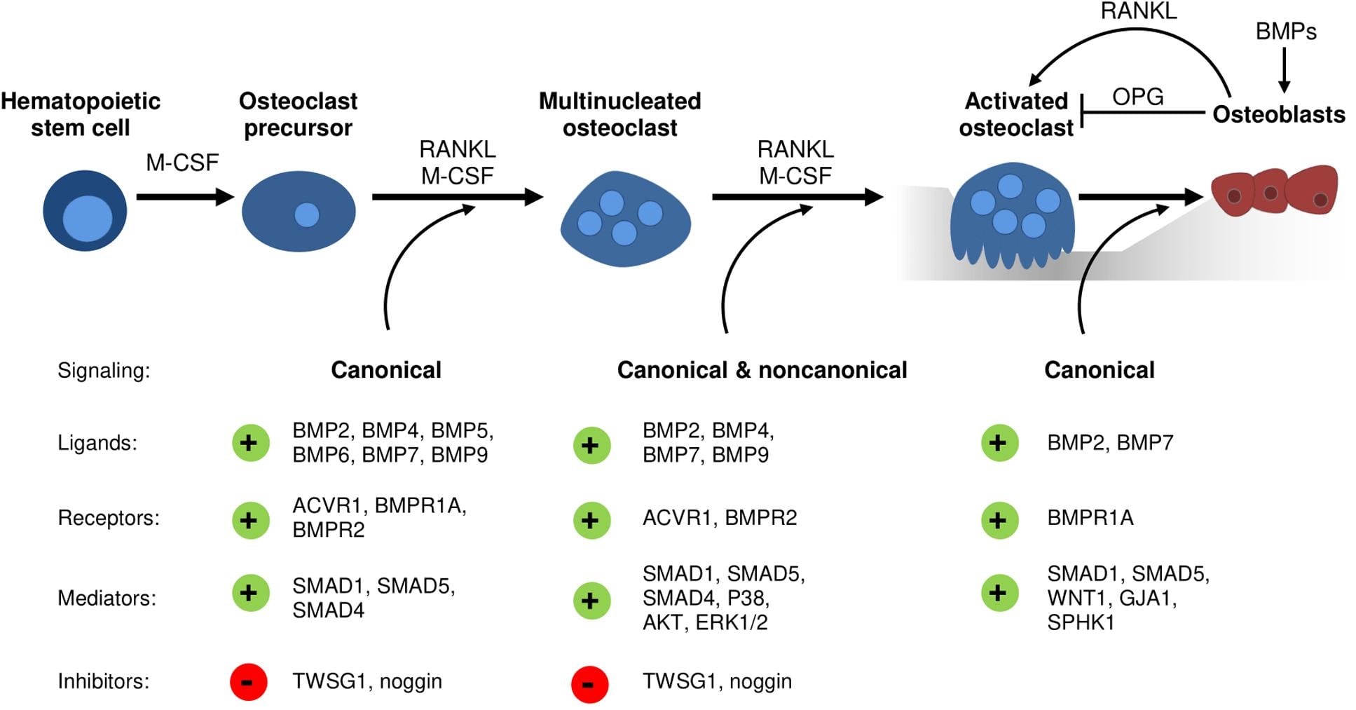
The family. More than 20 BMPs have been identified, divided into subgroups on structure and function. One subgroup induces bone, one opposes it, and the rest belong to tendon, joint and embryonic development.
- Key Members
- BMP-2, BMP-4, BMP-6, BMP-7, BMP-9
- Primary Function
- Bone and cartilage induction
- Clinical Relevance
- BMP-2 FDA-approved; BMP-7 humanitarian device exemption
- Key Members
- BMP-3 (osteogenin)
- Primary Function
- Negative regulator of bone formation
- Clinical Relevance
- Not used for clinical osteoinduction
- Key Members
- GDF-5 (BMP-14), GDF-6, GDF-7
- Primary Function
- Joint, tendon and ligament development
- Clinical Relevance
- Experimental tendon and ligament healing
- Key Members
- BMP-5, BMP-8, BMP-10
- Primary Function
- Embryonic patterning, heart
- Clinical Relevance
- Limited orthopaedic application
BMP-3 is the exception. Also called osteogenin, it is a negative regulator of bone formation rather than an inducer of it.
The two that reached the clinic. BMP-2 and BMP-7 were the two developed for clinical use, although neither is the most potent osteoinductor in the laboratory. Of the pair, BMP-2 has the greater osteoinductive potency and the higher complication rate.
In vitro osteogenic potency: BMP-9 greater than BMP-2 greater than BMP-6 greater than BMP-4 greater than BMP-7, BMP-9 exceeding BMP-2 by 10-fold in some assays. BMP-2 dominates clinical use regardless, on established manufacturing, an FDA approval pathway and extensive clinical experience; cost, manufacturing complexity and regulatory hurdles have kept the more potent molecules in the laboratory.
Receptor preference. Each group of ligands binds a characteristic set of type I receptors.
- Preferred Type I Receptor
- ALK-3 (BMPR-IA), ALK-6 (BMPR-IB)
- Biological Outcome
- Strong osteogenic, chondrogenic
- Preferred Type I Receptor
- ALK-2, ALK-3, ALK-6
- Biological Outcome
- Osteogenic, broader tissue effects
- Preferred Type I Receptor
- ALK-1, ALK-2
- Biological Outcome
- Endothelial, vascular, osteogenic
- Preferred Type I Receptor
- ALK-6 predominant
- Biological Outcome
- Tendon, ligament, cartilage
The product. rhBMP-2 is sold as InFuse Bone Graft (Medtronic) and delivered on an absorbable collagen sponge.
Approved indications.
- Single-level anterior lumbar interbody fusion (ALIF), L4-S1
- Open tibial shaft fractures - the acute fracture, not an established nonunion
- Oral and maxillofacial reconstruction: sinus augmentation and ridge preservation
Off-label. Posterior lumbar fusion, long bone nonunion and revision arthroplasty with bone loss are all used off-label. So is cervical fusion, and that is the one off-label use with a documented risk to life: airway swelling. See ACDF.
BMP Signalling Pathways
The route in. A BMP dimer assembles its receptors at the cell surface, the type II receptor phosphorylates the type I receptor, and from there the signal divides into a canonical Smad arm and a non-canonical MAPK arm.
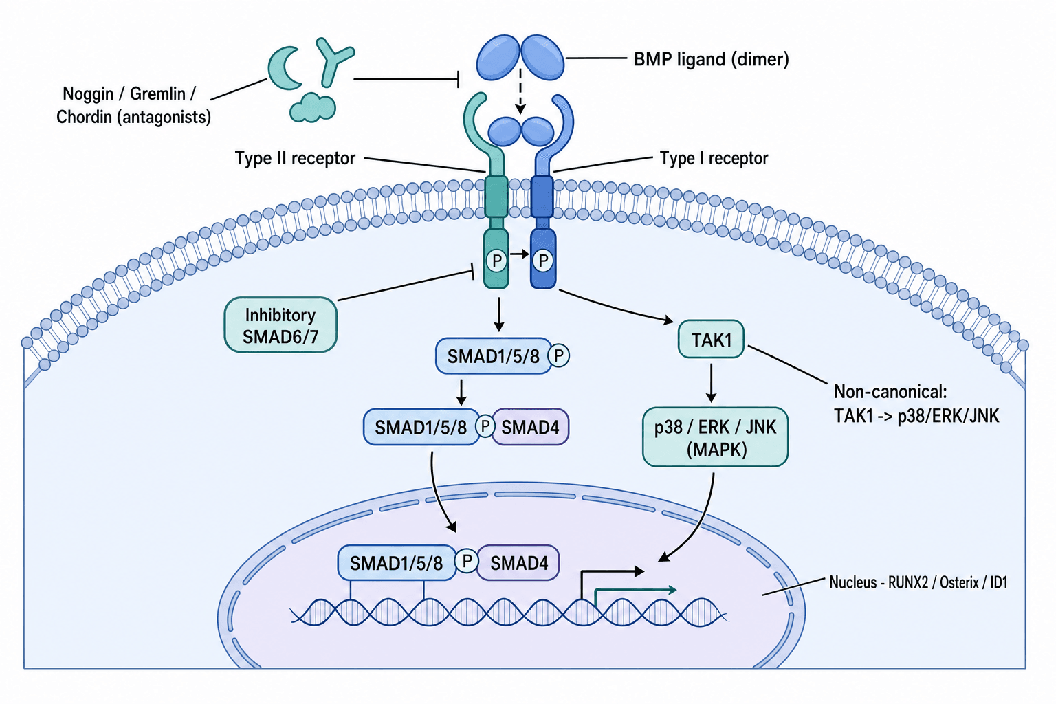
The canonical route runs through the Smad proteins, named for the Drosophila gene mothers against decapentaplegic.
BMP-Smad Signalling Cascade
The BMP homodimer binds the type II serine/threonine kinase receptor (BMPR-II, ActR-II), which is constitutively active.
The type II receptor recruits and phosphorylates a type I receptor (ALK-2, ALK-3, ALK-6), which becomes an active kinase.
The activated type I receptor phosphorylates the receptor-regulated Smads - Smad1, Smad5 and Smad8 - at C-terminal serine residues.
Phosphorylated Smad1/5/8 dissociates from the receptor and binds the common mediator Smad4, forming a heteromeric complex.
Importins carry the Smad1/5/8-Smad4 complex into the nucleus.
The complex binds Smad-binding elements, recruits the co-activators p300 and CBP, and transcribes Runx2, osterix, osteocalcin and alkaline phosphatase.
Runx2 (Cbfa1) is the master transcription factor for osteoblast differentiation, and BMP-Smad signalling activates its gene directly. Runx2 then induces the downstream osteoblast genes - osterix, osteocalcin, bone sialoprotein and type I collagen. Runx2 mutations cause cleidocranial dysplasia: absent clavicles and delayed skull ossification.
Investigations
Radiographs follow the healing, taken at 6, 12 and 24 weeks, looking for bridging bone, for the fusion mass in spinal cases, and for ectopic bone appearing where it should not be.
CT is the standard for judging fusion. Fine-cut images with three-dimensional reconstruction show the trabecular bridging and cortical continuity that plain films cannot resolve, and it is the study that settles complex anatomy.
Fusion rates assessed by CT come out 20-30% lower than by radiograph alone, which means plain films overestimate success. A successful fusion is bridging trabecular bone with no lucency at the graft-host interface, and that is a CT judgement.
Clinically, healing shows as pain settling at the fracture or fusion site, stability on examination and the return of weight-bearing. Afterwards the watch list is soft-tissue swelling, a neurological examination for the effects of ectopic bone, and dysphagia screening in the cervical spine - where BMP should not have been used in the first place.
Laboratory markers are research tools rather than routine clinical tests.
- What It Measures
- Osteoblast activity
- Clinical Relevance
- May rise during active bone formation
- What It Measures
- Bone formation marker
- Clinical Relevance
- Indicates BMP-induced osteogenesis
- What It Measures
- Collagen synthesis rate
- Clinical Relevance
- Reflects bone matrix production
- What It Measures
- Immune response to rhBMP
- Clinical Relevance
- Rare clinical concern, monitored in trials
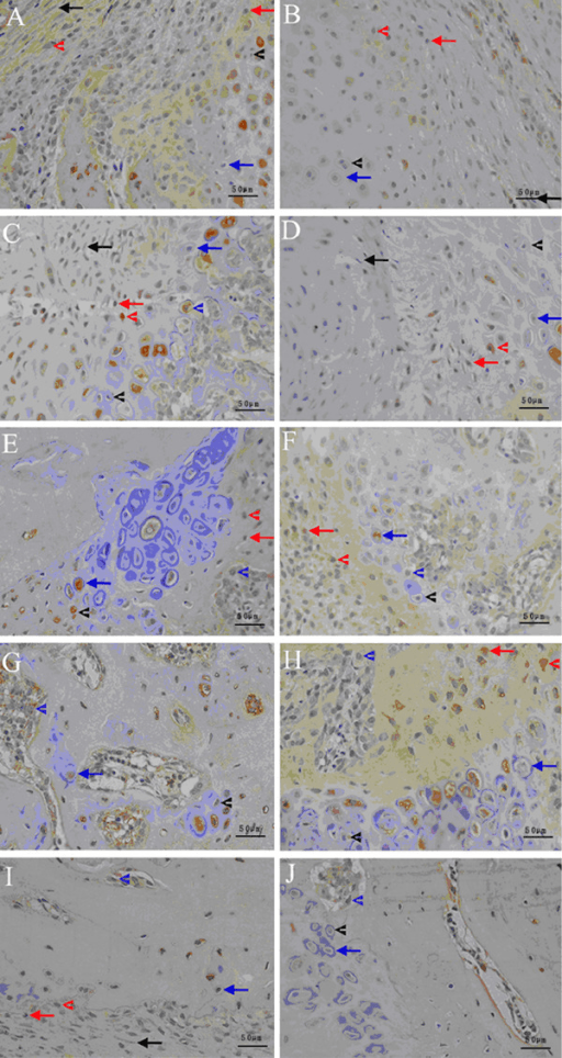
Histology, equally a research tool, means bone biopsy with immunohistochemistry for Runx2 and osterix, assessment of osteoblast differentiation markers, quantification of new bone formation and evaluation of how far the carrier has degraded.
Neutralising antibodies against rhBMP-2 can develop after exposure. Clinical trials monitor for them, but rates are low (under 5%) and the clinical significance is unclear. Repeat BMP use carries a theoretical risk of an immune response, though this has not been shown to affect outcomes.
Before using BMP the workup is the ordinary one: routine preoperative bloods (FBC, UEC, coagulation), nutritional assessment with vitamin D and calcium where bone healing is a concern, and inflammatory markers (CRP, ESR, WCC) where infection is. No BMP-specific test is required.
Management
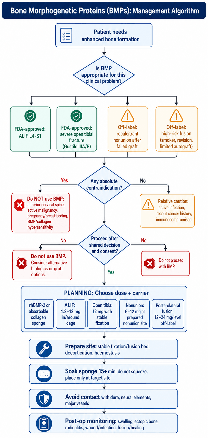
The decision starts with whether the problem genuinely needs enhanced bone formation, and then with whether the indication is an approved one or an off-label one that has to be justified.
When BMP is reasonable.
- ALIF at L4-S1 - the approved spinal indication
- Severe open tibial fractures, Gustilo IIIA/B
- Recalcitrant nonunion after autograft has failed
- High-risk fusion: smokers, revision surgery
Patient factors that push the decision that way are inadequate autograft quantity, concern about donor-site morbidity, and metabolic bone disease.
When not to. BMP is not used at all in these situations:
- Anterior cervical spine - not approved, airway swelling
- Active malignancy
- Pregnancy or breastfeeding
- Known hypersensitivity to BMP or to bovine collagen
Active infection, a recent cancer history and immunocompromise are relative contraindications.
Dose follows indication, and the off-label doses are the higher ones.
- Dose
- 12 mg (small kit) or 4.2 mg (XS kit)
- Carrier
- Absorbable collagen sponge (ACS)
- Notes
- FDA-approved indication
- Dose
- 12 mg
- Carrier
- ACS placed within IM nail cavity
- Notes
- FDA-approved indication
- Dose
- 12-24 mg per level
- Carrier
- ACS with local bone or allograft
- Notes
- Higher dose, increased complications
- Dose
- 6-12 mg
- Carrier
- ACS packed into nonunion site
- Notes
- Combined with stable fixation
The carrier is not packaging. The absorbable collagen sponge is a bovine type I collagen matrix that absorbs the BMP solution and delivers it slowly, degrading over 4-8 weeks and acting as an osteoconductive scaffold while it does. It is the only FDA-approved carrier for BMP-2.
Surgical Technique
Preparation. Improper preparation reduces osteoinductive efficacy, so the sequence is prescribed.
BMP Preparation and Application
Open the kit sterilely and add sterile water to the lyophilised BMP vial. Let it dissolve completely without shaking. The final concentration for BMP-2 is 1.5 mg/mL.
Pour the solution evenly over the absorbable collagen sponge and leave it to absorb for at least 15 minutes. Do not wring, squeeze or handle it more than necessary.
Prepare the fusion bed or fracture site, decorticating the surfaces where applicable and clearing soft tissue from the target area.
Place the soaked sponge at the target: within the cage for ALIF, its dimensions matched to the cage size, or packed into the site for a nonunion.
Close in layers without excess tension. A drain is controversial, since it may remove the BMP. Monitor for swelling afterwards.
- BMP Placement
- Inside cage and/or anterior to cage
- Dose
- 12 mg or 4.2 mg
- Key Technical Points
- Contained within disc space, avoid retraction on vessels
- BMP Placement
- Within intramedullary canal around nail
- Dose
- 12 mg
- Key Technical Points
- Apply after reaming, before final nail insertion
- BMP Placement
- Over decorticated transverse processes
- Dose
- 12-24 mg/level
- Key Technical Points
- Contain within paraspinal gutters, avoid neural elements
- BMP Placement
- Packed into nonunion site after debridement
- Dose
- 6-12 mg
- Key Technical Points
- Combine with stable fixation, fresh bleeding bone ends
Over-application and poor containment are the main technical causes of complications.
- Containment - keep the sponge away from neural elements, or the ectopic bone it induces will compress them
- Haemostasis - bleeding dilutes the BMP and increases the risk of haematoma
- Dura - BMP near dura can cause epidural fibrosis and ectopic bone
- Position - make sure the sponge stays where it was put during closure
- Dose - the minimum effective dose, particularly for off-label applications
Combining BMP with graft is off-label but common. Allograft chips soaked in BMP solution, or a BMP sponge layered with allograft, add structural support and osteoconduction to the osteoinductive signal; demineralised bone matrix and synthetic ceramics (TCP, hydroxyapatite) are used the same way, and the combination may reduce the BMP dose needed. Local autograft from the decompression, mixed with the BMP sponge, brings osteogenic cells as well and spares an iliac crest harvest.
Complications and Controversies
Supraphysiologic dosing and off-label use are what lie behind the recognised complications and the arguments that still surround them.
- Incidence
- 10-30% posterior fusion
- Mechanism
- BMP diffusion into surrounding soft tissues
- Management
- Observation if asymptomatic, excision if nerve compression
- Incidence
- 10-50% anterior cervical (CONTRAINDICATED)
- Mechanism
- Cytokine release, oedema in confined space
- Management
- Airway monitoring, intubation if severe
- Incidence
- 3-8% ALIF
- Mechanism
- Inflammation, ectopic bone near nerve roots
- Management
- NSAIDs, neuropathic agents, decompression if severe
- Incidence
- 5-15% spinal fusion
- Mechanism
- Inflammatory osteoclast activation
- Management
- Observation, revision if structural concern
- Incidence
- Variable
- Mechanism
- Ectopic induction in muscle/soft tissue
- Management
- Prophylaxis with NSAIDs (if used), excision if symptomatic
- Incidence
- 5-10%
- Mechanism
- Fluid accumulation at BMP site
- Management
- Aspiration if large, observation
Clinical BMP doses are 100-1000 times physiologic levels. Normal fracture healing involves nanogram amounts; clinical use involves milligrams, and 12 mg is 12,000,000 nanograms. That excess is what produces the off-target effects - inflammation, ectopic bone, osteolysis. Dose-reduction strategies are under investigation and not yet validated.
Retrograde ejaculation is the BMP-specific complication that the technique viva and the FDA MAUDE signal both raise and the complications table omits. Antegrade ejaculation is lost, semen passing retrograde into the bladder, through dysfunction of the superior hypogastric plexus - the sympathetic network draped over the L5-S1 disc and sacral promontory, exactly where the anterior approach dissects and where the BMP sponge sits.
The association. Both independent re-analyses in this topic's evidence base report higher retrograde ejaculation and urogenital problems with rhBMP-2 in ALIF than with autograft (Carragee 2011; Fu 2013, where the increase was present but not statistically significant). The proposed mechanism is a BMP-driven local inflammatory reaction injuring the adjacent autonomic plexus, additive to the dissection itself.
Reducing the risk means blunt rather than monopolar dissection over the promontory, a left-sided retroperitoneal approach, and containing the sponge within the cage away from the plexus - or a posterior approach, or autograft, in a young man who wishes to father children.
Retrograde ejaculation affects men, is often permanent and matters for fertility, so it is a specific, documented consent item for any man of reproductive age undergoing ALIF with rhBMP-2.
How much of this is off-label. Studies suggest over 50% of BMP use is off-label - posterior fusion, cervical spine, nonunion - and the absence of approval for those indications creates medico-legal exposure. Evidence quality across them is variable, and industry-sponsored trials dominate the literature.
The malignancy signal. The independent YODA / Annals individual-patient-data meta-analysis (Fu 2013) found a statistically significant increase in new cancers at 24 months (RR 3.45, 95% CI 1.98-6.00), and the concern was dose-dependent - most prominent in the high-dose rhBMP-2 ("Amplify", around 40 mg) posterolateral-fusion programme, which the FDA did not approve.
Why it is not accepted as a proven causal risk. The event numbers were small and the tumour types heterogeneous, no single cancer dominating; follow-up was short; and harm ascertainment in the source industry trials was poor. Larger and longer administrative-database and registry studies at standard clinical doses have not confirmed a consistent causal increase.
The practical position. There is no proven causal cancer risk at standard doses, but the concern is biologically plausible, BMPs being mitogenic, and the signal was dose-dependent. So rhBMP-2 is avoided in active malignancy and recent cancer, and high off-label doses are discouraged. That nuanced synthesis, rather than a flat "safe" or "carcinogenic", is the exam-ready answer.
Postoperative Care
Follow-up after BMP watches two things at once: the healing, and the complications that belong to the BMP rather than to the operation.
Postoperative Care Timeline
Monitor for soft-tissue swelling, assess neurology after a spinal procedure, inspect the wound for haematoma and manage pain to protocol.
Wound check at two weeks, ask about radiculitis symptoms, check inflammatory markers if there is concern, and mobilise to the surgical protocol.
First radiographs to assess fusion or healing, activity restrictions continued, physiotherapy as appropriate, and a look for symptoms of ectopic bone.
CT to confirm fusion in spinal cases, progressive return to activity, final radiographs at twelve months and discharge once healed.
- Presentation
- Radicular leg pain, numbness
- Timing
- 1-6 weeks postop
- Management
- NSAIDs, gabapentin, rarely decompression
- Presentation
- New pain, stiffness, nerve symptoms
- Timing
- 6-24 weeks postop
- Management
- Observation if asymptomatic, excision if symptomatic
- Presentation
- Lucency on imaging, graft subsidence and cage settling or migration
- Timing
- 3-12 months postop
- Management
- Observation, revision if progressive instability
- Presentation
- Swelling at surgical site
- Timing
- 1-4 weeks postop
- Management
- Observation, aspiration if large
- Presentation
- Persistent pain, motion on imaging
- Timing
- 6-12 months postop
- Management
- Revision fusion with autograft or repeat BMP
Ectopic bone is commonest in posterolateral fusion, where BMP can migrate towards neural elements. New neurological symptoms after BMP use mean an urgent MRI or CT to look for it, and surgical decompression if neural compression is found; early intervention prevents permanent deficit. Document what happened for the consent discussion in future cases.
Returning to activity after spinal fusion means walking on day one, light activities at 6 weeks, desk work at 6-8 weeks, physical work at 3-6 months and contact sport at 6-12 months, with fusion confirmed on CT before unrestricted activity.
After a long bone or nonunion, weight-bearing is protected for 6-12 weeks, progressive from 12-16 weeks, and unrestricted only after radiographic union, with serial radiographs and the callus guiding the progression.
Outcomes
The evidence pendulum. The early industry-sponsored randomised trials were favourable - Burkus in ALIF, Govender in open tibial fractures. The independent re-analyses that followed, Carragee in 2011 and the YODA individual-patient-data analysis by Fu in 2013, found markedly higher harms and no proven advantage over autograft. That tension is the heart of any BMP discussion.
Fusion rates. Reported ALIF fusion with rhBMP-2 runs 94-100%, against 85-90% for autograft. Off-label posterolateral fusion runs 85-95% and varies between series, with mixed evidence against autograft and higher complication rates.
What patients report. ODI improvement, VAS pain scores and return-to-work times are equivalent to autograft, and satisfaction is high wherever fusion is achieved. At 5-10 years the fusion is maintained, adjacent segment disease is no different, and the cancer concern has not been borne out as a causal risk at standard doses.
Reoperation overall is similar to autograft. It may be higher where ectopic bone has to be excised, and revision for pseudarthrosis is no different.
Cost is the argument against. BMP costs thousands of dollars per dose, the saving from avoiding a harvest is offset by that price and by the cost of managing complications, and QALY analyses show marginal benefit at best.
- BMP Advantage
- None (BMP costs thousands)
- Autograft Advantage
- Minimal additional cost
- BMP Advantage
- Shorter (no harvest)
- Autograft Advantage
- Longer with iliac crest harvest
- BMP Advantage
- Eliminated
- Autograft Advantage
- Chronic donor-site pain
- BMP Advantage
- Higher with off-label use
- Autograft Advantage
- Lower overall
- BMP Advantage
- Similar or slightly higher
- Autograft Advantage
- Gold standard comparison
Most of the randomised evidence was funded by the manufacturer (Medtronic for BMP-2), with concerns about publication bias, ghostwriting and selective outcome reporting, and independent reviews consistently show less favourable results; the manufacturer's marketing practices drew regulatory scrutiny. Off-label indications have limited high-quality evidence of any provenance. Acknowledge those limits when counselling a patient.
Clinical Applications
rhBMP-2 is the most widely used osteobiologic in orthopaedic surgery, controversy over safety and off-label use notwithstanding.
The approved indication is single-level anterior lumbar interbody fusion, L4-S1. In the pivotal randomised trial fusion was achieved in 94.5% of rhBMP-2 patients against 88.7% with iliac crest autograft at 24 months, with equivalent clinical outcomes on ODI and VAS, a shorter operation and no donor site to hurt afterwards.
Posterolateral fusion is off-label. It is not an approved approach, uses higher doses per level, and carries the complications of BMP laid in an open paraspinal gutter from which it can migrate towards neural elements: ectopic bone compressing nerve roots, osteolysis with screw loosening, and radiculitis. Evidence on fusion rates against autograft is mixed.
Cervical use is different in kind - the one off-label application with a documented risk to life.
The FDA has warned specifically against rhBMP-2 in anterior cervical fusion, which is not an approved indication. The enclosed prevertebral space amplifies inflammatory swelling into airway oedema, haematoma, dysphagia and dysphonia; the reported consequences include prolonged intubation, emergency tracheostomy and death.
Guidelines, Registries & Global Practice
Global Epidemiology and Regulatory Landscape
rhBMP-2 use peaked in the United States in the late 2000s (used in roughly a quarter to a half of lumbar fusions at its height) and has since declined substantially following the 2011 Spine Journal critique (Carragee) and the 2013 independent YODA/Annals re-analysis (Fu). Uptake has always been far lower in Europe, the UK, Australia and Asia, driven by cost and a more conservative stance toward off-label use. The vast majority of BMP exposure worldwide is now in anterior or posterolateral lumbar fusion and selected long-bone trauma/nonunion.
- Regulator
- FDA (PMA)
- Approved Indications
- rhBMP-2: single-level ALIF (LT-CAGE), acute open tibial fracture with IM nail, sinus/alveolar ridge augmentation; rhBMP-7: HDE only
- Practice Note
- Black-box-equivalent safety communication for anterior cervical use
- Regulator
- CE mark (medicinal product, dibotermin alfa)
- Approved Indications
- rhBMP-2: single-level lumbar interbody fusion, acute tibial fracture; rhBMP-7 (eptotermin alfa) historically for tibial nonunion
- Practice Note
- OP-1 / InductOs availability has contracted; cervical use contraindicated
- Regulator
- MHRA + NICE
- Approved Indications
- InductOs (rhBMP-2) for acute tibial fracture and lumbar fusion within licence
- Practice Note
- NICE has not endorsed routine biologic use over autograft; cost-effectiveness scrutiny
- Regulator
- TGA (ARTG)
- Approved Indications
- rhBMP-2 registered for spinal fusion and tibial fracture indications mirroring FDA labelling
- Practice Note
- Use reserved for high-risk cases; cost borne by hospital or insurer
Key drivers of variation:
- Cost - thousands of dollars/pounds/euros per kit; the dominant barrier outside the US private system
- Evidence shift - independent re-analyses (Carragee 2011; Fu 2013) showed no proven advantage over autograft and higher harms, reducing enthusiasm
- Off-label exposure - historically more than half of US use was off-label; far more restrained internationally
- Reserved indications - revision fusion, recalcitrant nonunion, smokers, metabolic bone disease, inadequate autograft
Across all systems autograft (iliac crest or local bone) remains the reference standard for primary fusion.
MCQ Practice Points
Q: What is the mechanism of action of BMP-2 and BMP-7 in bone healing?
A: Osteoinduction - BMPs induce differentiation of mesenchymal stem cells (MSCs) into osteoblasts. They signal through SMAD pathway (receptor binding → SMAD phosphorylation → gene transcription). BMP-2 and BMP-7 are the only clinically approved BMPs. Distinct from osteoconduction (scaffold providing surface for bone growth) and osteogenesis (cells directly forming bone).
Q: What are the approved clinical indications for recombinant BMP-2 (rhBMP-2, Infuse)?
A: (1) Anterior lumbar interbody fusion (ALIF) in titanium cage - FDA approved. (2) Open tibial fractures with IM nail - FDA approved. Off-label use in many other applications (posterolateral fusion, nonunion, spinal deformity). Note: NOT approved for posterior cervical spine due to swelling risk (dysphagia, airway compromise). Dose: 1.5mg/mL on absorbable collagen sponge.
Q: What are the potential complications of BMP-2 use in spinal surgery?
A: (1) Heterotopic ossification (can cause neural compression), (2) Radiculitis/nerve inflammation, (3) Osteolysis adjacent to cage, (4) Increased cancer risk (controversial - early studies suggested, later refuted), (5) Swelling - especially concerning in anterior cervical spine (dysphagia, airway compromise). Use in posterior cervical spine discouraged. Cost: approximately $5000-$10000 per application.
Q: How does BMP-7 (OP-1) differ from BMP-2 in clinical applications?
A: BMP-7 (OP-1, Stryker) was approved for tibial nonunion and posterolateral spinal fusion under Humanitarian Device Exemption (HDE). Lower osteoinductive potency than BMP-2 but possibly fewer inflammatory complications. BMP-7 is no longer commercially available (discontinued 2014). BMP-2 remains the only clinically available recombinant BMP. Research continues on other BMPs and delivery systems.
Q: What is the role of the carrier/scaffold in BMP delivery and what carriers are used?
A: The carrier provides: (1) Sustained release of BMP, (2) Localization at target site, (3) Structural support. Absorbable collagen sponge (ACS) is the FDA-approved carrier for BMP-2. Other carriers: DBM, calcium phosphate ceramics, synthetic polymers. The carrier affects release kinetics - initial burst release followed by sustained release. BMP alone without carrier has poor retention at site.
Exam Viva Scenarios
Practise clinical reasoning and management decisions out loud
“How are BMPs classified and which are used clinically?”
“When would you consider using BMP in a patient undergoing spinal fusion?”
“Describe how you would use BMP-2 in an anterior lumbar interbody fusion.”
“What is the evidence for BMP-2 use in spinal fusion, and what are the controversies?”
“A patient in your public hospital requires spinal fusion. How do you decide whether to use BMP?”
BMP Biology Fundamentals
- BMPs: TGF-beta superfamily, secreted growth factors
- Over 20 BMP subtypes, BMP-2 and BMP-7 most osteogenic
- Osteoinductive: induce bone in non-skeletal sites (Urist 1965)
- BMP-2 more potent than BMP-7 but higher complications
Smad Signaling Pathway
- Type II receptor (BMPR-II) binds BMP, recruits type I (ALK-2/3/6)
- Type I phosphorylates Smad1/5/8 (R-Smads) at C-terminus
- Smad1/5/8 + Smad4 (Co-Smad) complex translocates to nucleus
- Activates Runx2 (master osteoblast TF) → Osterix, Osteocalcin, BSP, Collagen I
Clinical BMPs
- BMP-2 (InFuse): FDA-approved ALIF L4-S1, open tibial fractures
- BMP-7 (OP-1): HDE for recalcitrant long bone nonunion (limited availability)
- Dosing: 12 mg ALIF, 12-24 mg posterolateral (off-label)
- Supraphysiologic: 100-1000x normal healing levels
FDA-Approved Indications
- BMP-2: ALIF single-level L4-S1 (fusion rate 94-100%)
- BMP-2: Open tibial shaft fractures (44% fewer secondary interventions, BESTT)
- BMP-2: Oral maxillofacial (sinus augmentation, ridge preservation)
- BMP-7: HDE only - recalcitrant nonunion after failed autograft
Complications
- Ectopic bone: 10-30% posterolateral (nerve compression risk)
- Osteolysis/cysts: 5-15% (screw loosening concern)
- Radiculitis: 3-8% ALIF (inflammation near nerve roots)
- Cervical CONTRAINDICATED: airway edema, life-threatening - NOT approved
Key Exam Points
- Urist 1965: demineralized bone matrix subcutaneous → ectopic bone
- Osteoinduction (active MSC recruitment) vs osteoconduction (passive scaffold)
- Runx2 knockout: cleidocranial dysplasia (absent clavicles)
- Off-label use over 50%: posterolateral fusion, nonunion, revision surgery
Evidence Base
Bone Formation by Autoinduction - The Founding Observation
- Demineralised (decalcified) bone matrix implanted into non-skeletal sites induced new bone via autoinduction
- Host mesenchymal cells of the recipient bed were recruited and differentiated into bone-forming cells
- Established the concept of an osteoinductive substance latent within bone matrix
- Coined the framework that later led to identification of bone morphogenetic protein
Molecular Cloning of BMPs - Defining the Family
- Isolated human cDNA clones for three BMPs (BMP-1, BMP-2A, BMP-3) from purified bovine bone extract
- Each recombinant protein independently induced cartilage formation in vivo
- BMP-2A and BMP-3 identified as new members of the TGF-beta supergene family
- Enabled recombinant production of BMPs and the subsequent clinical biologics
rhBMP-2 vs Autograft in ALIF - Pivotal RCT
- 279 patients randomised: rhBMP-2/absorbable collagen sponge (143) vs iliac crest autograft (136), tapered threaded cages
- Fusion rate at 24 months: rhBMP-2 94.5% vs autograft 88.7%
- Shorter operative time and lower blood loss with rhBMP-2 (no harvest)
- 32% of autograft patients reported persistent iliac crest donor-site discomfort at 24 months
rhBMP-2 in Open Tibial Fractures - BESTT Trial
- 450 open tibial shaft fractures randomised to standard IM nail care vs rhBMP-2 (0.75 or 1.5 mg/mL) on collagen sponge
- 1.5 mg/mL group: 44% reduction in risk of secondary intervention (RR 0.56, 95% CI 0.40-0.78, p=0.0005)
- Significantly faster fracture and wound healing; fewer infections in Gustilo type-III injuries (p=0.0219)
- Dose-dependent effect (1.5 mg/mL superior to 0.75 mg/mL)
Emerging Safety Concerns - Critical Review of Industry Trials
- Systematic review comparing 13 industry-sponsored rhBMP-2 publications (780 patients) with FDA data and follow-up studies
- Original trials reported 0% rhBMP-2-associated adverse events; true rate estimated at 10-50% depending on approach
- Anterior cervical fusion: ~40% increased early adverse-event risk including life-threatening airway events
- Higher rhBMP-2 doses associated with greater apparent risk of new malignancy; methodological bias against autograft controls
Independent IPD Meta-Analysis (YODA Project)
- Individual-patient-data analysis of 17 industry studies (13 RCTs, 31 cohorts) via the Yale Open Data Access project
- For lumbar fusion, rhBMP-2 and iliac crest autograft were similar in fusion and overall success (77-93% at 24 months)
- Anterior cervical fusion: increased wound complications and dysphagia with rhBMP-2
- Increased 24-month cancer risk with rhBMP-2 (RR 3.45, 95% CI 1.98-6.00), though event rates were low and heterogeneous