Brooker Classification of Heterotopic Ossification
Examiners frequently ask you to grade a radiograph using the Brooker system and then state the prophylaxis options. Know that indomethacin and radiotherapy are equally effective for prophylaxis; the choice depends on patient factors (avoid NSAIDs in renal impairment or elderly patients; avoid radiotherapy in women of childbearing age and around cementless stems in early integration). Grade IV (apparent ankylosis) is rare but devastating — the hip is fused by heterotopic bone and functional outcome is poor without excision.
Risk Factors for Heterotopic Ossification After THA
Identifying high-risk patients is critical because prophylaxis is far more effective than treatment of established HO.
- Relative Risk
- Highest
- Prophylaxis Recommendation
- Strongly recommended — indomethacin OR radiotherapy
- Relative Risk
- Moderate–High
- Prophylaxis Recommendation
- Prophylaxis recommended
- Relative Risk
- Moderate–High
- Prophylaxis Recommendation
- Prophylaxis recommended
- Relative Risk
- Moderate
- Prophylaxis Recommendation
- Prophylaxis recommended
- Relative Risk
- Moderate (men greater risk than women)
- Prophylaxis Recommendation
- Consider in presence of additional risk factors
- Relative Risk
- Moderate
- Prophylaxis Recommendation
- Consider prophylaxis; posterior approach has lower risk
- Relative Risk
- Moderate
- Prophylaxis Recommendation
- Prophylaxis recommended
- Relative Risk
- Moderate–High
- Prophylaxis Recommendation
- Prophylaxis recommended
PHAMPLD — Prior HO, Hypertrophic OA, AS, Male, Post-traumatic, Lateral approach, DISH/PagetHigh-risk patients for HO prophylaxis
Hook:The more letters in PHAMPLD that apply, the stronger the indication for prophylaxis.
Pathophysiology of Heterotopic Ossification
Understanding the biology helps explain why prophylaxis works and why timing of excision matters:
- HO is the formation of mature lamellar bone in soft tissues where bone does not normally exist — it is NOT calcification (dystrophic or metastatic) and it is NOT ossification from periosteum.
- The trigger is thought to be osteoprogenitor cells (likely derived from mesenchymal stem cells) that are incorrectly signalled to differentiate along the osteoblastic lineage in response to inflammation and tissue injury.
- Key molecular mediators: Bone morphogenetic proteins (BMP-2, BMP-4), prostaglandin E2, and inflammatory cytokines released at the time of surgery activate the process. This is why COX inhibition (indomethacin) is effective prophylaxis.
- Three phases of HO formation:
- Early (0–4 weeks): Inflammatory; soft-tissue swelling, pain, decreased range of motion. Radiographs may be normal. Bone scan shows increased uptake.
- Intermediate (4–16 weeks): Osteoid formation; mineralisation begins. Radiographs show faint, fluffy ossification. Alkaline phosphatase is elevated.
- Mature (4–12+ months): Organised lamellar bone with cortical and cancellous patterns. Radiographs show well-defined bone. Bone scan uptake decreases. This is the safe window for excision.
HO is not just a hip-replacement problem, and the topic's "don't confuse it with myositis ossificans" warning deserves an answer:
- Post-surgical / post-traumatic — also after acetabular-fracture fixation and, very commonly, after elbow trauma/fracture-dislocation (the elbow is the classic peripheral site; functional elbow HO is graded by the Hastings and Graham system).
- Neurogenic HO — after traumatic brain injury, spinal cord injury, or burns; it forms around large joints (often below the neurological level), tends to be more florid, and relates to injury severity and spasticity.
- Myositis ossificans (traumatica) — heterotopic bone within muscle after a discrete injury (e.g. quadriceps). The hallmark is zonal maturation: a mature ossified peripheral rim around an immature centre (the reverse of an osteosarcoma, which is densest centrally) with a lucent cleft from the parent bone. It matures and can regress — do not biopsy early, as it can mimic sarcoma histologically.
- Fibrodysplasia ossificans progressiva (FOP) — a rare genetic disorder (ACVR1/ALK2 gain-of-function), with a congenital great-toe malformation and progressive HO in a characteristic axial-to-appendicular pattern; trauma, biopsy and intramuscular injections trigger flares, so these are avoided.
- Dystrophic / metastatic calcification — amorphous calcium deposition without trabeculae, cortex or marrow (dystrophic in damaged tissue with normal serum calcium; metastatic in hypercalcaemia/renal failure).
The unifying discriminator: HO and myositis ossificans are TRUE bone (trabeculae, cortex, marrow) that matures peripherally, whereas calcification is amorphous mineral with no bony architecture.
The Brooker Classification (Grades I–IV)
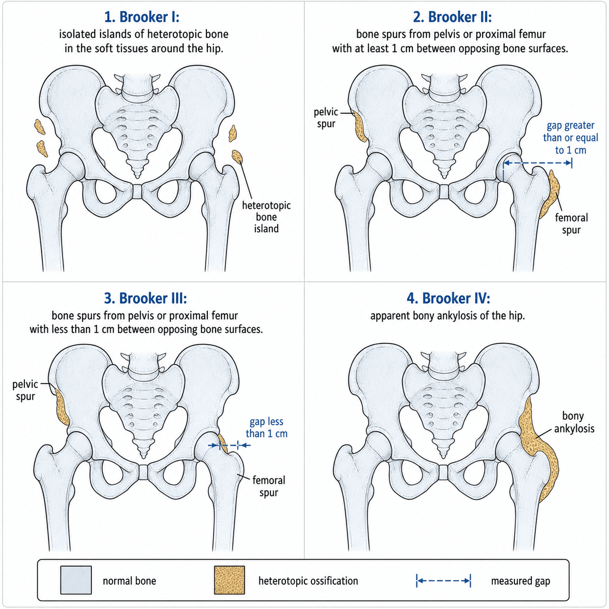
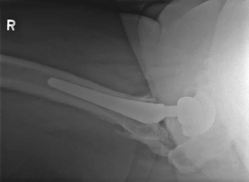
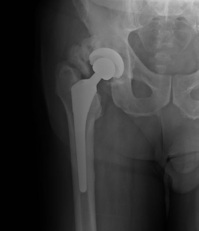
The classification is assessed on a single AP radiograph of the hip (typically a supine or standing AP pelvis). It describes the most severe heterotopic bone formation visible in the soft tissues between the greater trochanter and the femoral shaft/pelvis.
- Radiographic Appearance
- Islands of bone within the soft tissues about the hip
- Gap Between Surfaces
- No bridging — no gap to measure
- Clinical Significance
- Usually asymptomatic; no treatment needed
- Radiographic Appearance
- Bone spurs from the pelvis or proximal end of the femur
- Gap Between Surfaces
- At least 1 cm between opposing bone surfaces
- Clinical Significance
- May cause mild stiffness; rarely needs intervention
- Radiographic Appearance
- Bone spurs from the pelvis or proximal femur
- Gap Between Surfaces
- Less than 1 cm between opposing surfaces
- Clinical Significance
- Clinically significant stiffness; may warrant excision
- Radiographic Appearance
- Apparent ankylosis — complete bridging bone
- Gap Between Surfaces
- No gap — continuous bone mass
- Clinical Significance
- Severe functional limitation; excision with prophylaxis
Islands, Spurs-gap, Spurs-narrow, AnkylosisBrooker Grades in Order
Hook:Think of the gap shrinking from wide to none as the grade worsens — islands, then spurs with a gap, then spurs nearly touching, then ankylosis.
The Brooker classification only uses the AP radiograph. It does not require CT or lateral views. However, if surgical excision is planned, a CT scan is essential to map the three-dimensional extent of the HO and its relationship to neurovascular structures (particularly the sciatic nerve).
Differential Classification: Brooker vs Other Systems
The Brooker system is the most widely used, but other classifications exist:
- Hattrick and Harris (modified Brooker): Adds a grade 0 (no HO) and refines the gap measurement — sometimes used in research trials for greater granularity.
- DeLee classification: Uses CT-based assessment for pre-operative planning of excision; maps the anatomic zone of HO (anterior, posterior, medial, lateral) — more useful for surgical planning than the Brooker AP-only assessment.
- Santiago classification: A CT-based system that grades HO by the quadrant involved and the volume of heterotopic bone — provides three-dimensional information the Brooker system lacks.
In clinical practice and exams, Brooker remains the standard. Know that it has moderate inter-observer reliability (especially distinguishing Grade II from Grade III when the gap is close to 1 cm) and that CT is the investigation of choice for surgical planning.
Prophylaxis and Treatment
Prophylaxis Options
Two strategies have Level I evidence for HO prophylaxis after THA:
- Indomethacin
- 75 mg daily
- Radiotherapy (Single Fraction)
- 7 Gy in a single fraction
- Indomethacin
- 3–6 weeks, starting within 24 hours post-op
- Radiotherapy (Single Fraction)
- Within 72 hours of surgery (pre-op also effective)
- Indomethacin
- COX inhibition blocks prostaglandin-mediated osteogenesis
- Radiotherapy (Single Fraction)
- Inhibits osteoprogenitor differentiation in irradiated field
- Indomethacin
- Reduces Grade III–IV HO from about 10–20 percent to 1–3 percent
- Radiotherapy (Single Fraction)
- Reduces Grade III–IV HO to a similar degree
- Indomethacin
- No specialised equipment; inexpensive; systemic effect
- Radiotherapy (Single Fraction)
- One-time treatment; no GI or renal side effects
- Indomethacin
- GI toxicity, renal impairment, bleeding risk; contraindicated in elderly or renal disease
- Radiotherapy (Single Fraction)
- Requires radiotherapy department; avoid near cementless stems (early integration); contraindicated in pregnancy
Never offer both indomethacin and radiotherapy simultaneously as routine prophylaxis. Combined therapy has not shown a meaningful additional benefit over either alone in most trials, and the additive side-effect burden (GI bleed risk plus radiation exposure) is unjustified. Reserve combination therapy for patients with recurrent HO despite adequate single-agent prophylaxis.
This is asked repeatedly and the honest answer is a theoretical concern that clinical evidence has not borne out for short courses:
- The theory: the very mechanism that makes NSAIDs effective against HO — inhibition of prostaglandin-mediated bone formation — raises the worry that they could also blunt the bone formation needed for osseointegration of a press-fit (cementless) implant.
- Laboratory/animal data do show that NSAIDs (particularly non-selective agents) can impair early bone ingrowth and fracture healing in models.
- But the clinical data are reassuring: cohort and trial data have not demonstrated a clinically meaningful increase in cementless stem loosening, migration, or revision with a short post-operative course of NSAID prophylaxis. For short courses the concern remains largely theoretical.
- Practical resolution: mitigate the theoretical risk with a short course (rather than prolonged) and/or a COX-2-selective agent; if you remain concerned or NSAIDs are contraindicated, use single-fraction radiotherapy instead — but note radiotherapy has its own theoretical effect on an ingrowth surface, so the porous-coated portion of a cementless implant should be kept out of (or shielded from) the high-dose field.
- Bottom line: do not withhold effective prophylaxis from a genuinely high-risk patient over an unproven ingrowth concern — choose the modality that best fits the patient.
Surgical Excision
- Indication: Brooker Grade III–IV HO causing functional limitation (restricted flexion less than 60 degrees, impaired gait, difficulty with sitting or hygiene).
- Timing: Delay excision until the HO is mature — typically 12–18 months after index surgery. Maturity is confirmed by a stable Brooker grade on serial radiographs, normalising alkaline phosphatase, and a 'cold' or decreasing uptake on bone scan.
- Approach: Excision through the original surgical approach where possible. Meticulous haemostasis is critical because haematoma promotes recurrence.
- Peri-operative prophylaxis is MANDATORY: Either indomethacin or radiotherapy must be given at the time of excision (typically radiotherapy within 48 hours post-excision) — recurrence rates without prophylaxis are high (up to the original grade or worse).
12–18 months, Mature bone, Normal ALP, Cold scanTiming of HO excision
Hook:Wait a year — premature excision feeds the fire. Mature bone, normal enzyme, quiet scan, then operate.
Guidelines, Registries and Global Practice
Global Epidemiology
- HO after THA has an overall incidence of 8–50 percent on radiographs, but only 1–5 percent develop functionally significant (Brooker Grade III–IV) ossification.
- Male patients are 2–3 times more affected than female patients. Bilateral THA in a patient who developed HO after the first side has a recurrence rate over 80 percent without prophylaxis.
- Higher rates reported with lateral and anterolateral surgical approaches compared with the posterior approach, likely due to greater soft-tissue dissection and gluteal muscle trauma.
Guidelines Comparison
- Prophylaxis Recommendation
- Recommend prophylaxis for high-risk patients
- Regimen
- Indomethacin 75 mg daily for 3–6 weeks OR single 7 Gy radiotherapy fraction
- Prophylaxis Recommendation
- Prophylaxis for patients with prior HO or high-risk factors
- Regimen
- Indomethacin preferred first-line unless contraindicated; radiotherapy as alternative
- Prophylaxis Recommendation
- Risk-stratified approach
- Regimen
- Indomethacin for moderate risk; radiotherapy for very high risk or recurrent HO
- Prophylaxis Recommendation
- Single-agent prophylaxis (not combined) for high-risk THA
- Regimen
- Either modality; choice based on renal function, age, and cementless vs cemented stem
Key Practice Variation
- US practice tends toward radiotherapy more commonly for high-risk patients, partly driven by radiotherapy infrastructure availability and the higher rate of cementless stems (where early NSAID use may theoretically impair bony ingrowth, though clinical evidence for this is weak).
- European and UK practice tends to favour indomethacin as first-line prophylaxis due to cost-effectiveness and ease of administration, reserving radiotherapy for recurrent HO or NSAID-intolerant patients.
- Japanese and East Asian registries report slightly lower HO incidence overall, which may reflect differences in surgical approach preferences or genetic factors, though data are limited.
Viva practice
Exam Viva
Practise clinical reasoning and management decisions out loud
“You are shown an AP pelvis radiograph of a 62-year-old man 6 months after a right cementless total hip replacement. There is a continuous bridge of bone between the greater trochanter and the ilium, with no visible gap. The patient reports stiffness and difficulty with footwear. Describe what you see and how you would manage this.”
“A 55-year-old man with ankylosing spondylitis is listed for a bilateral staged cementless total hip replacement. He had Brooker Grade III HO after a previous left hip replacement 3 years ago, which was treated conservatively. Outline your peri-operative strategy for his upcoming right THA.”
Exam cheat sheet
The four grades (AP radiograph)
- Grade I: islands of bone in soft tissues — no gap
- Grade II: bone spurs from pelvis or femur with gap of 1 cm or more
- Grade III: bone spurs with gap less than 1 cm
- Grade IV: apparent ankylosis — continuous bridging bone, no gap
Prophylaxis for high-risk patients
- Indomethacin 75 mg daily for 3–6 weeks (avoid if renal impairment, elderly, or peptic ulcer disease)
- Radiotherapy: single 7 Gy fraction within 72 hours of surgery (avoid in pregnancy, caution near cementless stems)
- Both equally effective — choose based on patient factors, never combine routinely
- High-risk = prior HO, hypertrophic OA, AS, DISH, Paget, male, lateral approach
Surgical excision of mature HO
- Indicated for Brooker Grade III–IV with functional limitation
- Delay 12–18 months until mature: stable X-ray grade, normalising ALP, cold bone scan
- Mandatory peri-operative prophylaxis at time of excision to prevent recurrence
- CT for pre-operative planning; be wary of sciatic nerve in posterior/medial HO
Evidence
Ectopic ossification following total hip replacement: incidence and a method of classification
- The original paper proposing the four-grade (I–IV) classification of ectopic (heterotopic) bone after total hip replacement.
- Graded the most severe ossification between the proximal femur and pelvis on the AP radiograph — no special views required.
- Distinguished clinically trivial low grades from the bridging/ankylosing high grades that limit function.
Surgical treatment of severe heterotopic ossification after total hip arthroplasty over the last 25 years: a systematic review and a new case series
- Systematic review (7 studies, 41 patients) of excision for Brooker grade III/IV HO; perioperative prophylaxis was irradiation alone (10), irradiation + indomethacin (10), or indomethacin alone (21).
- At a mean follow-up of ~15 months, range of motion improved but pain relief was inconsistent; there were only 2 recurrences, 1 dislocation and 1 GI complication.
- Low-dose irradiation prevented recurrence; the authors concluded there is insufficient good-quality data and that multicentre studies are needed.
According to PubMed, the classification is from Brooker et al. 1973 (J Bone Joint Surg Am 1973;55(8):1629-32; PMID 4217797). Prophylaxis evidence comes from the randomised Knelles et al. 1997 (indomethacin and 7 Gy effective, aspirin/5 Gy inferior; DOI 10.1302/0301-620x.79b4.6829) and Hedley et al. 1989 (single-fraction radiotherapy; DOI 10.1016/s0883-5403(89)80033-6), with the contemporary NSAID network meta-analysis (etoricoxib 90 mg/d optimal) by Xie et al. 2025 (DOI 10.1186/s12891-025-09277-5). Surgical excision outcomes are reviewed by Lachiewicz et al. 2024 (DOI 10.1016/j.arth.2024.02.017).