Subacute Localised Osteomyelitis | Metaphyseal Lucency | Sclerotic Rim
- Brodie abscess = subacute localised pyogenic osteomyelitis with central cavitation and surrounding reactive sclerosis
- Classic imaging: well-circumscribed metaphyseal lucency with thick sclerotic rim (likely benign but must exclude tumour)
- Penumbra sign on T1 MRI: high-signal rim of granulation tissue distinguishes abscess from tumour
- Staphylococcus aureus cultured in roughly 50-70 percent; many are culture-negative despite histological confirmation
- Definitive treatment: thorough curettage, bone grafting of cavity, and targeted antibiotics for 4-6 weeks
- “Brodie abscess is a diagnosis of exclusion — must rule out osteoid osteoma, Ewing sarcoma, and Langerhans cell histiocytosis
- “Penumbra sign on T1 MRI is the key POSITIVE discriminator - specificity ~96% but sensitivity only ~27%, so absence does NOT exclude it
- “Always send tissue for both microbiology AND histopathology (to exclude malignancy)
- “Children and adolescents with vague metaphyseal pain and night symptoms: think Brodie abscess and osteoid osteoma
Overview and Epidemiology
A Brodie abscess is defined by the company it keeps. It is the walled-off, subacute form of osteomyelitis - the host contained the infection instead of clearing it - which is why it sits between acute paediatric osteomyelitis at one end and the biofilm-protected chronic disease on biofilm formation at the other. Its whole clinical difficulty is that it does not look like infection: the two lesions it is mistaken for are osteoid osteoma, which shares the metaphyseal site and the night pain, and Ewing sarcoma, which shares the permeative appearance and the raised inflammatory markers. Multifocal, culture-negative disease in a child should also raise CRMO, which is not an infection at all.
The name. Sir Benjamin Brodie described the lesion in 1832, a tibial abscess in a man treated by amputation. What he described is what the term still means: subacute localised pyogenic osteomyelitis, a contained abscess cavity within bone surrounded by granulation tissue and reactive sclerosis.
Who gets one. Presentation peaks in the second decade, in children and adolescents, with a male-to-female ratio of roughly 2:1. The distal tibial metaphysis is the commonest site, followed by the distal femur and the proximal tibia.
What grows. Staphylococcus aureus is isolated in 50-70 percent of culture-positive cases, and up to 50 percent of abscesses are culture-negative despite adequate tissue sampling.
What is at stake. The lesion mimics a bone tumour, and failure to distinguish a benign abscess from an aggressive malignancy has serious consequences. The penumbra sign on MRI and tissue biopsy at curettage are the critical diagnostic tools.
Pathophysiology
How the abscess forms. A bacterial focus, typically S. aureus, reaches the metaphysis of a long bone haematogenously, or is inoculated directly at trauma or surgery. In the subacute form the host response is enough to contain the infection but not to eliminate it, leaving a central abscess cavity of pus or serous fluid, lined by granulation tissue and surrounded by a thick rim of reactive sclerotic bone (the "cloaca", or wall).
Why the metaphysis. The metaphyseal arterial loops are terminal, slow-flowing and lack phagocytic lining cells, so bacteria lodge there, evade immune clearance and multiply within cancellous bone. In children the abscess is therefore typically juxtaphyseal, sitting on the metaphyseal side of the growth plate, which acts as a barrier preventing epiphyseal spread in most cases; in adults the lesion may occur at any metaphyseal or meta-diaphyseal location. Minor trauma may contribute, causing local microvascular injury that creates a favourable site for bacterial seeding.
Why the course is indolent. Partial containment by granulation tissue and reactive sclerosis prevents systemic dissemination, so what the patient experiences is chronic low-grade inflammation with intermittent flares, and a lesion may stay quiescent for years before reactivating. Inside the cavity the oxygen tension is low and the acidity high, which slows bacterial replication but also limits immune cell function.
Why antibiotics alone are not enough. The abscess cavity is avascular, and S. aureus can form biofilm within it, resisting both immune attack and antibiotic penetration.
- Brodie Abscess
- Insidious over weeks to months
- Acute Osteomyelitis
- Acute over days
- Chronic Osteomyelitis
- Persistent or relapsing over months to years
- Brodie Abscess
- Minimal or absent
- Acute Osteomyelitis
- Fever, rigors, sepsis
- Chronic Osteomyelitis
- Intermittent flares, low-grade fever
- Brodie Abscess
- Mildly elevated or normal
- Acute Osteomyelitis
- Markedly elevated (CRP, ESR, WCC)
- Chronic Osteomyelitis
- Variable, often mildly elevated
- Brodie Abscess
- Well-defined lucency + sclerotic rim
- Acute Osteomyelitis
- Metaphyseal lucency + periosteal reaction
- Chronic Osteomyelitis
- Sequestra, involucrum, sinus tracts
- Brodie Abscess
- Curettage + bone graft + antibiotics
- Acute Osteomyelitis
- IV antibiotics + surgical drainage
- Chronic Osteomyelitis
- Sequestrectomy + dead space management + prolonged antibiotics
Classification and Types
Classification by Anatomical Site
- Frequency
- Most common (roughly 30-40 percent)
- Clinical Features
- Ankle pain, limp, local tenderness
- Key Considerations
- May abut physeal plate in skeletally immature
- Frequency
- Second most common
- Clinical Features
- Knee pain, referred thigh pain
- Key Considerations
- Must exclude distal femur bone tumours
- Frequency
- Common
- Clinical Features
- Proximal shin pain, limp
- Key Considerations
- Proximity to tibial tuberosity in adolescents
- Frequency
- Reported in children
- Clinical Features
- Heel pain, limping child
- Key Considerations
- Can mimic Sever disease clinically
- Frequency
- Less common
- Clinical Features
- Site-specific pain, may present late
- Key Considerations
- Higher risk of delayed diagnosis
Clinical Assessment
The history. Vague, deep, aching pain over the affected metaphysis, often worse at night, intermittent over weeks to months. A limp is common in children and may be the presenting complaint. Some patients report minor trauma before the symptoms began.
Systemic features are minimal. Patients are typically afebrile with no rigors, although mild malaise is common. Ask about NSAIDs: relief here is partial, which is the point of difference from osteoid osteoma, where it is dramatic.
On examination. Tenderness is localised over the affected metaphysis, with mild swelling if any and no significant soft tissue mass, unlike tumour, and warmth may be slightly increased. Range of motion is usually preserved because the joint is not typically involved, and lymphadenopathy is absent or minimal, unlike acute infection. An antalgic limp is common with lower limb lesions.
If any of the following features are present, suspect malignancy (Ewing sarcoma, osteosarcoma, lymphoma) and arrange urgent MRI and tissue diagnosis rather than assuming Brodie abscess:
- Rapidly worsening pain (days to weeks rather than months)
- Large soft tissue mass on examination or imaging
- Systemic symptoms: weight loss, night sweats, persistent fever
- Aggressive periosteal reaction (sunburst, Codman triangle, onion-skin)
- Cortical breakthrough with soft tissue extension on imaging
- Markedly elevated ESR greater than 50 mm/hr or CRP greater than 50 mg/L
Never assume a lytic bone lesion is a Brodie abscess without MRI and tissue confirmation.
- Age and Site
- Child or adolescent, metaphysis
- Key Discriminator
- Penumbra sign on T1 MRI, sclerotic rim, indolent course
- Definitive Test
- MRI + tissue culture and histology at curettage
- Age and Site
- Child or young adult, cortical nidus
- Key Discriminator
- Nidus less than 1.5 cm, dramatic NSAID response, central calcification
- Definitive Test
- CT to identify nidus; marked osteoblastic uptake on bone scan
- Age and Site
- Child or adolescent, diaphysis or metaphysis
- Key Discriminator
- Aggressive periostitis, soft tissue mass, systemic symptoms, high ESR
- Definitive Test
- MRI + biopsy (tissue diagnosis mandatory)
- Age and Site
- Child, variable sites
- Key Discriminator
- Punched-out lytic lesion, may be multifocal, variable sclerosis
- Definitive Test
- Biopsy: CD1a and S-100 positive Langerhans cells
- Age and Site
- Adolescent, epiphysis
- Key Discriminator
- Epiphyseal location, chondroid matrix on CT, open growth plate
- Definitive Test
- MRI + biopsy: S-100 positive chondroblasts
- Age and Site
- Adolescent, metaphysis (same site as Brodie)
- Key Discriminator
- Aggressive: cortical destruction, soft tissue mass, osteoid matrix
- Definitive Test
- MRI + biopsy: malignant osteoid production
Osteoid osteoma and Brodie abscess are easy to confuse. Both produce night pain and a sclerotic bone lesion in a young patient, so separate them on size and on drug response: the osteoid osteoma nidus is under 1.5 cm, often with central calcification, and its pain is dramatically relieved by NSAIDs through a prostaglandin-mediated mechanism. A Brodie abscess is usually over 2 cm, responds only partially to NSAIDs, shows an abscess cavity on MRI where osteoid osteoma has none, and yields tissue showing chronic inflammation with organisms on culture. CT is the modality for finding a nidus; MRI with contrast is the one for an abscess.
OSTEOMADifferential Diagnosis of Brodie Abscess
Hook:OSTEOMA — run through the differential before committing to Brodie abscess diagnosis!
The differential above is dominated by tumours, but in endemic regions and in immunocompromised hosts a tuberculous (or other granulomatous) osteomyelitis can produce a metaphyseal or epiphyseal abscess that looks like a pyogenic Brodie abscess - and missing it leads to failed pyogenic-antibiotic treatment.
The physis is a key discriminator. Pyogenic subacute osteomyelitis typically respects it, whereas TB and other granulomatous or atypical infections can cross it into the epiphysis and even the joint, so a transphyseal abscess should raise TB, fungal or atypical mycobacterial infection. The course and the host fit the same story: a more indolent, "cold" abscess, often with constitutional symptoms such as weight loss and night sweats, in a patient from an endemic area, with HIV, on immunosuppression, or with multifocal disease.
Do not rely on routine pyogenic cultures, which are typically negative. Send tissue for:
- AFB smear (Ziehl-Neelsen), mycobacterial culture and GeneXpert/PCR
- Histology looking for caseating granulomas
- Fungal stains and culture
The diagnosis changes the treatment entirely. Confirmed skeletal TB needs prolonged multi-drug anti-tuberculous chemotherapy, with surgery reserved for drainage of large abscesses, debridement or instability. So a "Brodie abscess" that crosses the physis, is culture-negative on routine pyogenic cultures, or arises in an endemic or immunocompromised setting is tuberculous or granulomatous osteomyelitis until proven otherwise, and gets anti-TB chemotherapy rather than a short anti-staphylococcal course.
Investigations
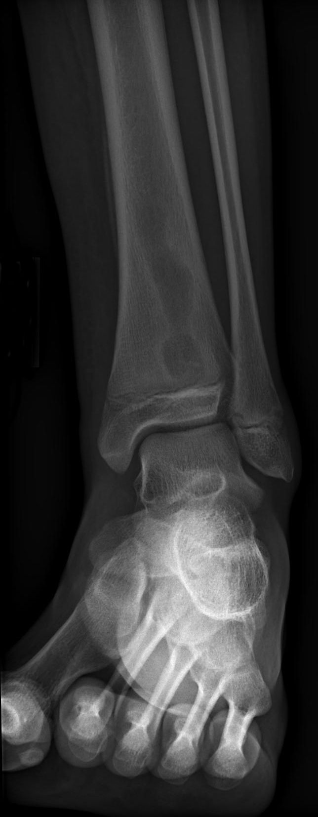
Radiographs first. AP and lateral of the affected bone, including the joint above and below. The classic lesion is a well-circumscribed lytic lesion in the metaphysis with a surrounding zone of dense reactive sclerosis blending into normal bone - the "target" or "bull's-eye" appearance - with no aggressive periosteal reaction, no soft tissue mass and no cortical destruction.
What the radiograph misses. An early lesion may show only subtle metaphyseal rarefaction, and the sclerosis can be read as osteoid osteoma or tumour.
MRI is the essential study, taken as T1, T2, STIR and post-gadolinium T1. The cavity is low signal on T1 and high signal on T2 and STIR, the abscess wall enhances after contrast, and on unenhanced T1 a high-signal rim of granulation tissue lines the cavity: the penumbra sign. MRI earns its place beyond that sign as well: it excludes a soft tissue mass (ruling out malignancy), defines the extent of the lesion, identifies sinus tracts and shows the relationship to physis and joint.
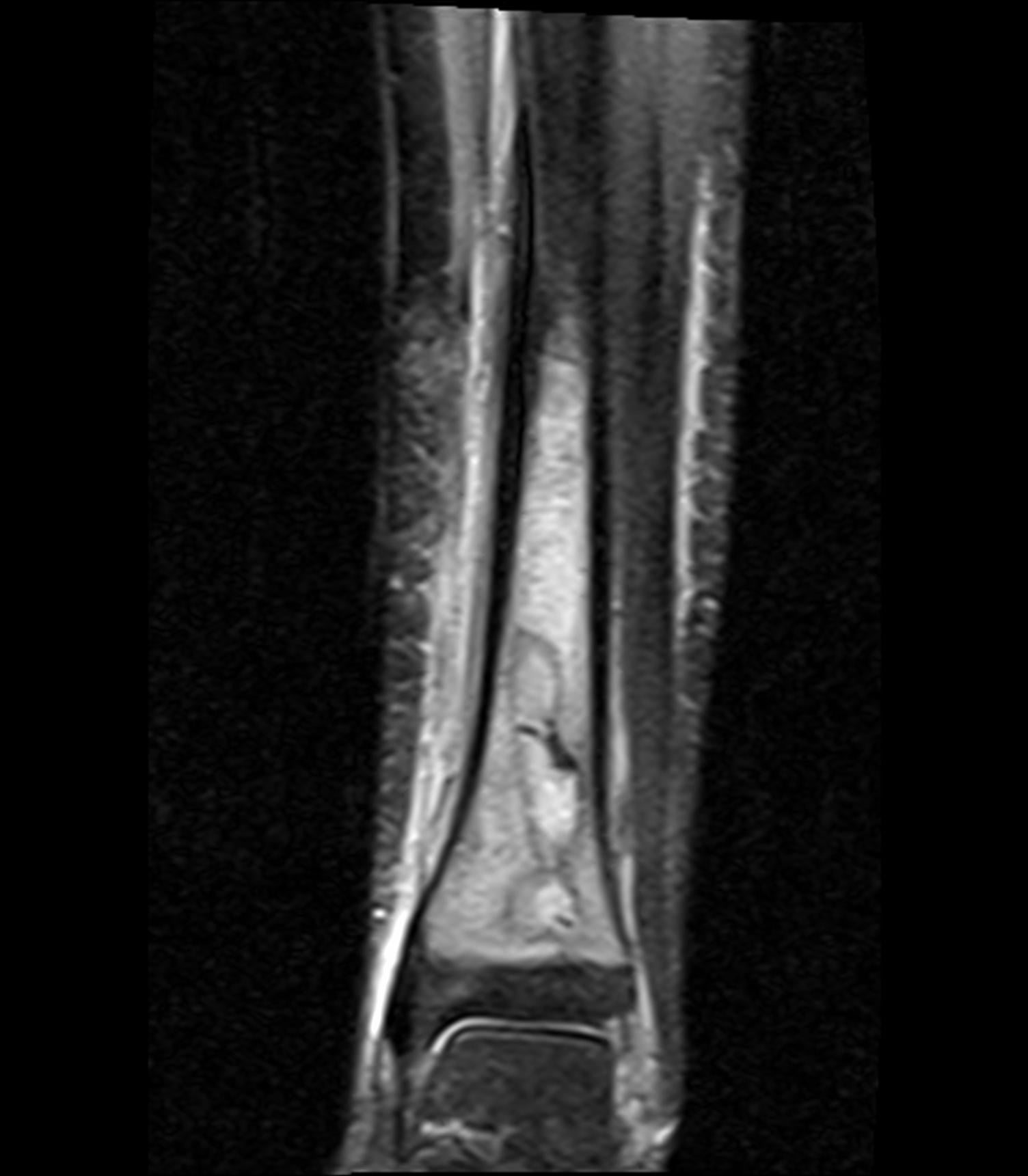
The penumbra sign is the single most useful POSITIVE finding, a high-signal-intensity ring of granulation tissue around the low-signal cavity, standing out against the low-signal sclerotic bone. Its operating characteristics are asymmetric: specificity about 96 percent, sensitivity only about 27 percent (McGuinness). Its presence, against a fitting clinical picture, strongly supports Brodie abscess over tumour, while its absence is close to uninformative, since roughly three-quarters of proven infections never show it. Never let a missing penumbra sign argue you out of an infection, and never treat imaging as definitive - tissue goes for histopathology at curettage.
The channel, or serpentine tract, sign is a tortuous lucent tract running from the metaphyseal abscess cavity toward the physis, or toward the cortex, and represents the route of the original infective or drainage tract. Bone tumours do not produce such a connecting channel, so a lesion "pointing to the growth plate" through a serpiginous lucency is relatively specific for subacute osteomyelitis.
A sequestrum may appear in the cavity. A small central focus of dense, dead bone is more typical of chronic osteomyelitis, but it can occur in subacute disease; an intracavitary calcific focus should prompt infection rather than be mistaken for the central calcified nidus of an osteoid osteoma, which is cortical and has a much smaller lucent nidus.
When you argue that a metaphyseal lytic lesion is infective rather than neoplastic, cite the penumbra sign PLUS a serpentine channel pointing to the physis, together with the absence of a soft-tissue mass and aggressive periostitis. The channel/tract sign is the often-forgotten second discriminator.
CT answers a narrower question. Thin slices through the metaphysis are the way to exclude the nidus of an osteoid osteoma, and CT also delineates the sclerotic rim and any internal calcification or sequestrum within the cavity. For surgical planning it defines cortical thinning and proximity to the physis.
What the bloods show. CRP and ESR are mildly elevated or normal, and a markedly high ESR should push you towards an alternative diagnosis; the white cell count is usually normal and blood cultures are rarely positive in a subacute presentation. Normal inflammatory markers do NOT exclude a Brodie abscess.
Management Algorithm
Treatment follows the symptoms and the certainty of the diagnosis. A symptomatic lesion is curetted, grafted and treated with targeted antibiotics; a truly asymptomatic incidental lesion may be observed with serial imaging; and an adult whose old lesion has reactivated needs the differential reopened before anything is planned.
- Diagnosis
- Metaphyseal lucency with sclerotic rim on X-ray, penumbra sign on MRI
- Treatment
- Curettage + bone graft + 4-6 weeks antibiotics
- Key Pearl
- Most common site: distal tibia metaphysis
- Diagnosis
- Small well-defined lesion, no periostitis, no soft tissue mass
- Treatment
- Observe with serial imaging if truly asymptomatic
- Key Pearl
- Ensure tumour markers, MRI and ESR/CRP are normal
- Diagnosis
- Expanding lucency, new periosteal reaction, rising inflammatory markers
- Treatment
- Aggressive curettage + prolonged IV antibiotics
- Key Pearl
- Consider alternative diagnoses including malignancy
Surgical Management: Curettage and Bone Grafting
The goal is complete evacuation of the abscess cavity, tissue for microbiology and histopathology, and obliteration of the dead space with bone graft.
Surgical Protocol
Review the MRI to define the extent of the lesion and plan the approach. Consent covers pathological fracture, non-union, growth disturbance where the lesion is physeal, and recurrence.
Hold the antibiotics. Do NOT give pre-operative antibiotics until tissue cultures are obtained, unless the patient is septic, because they reduce culture yield and may mask the causative organism. If antibiotics have already been started, stop them 48 hours before surgery if that is safe.
A direct approach to the metaphyseal lesion, planned on the imaging, and then in order:
- Create a cortical window over the abscess, oval or rectangular, preserving structural bone
- Evacuate all pus and necrotic material, and curette the abscess wall until healthy bleeding bone is encountered
- Send pus and tissue for microbiology - Gram stain with aerobic, anaerobic and fungal cultures - AND histopathology to exclude malignancy
- Irrigate the cavity copiously with saline
- Pack the cavity with autograft (iliac crest), allograft bone chips or bone substitute
- Close over drains if needed
A cast or brace for 4-6 weeks protects the bone graft and guards against pathological fracture, with weight-bearing withheld or protected depending on the site and the size of the graft.
IV antibiotics start once cultures have been taken and switch to oral when sensitivities are known: 4-6 weeks in total, around 2 weeks IV then 2-4 weeks oral, guided by the organism and the clinical response.
Serial radiographs at 6 weeks, 3 months, 6 months and 12 months confirm graft incorporation and resolution of the cavity, with CRP and ESR at each visit to track the response.
Return to activity is gradual once the graft incorporates and the pain resolves, typically 3-6 months. Recurrent symptoms mean a repeat MRI and consideration of repeat curettage.
Complications
- Incidence
- 10-20 percent
- Risk Factors
- Incomplete curettage, inadequate antibiotics, resistant organism
- Management
- Repeat MRI, repeat curettage with prolonged antibiotics
- Incidence
- Rare but reported
- Risk Factors
- Large metaphyseal defect, inadequate grafting, early weight-bearing
- Management
- Protected weight-bearing 6-8 weeks; internal fixation if displaced
- Incidence
- Risk in skeletally immature
- Risk Factors
- Curettage across or near the physis
- Management
- Monitor leg length; epiphysiodesis of contralateral side if discrepancy develops
- Incidence
- Uncommon with adequate treatment
- Risk Factors
- Incomplete debridement, immunocompromise, non-compliance
- Management
- Sequestrectomy, dead space management, prolonged IV antibiotics
- Incidence
- Rare but catastrophic
- Risk Factors
- Diagnosing Brodie abscess without tissue biopsy
- Management
- Always send tissue for histopathology at index procedure
The most devastating complication is missed malignancy. Ewing sarcoma and lymphoma can produce a lytic metaphyseal lesion with surrounding sclerosis that mimics a Brodie abscess on imaging, so every curettage specimen goes to histopathology as well as microbiology. A "surprise" tumour diagnosis after Brodie abscess curettage is a recognised and avoidable catastrophe.
Outcomes and Prognosis
- Cure Rate
- 85-95 percent
- Complications
- Recurrence 10-15 percent, rare fracture
- Return to Activity
- 3-6 months
- Cure Rate
- 30-50 percent
- Complications
- High recurrence, prolonged treatment, risk of chronicity
- Return to Activity
- Variable, often limited by recurrence
- Cure Rate
- Under 50 percent
- Complications
- Recurrence, progression to chronic osteomyelitis
- Return to Activity
- Often requires reoperation
What predicts a good result. Complete curettage back to healthy bleeding bone, an organism identified on culture, 4-6 weeks of targeted antibiotics, and a compliant patient. Recurrence in that group is quoted at 10-15 percent, against the 10-20 percent carried for the condition as a whole.
What predicts a poor one. Culture-negative disease treated empirically, an immunocompromised host, inadequate initial debridement, and proximal femoral or spinal lesions where surgical access is difficult.
Guidelines, Registries & Global Practice
- Worldwide distribution — no geographic predilection
- Peak incidence: Children and adolescents aged 5-15 years globally
- Organism variation: S. aureus predominates worldwide; Kingella kingae is increasingly recognised in children under 4 in developed countries with improved PCR diagnostics
- Resource-limited settings: Brodie abscess may present later and at larger size due to delayed imaging access; higher rates of culture-negative cases where pre-treatment with empirical antibiotics is common
- High-resource: MRI readily available, penumbra sign assessment routine, image-guided biopsy if atypical features, dedicated paediatric orthopaedic and infectious disease teams
- Limited-resource: Diagnosis often relies on plain radiographs and clinical assessment; CT may be used when MRI unavailable; longer courses of empirical antibiotics may be given when surgery is delayed
- Universal principle: Tissue sampling for both micro and histo remains the gold standard regardless of resource setting
- Diagnosis emphasis
- MRI with penumbra sign; tissue biopsy mandatory at surgery
- Treatment approach
- Curettage + graft + targeted antibiotics for symptomatic lesions
- Duration of antibiotics
- 4-6 weeks total (IV to oral switch guided by clinical response)
- Diagnosis emphasis
- MRI mandatory before surgery; CT if osteoid osteoma in DDx
- Treatment approach
- Surgical curettage is standard; antibiotics alone reserved for small asymptomatic lesions
- Duration of antibiotics
- Minimum 4 weeks targeted therapy; 6 weeks if extensive disease
- Diagnosis emphasis
- MRI with contrast; biopsy if any atypical features
- Treatment approach
- Curettage and bone grafting; percutaneous drainage in select cases
- Duration of antibiotics
- 4-6 weeks; transition from IV to oral based on clinical and lab improvement
- Diagnosis emphasis
- Step-wise imaging: X-ray, MRI, then CT for surgical planning
- Treatment approach
- Thorough curettage with bone graft; structural graft for large defects
- Duration of antibiotics
- 4-6 weeks targeted antibiotics; empirical flucloxacillin if culture-negative
There is no dedicated registry for Brodie abscess outcomes. The evidence base consists of retrospective case series and expert opinion — no randomised controlled trials exist comparing surgical vs non-surgical management. Current guidance is principle-based: MRI for diagnosis, tissue sampling at surgery for both micro and histo, curettage and grafting for symptomatic lesions, and 4-6 weeks of targeted antibiotics.
Record in every suspected Brodie abscess:
- MRI findings (penumbra sign present or absent; any atypical features)
- Differential diagnosis considered (specifically osteoid osteoma, Ewing sarcoma, LCH, chondroblastoma)
- Tissue sent for both microbiology AND histopathology
- Antibiotics started only after cultures obtained
- Follow-up plan with serial imaging and inflammatory markers
A missed malignancy after treating a lytic bone lesion as Brodie abscess without histopathology is a critical and avoidable error.
Controversies & Areas of Uncertainty
Small asymptomatic lesions discovered incidentally may resolve with antibiotics alone in some series, but reported success rates vary widely (30-70 percent). Most experts recommend surgery for definitive management because curettage provides tissue diagnosis (excluding malignancy) and mechanical debridement of the biofilm-lined cavity. Antibiotics alone are reserved for patients unfit for surgery or very small asymptomatic lesions with characteristic MRI findings.
No RCTs define the optimal duration. Recommendations range from 3-6 weeks, with 4-6 weeks being the most common guidance. IV-to-oral switch timing is also debated — most centres switch when inflammatory markers trend downward and clinical improvement is evident, typically at 2 weeks.
Autologous iliac crest bone graft is the traditional gold standard for filling the cavity, but allograft chips, demineralised bone matrix, and calcium phosphate bone substitutes are increasingly used. No head-to-head trials exist. Autograft has osteoinductive and osteoconductive properties but carries donor site morbidity.
While most guidance advises withholding antibiotics until intra-operative cultures are obtained, this is sometimes impractical when patients have already been started on oral antibiotics by referring clinicians. Stopping antibiotics 48 hours before surgery may improve culture yield, but this must be balanced against the risk of disease progression in the interim.
MCQ Practice Points
Q: What is the penumbra sign and in which MRI sequence is it seen? A: The penumbra sign is a high-signal-intensity rim seen on unenhanced T1-weighted MRI surrounding a low-signal abscess cavity. It represents a layer of granulation tissue lining the abscess wall. It is the most useful POSITIVE radiological discriminator between Brodie abscess and bone tumour — specificity about 96 percent, but sensitivity only about 27 percent, so it rules infection IN and does not rule it out. On T2 and STIR sequences, the central cavity is high-signal (fluid/pus) and the sclerotic rim remains low-signal. Post-gadolinium images show rim enhancement.
Q: How do you distinguish Brodie abscess from osteoid osteoma? A: Both cause night pain and a sclerotic bone lesion in a young patient. Osteoid osteoma has a nidus less than 1.5 cm (often with central calcification on CT), pain is dramatically relieved by NSAIDs, and CT is the best modality to identify the nidus. Brodie abscess is typically larger (usually greater than 2 cm), has the penumbra sign on T1 MRI, pain is only partially NSAID-responsive, and tissue at curettage shows chronic inflammation with organisms on culture.
Q: What is the most common causative organism of Brodie abscess? A: Staphylococcus aureus is the most common organism, isolated in roughly 50-70 percent of culture-positive cases. However, up to 30-50 percent of Brodie abscesses are culture-negative despite adequate tissue sampling. In children under 4 years, Kingella kingae should also be considered. In immunocompromised patients, consider atypical organisms including fungi and mycobacteria.
Q: What is the definitive management of symptomatic Brodie abscess? A: Thorough curettage + bone grafting + targeted antibiotics for 4-6 weeks. The abscess cavity is evacuated through a cortical window, the walls are curetted to bleeding bone, and the dead space is packed with autograft or allograft. Tissue is sent for both microbiology and histopathology (to exclude malignancy). Antibiotics are started after cultures are obtained: initially IV, then oral guided by sensitivities. Small asymptomatic lesions discovered incidentally may be observed with serial imaging.
Q: What is the most common site of Brodie abscess? A: The distal tibial metaphysis is the most common site, followed by the distal femur and proximal tibia. In children, the lesion is typically juxtaphyseal (abutting the growth plate) because the slow-flowing metaphyseal vascular loops are prone to bacterial trapping. The physis acts as a barrier preventing epiphyseal spread.
Q: What is the most important step at the time of curettage for a suspected Brodie abscess? A: Sending tissue for both microbiology and histopathology. Histopathology is essential to exclude malignancy (Ewing sarcoma and lymphoma can mimic Brodie abscess on imaging). Microbiology guides targeted antibiotic therapy. The biggest error is sending tissue for culture only and missing a tumour diagnosis.
Exam Viva Scenarios
Practise clinical reasoning and management decisions out loud
“A 12-year-old boy presents with a 6-week history of vague right ankle pain and an intermittent limp. He is afebrile and systemically well. Examination reveals tenderness over the distal tibial metaphysis with no soft tissue mass. Blood tests show CRP 12 mg/L, ESR 22 mm/hr, WCC normal. A plain radiograph demonstrates a well-circumscribed 2 cm lytic lesion in the distal tibial metaphysis with a surrounding sclerotic rim. Discuss your differential diagnosis, investigations, and management plan.”
“A 35-year-old man presents with 3 months of progressive left thigh pain, now causing a limp. He has had intermittent low-grade fevers. Plain radiographs show a 4 cm diaphyseal lucent lesion in the distal femur with cortical thickening and mild periosteal reaction. CRP is 35 mg/L and ESR is 48 mm/hr. MRI shows a lobulated intracortical and medullary lesion with surrounding marrow oedema but no convincing penumbra sign. Discuss your approach.”
Definition and Pathophysiology
- Subacute localised pyogenic osteomyelitis: contained abscess cavity with surrounding reactive sclerosis
- Haematogenous seeding of metaphyseal vascular loops; S. aureus most common (50-70 percent); 30-50 percent culture-negative
- Host partially contains infection with granulation tissue wall and reactive bone — explains indolent course
- First described by Sir Benjamin Brodie in 1832
Clinical Features
- Peak age: second decade (children and adolescents), male 2:1
- Most common site: distal tibia metaphysis (followed by distal femur, proximal tibia)
- Vague metaphyseal pain, worse at night, mild limp, typically afebrile
- Normal or mildly elevated CRP and ESR — marked elevation suggests alternative diagnosis
Imaging and Investigations
- X-ray: well-circumscribed metaphyseal lucency with sclerotic rim ('target' or 'bull's-eye')
- MRI: penumbra sign (high-signal T1 rim = granulation tissue), low T1/high T2 cavity, rim enhancement post-gadolinium
- CT: for excluding osteoid osteoma nidus and surgical planning
- Always MRI before surgery; biopsy if any atypical features (diaphyseal, soft tissue mass, aggressive periostitis)
Differential Diagnosis
- Osteoid osteoma: nidus less than 1.5 cm, dramatic NSAID response, CT shows nidus
- Ewing sarcoma: diaphyseal, aggressive periostitis, soft tissue mass, high ESR
- Langerhans cell histiocytosis: punched-out lytic lesion, CD1a/S-100 positive on biopsy
- Osteosarcoma: metaphyseal (same site), cortical destruction, osteoid matrix, soft tissue mass
Management
- Gold standard: curettage + bone grafting + targeted antibiotics 4-6 weeks
- Always send tissue for BOTH microbiology AND histopathology (exclude malignancy)
- Do NOT give pre-operative antibiotics before tissue cultures (unless septic)
- Antibiotics alone: reserved for small asymptomatic lesions or unfit patients; high failure rate
Complications and Follow-up
- Recurrence: 10-20 percent — repeat MRI and consider repeat curettage
- Missed malignancy: most devastating complication — prevented by routine histopathology
- Pathological fracture: protect with cast or brace for 4-6 weeks post-graft
- Follow up with serial X-rays and inflammatory markers at 6 weeks, 3 months, 6 months, 12 months
Evidence Base and Key Trials
Subacute osteomyelitis in children
- Classic paper classifying subacute osteomyelitis (Brodie abscess) by anatomical location and radiographic pattern
- Distal tibial metaphysis confirmed as the most common site
- Curettage with bone grafting yielded reliable healing in the majority
- Established the radiological classification framework still referenced in exams
The 'penumbra sign' on T1-weighted MR imaging in subacute osteomyelitis: frequency, cause and significance
- 32 patients referred to an orthopaedic oncology service but subsequently PROVEN to have osteomyelitis; the penumbra sign was present in 24 (75 percent)
- More sensitive than the corresponding T2/STIR double-line sign, which was evident in only 29 percent
- Histologically the penumbra was highly vascularised granulation tissue containing thick-walled arterioles; thickness 2-5 mm (unilocular in 46 percent, multilocular in 54 percent)
- The authors' own conclusion calls the sign characteristic but explicitly NOT pathognomonic
The penumbra sign on T1-weighted MRI for differentiating musculoskeletal infection from tumour
- 183 patients (57 infection, 126 neoplasm) - the study that actually QUANTIFIED the sign: specificity 96 percent (range 94-99) but sensitivity only 27 percent (range 21-34)
- So the penumbra sign is a strong RULE-IN sign for infection and a very poor RULE-OUT sign - it is absent in roughly three of every four proven infections
- Interobserver reliability moderate to good (average kappa 0.57); isolated soft-tissue infection had higher sensitivity (54 percent) at similar specificity (98 percent)
- Every true-positive penumbra sign identified by consensus was in subacute, chronic or acute-on-chronic infection - not acute
Infections simulating bone tumors: a review of subacute osteomyelitis
- Subacute osteomyelitis frequently mimics primary bone tumours on plain radiographs, including Ewing sarcoma and osteosarcoma
- Curettage provided both definitive diagnosis (via tissue sampling) and effective treatment
- S. aureus was the most common isolate, but a significant proportion were culture-negative
- Authors emphasised that histopathological examination is mandatory to exclude malignancy at every biopsy
Brodie's abscess revisited
- Comprehensive review of Brodie abscess imaging characteristics across plain radiography, CT, and MRI
- Confirmed the penumbra sign on T1-weighted MRI as a reliable indicator distinguishing abscess from bone tumour
- Highlighted the importance of MRI with gadolinium contrast in surgical planning and excluding aggressive pathology
- Emphasised that tissue diagnosis remains essential even when imaging appears characteristic