Recognise sterile recurrent bone inflammation, exclude dangerous mimics and treat disease burden
- CNO is a sterile autoinflammatory bone disease, not recurrent bacterial osteomyelitis.
- The usual presentation is recurrent bone pain, sometimes with swelling, limp or back pain.
- Blood tests may be normal or mildly inflammatory; no single blood test confirms the diagnosis.
- Whole-body MRI detects multifocal and silent lesions and is central for diagnosis and monitoring.
- Biopsy is appropriate when disease is unifocal, aggressive, atypical or not safely distinguished from infection or malignancy.
- “Do not label a solitary aggressive bone lesion as CRMO until malignancy and infection have been excluded.
- “Spinal disease matters because vertebral height loss can lead to kyphosis, scoliosis or neurological risk.
- “Antibiotics are not disease-modifying treatment for CNO unless bacterial infection is proven.
- “The orthopaedic role is recognition, safe biopsy when needed, fracture or deformity protection, and avoiding unnecessary operations.
- “Ask about psoriasis, palmoplantar pustulosis, inflammatory bowel symptoms, arthritis and family inflammatory disease.
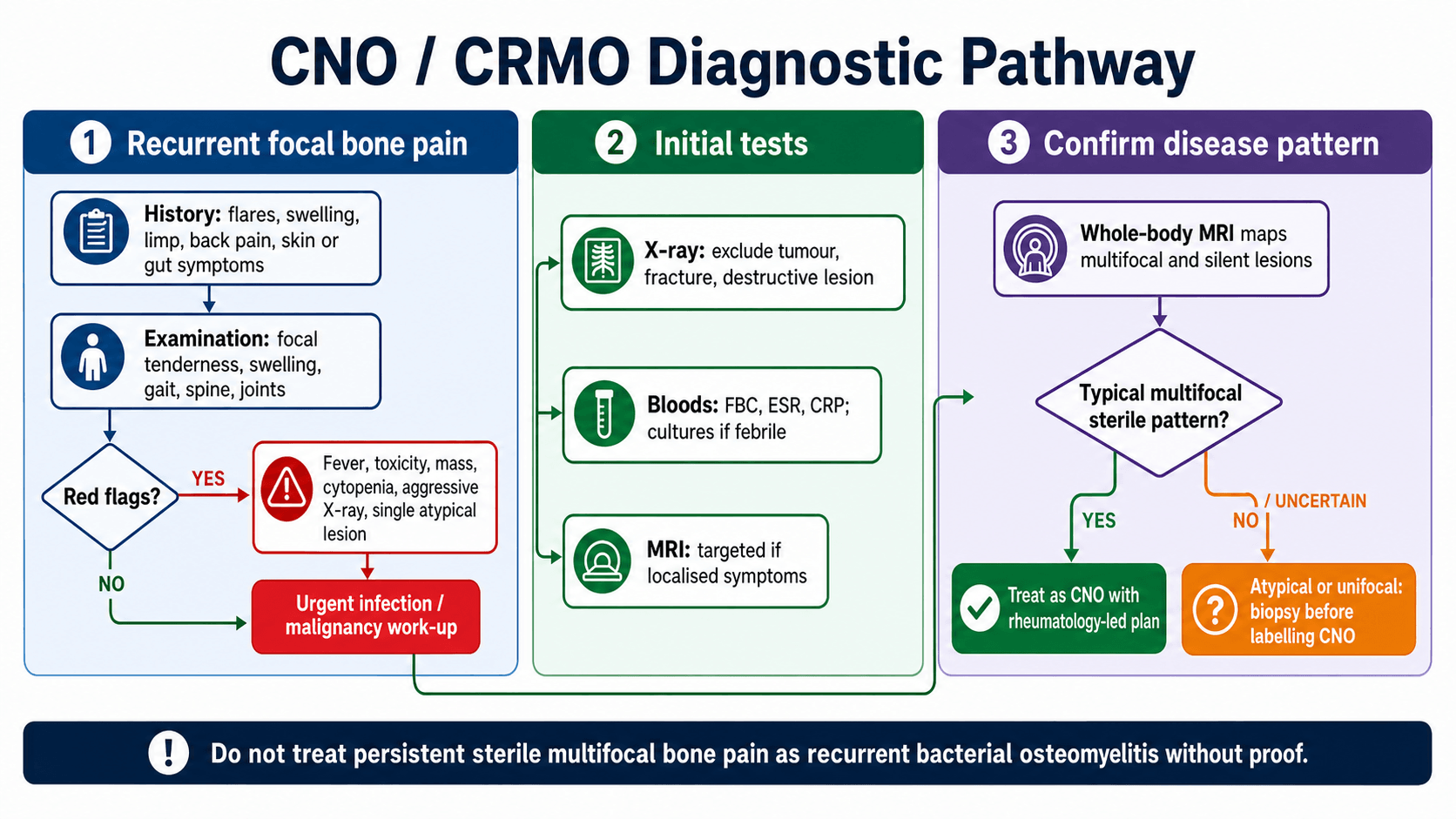
Chronic Nonbacterial Osteomyelitis and CRMO
Persistent or recurrent multifocal bone pain with negative cultures should not automatically lead to repeated antibiotic courses and repeated debridement. The dangerous balance is to recognise CNO early while still excluding infection, malignancy and other mimics when the pattern is atypical.
STERILECNO Safety Check
Hook:STERILE keeps the assessment focused on diagnosis safety and disease burden.
Overview and Epidemiology
Chronic nonbacterial osteomyelitis, or CNO, is a sterile autoinflammatory disorder of bone. Chronic recurrent multifocal osteomyelitis, or CRMO, describes the recurrent multifocal end of the same spectrum. The name is confusing because it contains the word osteomyelitis, but the disease is not primarily bacterial.
The practical clinical problem is a child or adolescent with recurrent bone pain, imaging evidence of osteitis and no microbiological proof of infection. Some children have a single symptomatic lesion. Others have multiple symptomatic and silent lesions in the long-bone metaphyses, clavicle, spine, pelvis, sternum, mandible or foot.
Recurrent focal bone pain, waxing and waning symptoms, multifocal MRI lesions, negative cultures, sterile biopsy inflammation and associated inflammatory conditions.
Ewing sarcoma, osteosarcoma, leukaemia, Langerhans cell histiocytosis, bacterial osteomyelitis, stress fracture, non-accidental injury and spinal infection.
Disease Burden and Risk
CNO is not classified by one universally used orthopaedic fracture-style system. It is more useful to describe disease by burden, risk and certainty of diagnosis.
- Meaning
- One apparent lesion.
- Clinical use
- Higher need for biopsy unless the clinical and imaging pattern is very typical.
- Meaning
- Several lesions on symptoms or MRI.
- Clinical use
- Supports CNO when infection and tumour are not otherwise suggested.
- Meaning
- Flares over time, often at changing sites.
- Clinical use
- Explains CRMO pattern and need for longitudinal treatment.
- Meaning
- MRI lesions without local symptoms.
- Clinical use
- Whole-body MRI can reveal disease burden missed by examination.
Pathophysiology
CNO is best understood as disordered innate immune regulation causing sterile bone inflammation. The exact pathway is not one single gene or one single cytokine for most children. Contemporary reviews describe imbalance between pro-inflammatory and anti-inflammatory signalling, inflammasome activation and osteoclast-mediated bone injury as important themes.
The pathology is osteitis without a proven bacterial driver. Bone biopsy, when performed, may show inflammatory infiltrate, chronic sterile osteomyelitis-like change, fibrosis or sclerosis, but it does not provide a pathognomonic signature. This is why the diagnosis is made from the whole clinical, laboratory, imaging and sometimes histological pattern.
- Meaning
- Innate immune dysregulation produces bone inflammation.
- Clinical implication
- Anti-inflammatory and immunomodulatory treatment is logical when infection is excluded.
- Meaning
- Cultures are negative unless there is a separate infection.
- Clinical implication
- Antibiotics do not treat CNO itself.
- Meaning
- Silent lesions may exist away from the painful site.
- Clinical implication
- Whole-body MRI can change both diagnosis and risk stratification.
- Meaning
- Lesions often involve metaphyseal, epiphyseal or apophyseal regions.
- Clinical implication
- Pain near growth plates should not be dismissed as growing pains when recurrent or focal.
- Meaning
- Vertebral inflammation may lead to height loss or deformity.
- Clinical implication
- Back pain or spinal lesions justify urgent MRI review and treatment escalation.
Although most CNO is polygenic, a few rare monogenic autoinflammatory syndromes cause CRMO-like sterile multifocal osteomyelitis and are classic exam fodder because they implicate the IL-1 axis:
- Majeed syndrome - autosomal recessive, LPIN2 gene; the triad of chronic recurrent multifocal osteomyelitis (early-onset, severe, lifelong), congenital dyserythropoietic anaemia, and a neutrophilic dermatosis (Sweet syndrome).
- DIRA (deficiency of the interleukin-1 receptor antagonist) - autosomal recessive, IL1RN gene; neonatal-onset sterile multifocal osteomyelitis with periostitis and pustulosis, dramatically responsive to IL-1 blockade (anakinra).
- Related conditions include cherubism and the broader autoinflammatory bone-disease group.
Why it matters: very early-onset, severe, familial or syndromic CRMO - especially with anaemia or neonatal onset - should prompt genetic evaluation, because identifying Majeed or DIRA changes prognosis and treatment (IL-1 blockade is specifically effective in DIRA).
Classification
CNO is best classified clinically by distribution, risk and diagnostic certainty. This is more useful than trying to force the condition into a fracture-style grading system.
- Meaning
- One apparent lesion.
- Use
- Treat the diagnosis as less secure unless the pattern is classic; biopsy is often needed.
- Meaning
- Several symptomatic or MRI-detected lesions.
- Use
- Supports CNO when infection and malignancy are not suggested.
- Meaning
- Recurrent multifocal disease with flares over time.
- Use
- Longitudinal treatment and monitoring are needed.
- Meaning
- MRI lesions without local symptoms.
- Use
- Explains why whole-body MRI changes diagnosis and follow-up.
SAPHO syndrome (Synovitis, Acne, Pustulosis, Hyperostosis, Osteitis) is widely regarded as the adult counterpart of CRMO and sits on the same chronic nonbacterial osteitis spectrum - a frequent exam pairing:
- The osteitis and hyperostosis classically involve the anterior chest wall (sternoclavicular joints, manubriosternal joint, medial clavicles), producing the "bull's head" sign on bone scintigraphy (increased uptake across the sternoclavicular joints and manubrium).
- The skin associations are palmoplantar pustulosis and severe acne (acne fulminans/conglobata).
- It may also involve the spine and sacroiliac joints, overlapping with the spondyloarthropathies.
- Like CNO it is sterile (though Cutibacterium acnes is sometimes cultured and debated as a trigger), and management mirrors CNO - NSAIDs first, then bisphosphonates, DMARDs or TNF inhibitors; antibiotics are not disease-modifying.
Take-home: an adult with anterior chest-wall pain, hyperostosis and palmoplantar pustulosis has SAPHO - the grown-up face of CRMO.
Clinical Presentation
Typical History
Children usually present with recurrent or persistent bone pain. The pain may flare, settle and recur at the same or different sites. Limp, activity limitation, focal swelling and local tenderness are common. Fever is absent or low-grade in many children; marked toxicity is a warning against uncomplicated CNO.
Important history points are not generic. They are chosen to separate CNO from infection, malignancy, stress injury and inflammatory disease:
- Ask about
- Focal recurrent bone pain, night pain, flare duration, rest pain, activity link and response to NSAIDs.
- Interpretation
- Recurrent flares fit CNO; progressive relentless pain or severe night pain needs tumour and infection exclusion.
- Ask about
- Fever, weight loss, anorexia, night sweats, lethargy or recurrent infection.
- Interpretation
- Toxicity, weight loss or cytopenia pattern should not be attributed to CNO without work-up.
- Ask about
- Psoriasis, palmoplantar pustulosis, acne, arthritis, enthesitis, inflammatory bowel symptoms and family history.
- Interpretation
- CNO may coexist with inflammatory skin, gut or joint disease.
- Ask about
- Back pain, stiffness, jaw pain, clavicular swelling, chest wall pain, foot pain or pelvic pain.
- Interpretation
- Spine, mandible, clavicle and pelvis are important CNO sites and may alter treatment urgency.
- Ask about
- Recent skin infection, puncture wound, bacteremia risk, immunosuppression, antibiotics and culture results.
- Interpretation
- A bacterial source changes the pathway completely.
- Ask about
- Sports load, recent increase in training, single-site mechanical pain and fracture history.
- Interpretation
- Stress injury can mimic CNO, especially in adolescents.
Examination
Examination should localise symptomatic lesions and look beyond the painful bone. Inspect gait, posture, spine, limb alignment, local swelling, warmth, tenderness, joint motion and neurovascular status. Examine skin and nails for psoriasis or pustulosis. Check jaw opening, clavicles, sternoclavicular joints and spine when symptoms suggest those regions.
Back pain in suspected CNO is not a minor symptom. Vertebral lesions can be silent or painful and may progress to height loss, kyphosis or scoliosis. Whole-body MRI should include adequate sagittal spinal assessment.
Imaging and Investigations
Plain Radiographs
Radiographs are often the first test because they are accessible and help exclude fracture, aggressive tumour, chronic infection and structural damage. Early radiographs may be normal. Established lesions may be lytic, sclerotic, mixed, expansile or show periosteal reaction. Radiographs are useful for structural complications, but they do not map disease burden.
Blood Tests
Order full blood count, ESR and CRP. Results can be normal, mildly raised or inflammatory. The absence of a marked inflammatory response does not exclude CNO. However, cytopenia, very high inflammatory markers, bacteremia, toxic appearance or persistent fever should push the assessment back toward infection, malignancy or systemic disease.
MRI
MRI is the central imaging test. Targeted MRI defines a symptomatic site, marrow oedema, periosteal reaction, soft-tissue inflammation, abscess-like appearances and structural damage. Whole-body MRI is preferred when CNO is suspected because it detects silent lesions and typical distribution patterns.
- Typical CNO finding
- Multifocal lesions, often metaphyseal or periphyseal, with clavicle, spine, pelvis, sternum or mandible possible.
- Why it matters
- Multifocal typical distribution supports CNO and may avoid biopsy in selected cases.
- Typical CNO finding
- Bright marrow signal at active lesions.
- Why it matters
- STIR whole-body MRI is sensitive for disease burden and follow-up.
- Typical CNO finding
- Vertebral body oedema, endplate involvement or height loss may be seen.
- Why it matters
- Spinal lesions require careful monitoring because collapse and deformity can occur.
- Typical CNO finding
- A large mass is not typical for uncomplicated CNO.
- Why it matters
- Mass, aggressive destruction or atypical unifocal disease should trigger biopsy.
- Typical CNO finding
- True pus collection is not expected in sterile CNO.
- Why it matters
- Abscess shifts the diagnosis toward bacterial infection until proven otherwise.
Biopsy
Biopsy is not required for every child, but it is mandatory when the diagnosis is not secure. The biopsy should be planned like a tumour or infection biopsy: correct compartment, safe tract, cultures and histology, and avoidance of contaminating future surgical planes.
Biopsy is appropriate for unifocal disease, aggressive radiographic appearance, soft-tissue mass, systemic illness, cytopenia, persistent fever, unusual age, atypical site, poor response to initial treatment or any case where infection or malignancy remains plausible.
Differential Diagnosis

- Clues against uncomplicated CNO
- Fever, toxicity, abscess, positive cultures, rising CRP or clear portal of infection.
- Action
- Culture and treat infection; drain pus when present.
- Clues against uncomplicated CNO
- Aggressive lesion, soft-tissue mass, constitutional symptoms or progressive destructive pattern.
- Action
- Urgent tumour-safe imaging and biopsy.
- Clues against uncomplicated CNO
- Bone pain with bruising, pallor, infection, cytopenia or systemic illness.
- Action
- Full blood count and haematology pathway.
- Clues against uncomplicated CNO
- Lytic skull, spine, pelvis or long-bone lesion; vertebra plana can overlap.
- Action
- Biopsy if the pattern is uncertain.
- Clues against uncomplicated CNO
- Single-site load-related pain with training history and fracture line.
- Action
- Activity modification and fracture management.
- Clues against uncomplicated CNO
- Dominant synovitis, enthesitis, inflammatory back pain or sacroiliitis pattern.
- Action
- Rheumatology assessment; CNO may coexist.
Management Principles
Management has two parallel aims: keep the diagnosis safe and control inflammation enough to preserve function and prevent structural damage. The child should usually be managed with paediatric rheumatology, paediatrics, radiology and orthopaedics rather than orthopaedics alone.
First-line Treatment
NSAIDs are commonly used as first-line anti-inflammatory treatment when safe and appropriate. They are not just analgesics in this setting; they may control inflammatory bone pain in a proportion of children. The child also needs activity modification, physiotherapy for gait and function, school and sport planning, and monitoring for flare recurrence.
First-line treatment is reasonable when the diagnosis is secure, the child is clinically well, there is no high-risk spinal collapse, no aggressive lesion, no fracture risk and symptoms are tolerable.
SPINETreatment Escalation
Hook:SPINE highlights the high-risk situations where watchful waiting may be unsafe.
Biopsy and Operative Details
When biopsy is required, plan it properly. A poorly planned biopsy can contaminate compartments, miss the lesion or create misleading results.
- Practical point
- Review radiographs, MRI and any suspected tumour pathway requirements.
- Reason
- A biopsy tract must be placed where it can be excised if malignancy is diagnosed.
- Practical point
- Target active representative lesion, preferably the most diagnostically useful site.
- Reason
- Necrotic or sclerotic tissue alone may be non-diagnostic.
- Practical point
- Send histology and microbiology, including bacterial cultures; add fungal or mycobacterial tests when clinically indicated.
- Reason
- The aim is to exclude infection and malignancy, not merely prove inflammation.
- Practical point
- Use image guidance or open biopsy depending site, safety and diagnostic need.
- Reason
- Accurate sampling matters more than making the smallest incision.
- Practical point
- Sterile inflammation supports CNO only when clinical and imaging context fits.
- Reason
- Histology alone is not pathognomonic.
Surgery is not a standard treatment for CNO inflammation. Operative intervention is reserved for diagnostic biopsy, fracture management, deformity correction, treatment of true infection if present, or rare structural complications. Repeated washouts without pus or culture-positive infection usually represent a management error.
ATYPICWhen to Biopsy
Hook:ATYPIC is the biopsy trigger: atypical, aggressive or unsafe-to-label disease.
Complications and Follow-up
Follow-up should track symptoms, function, inflammatory markers when useful, medication response, adverse effects and imaging activity. Whole-body MRI is used for disease burden and monitoring, but imaging should be interpreted with the clinical picture; persistent signal change does not always mean the child needs the same escalation if symptoms and structure are stable.
- Mechanism
- Persistent inflammatory bone pain and deconditioning.
- Follow-up focus
- Pain control, physiotherapy, school participation and mental health impact.
- Mechanism
- Active spinal osteitis weakens vertebral body.
- Follow-up focus
- Sagittal spinal MRI review, deformity monitoring and early escalation.
- Mechanism
- Destructive lesion or weakened bone.
- Follow-up focus
- Activity restriction, protection and fracture management.
- Mechanism
- Periphyseal inflammation and structural damage.
- Follow-up focus
- Limb length, alignment and joint function surveillance.
- Mechanism
- Misdiagnosis as recurrent bacterial osteomyelitis.
- Follow-up focus
- Reassess diagnosis when cultures are negative and disease is multifocal.
- Mechanism
- Overconfidence in CNO label.
- Follow-up focus
- Biopsy atypical, aggressive or unifocal disease.
Clinical Pitfalls
- Why it is wrong
- Stress fracture, tumour, infection and LCH can mimic it.
- Safer approach
- Use imaging pattern, labs and biopsy when atypical.
- Why it is wrong
- CNO is sterile and antibiotics do not control autoinflammation.
- Safer approach
- Reassess diagnosis and disease burden.
- Why it is wrong
- Spinal disease can cause collapse and deformity.
- Safer approach
- Whole-body MRI should include adequate spinal assessment.
- Why it is wrong
- Debridement does not treat sterile inflammatory osteitis.
- Safer approach
- Biopsy only for diagnosis or when another surgical indication exists.
- Why it is wrong
- Skin, gut and joint disease can coexist and change treatment.
- Safer approach
- Screen for psoriasis, pustulosis, IBD symptoms, arthritis and enthesitis.
Why It Matters
CNO is commonly delayed because the first X-ray can be normal, the symptoms can resemble injury or infection, and the child may look well between flares. Delay matters because untreated disease can cause chronic pain, functional limitation, pathological fracture, vertebral collapse, angular deformity, limb-length problems or unnecessary surgery.
The topic is important for orthopaedics because the child often enters the system through fracture clinic, tumour clinic, emergency department or infection pathway. The orthopaedic surgeon must know when to biopsy, when not to debride, when to protect a painful bone, and when to involve paediatric rheumatology early.
- Question to answer
- Is the clinical and MRI pattern typical, or are infection and malignancy still plausible?
- Why it matters
- A missed tumour or bacterial infection is the serious diagnostic failure.
- Question to answer
- Are there silent lesions on whole-body MRI, especially spine or pelvis?
- Why it matters
- Clinical symptoms underestimate burden in many children.
- Question to answer
- Is pain controlled, function preserved and MRI stable?
- Why it matters
- Spine, mandible, destructive lesions and NSAID failure justify escalation.
Guidelines, Registries and Global Practice
CNO and CRMO are recognised worldwide but remain rare, under-coded and without internationally validated diagnostic criteria, so epidemiology and practice vary by region and resource setting.
Global Epidemiology
- Typical finding
- School-age and early adolescence; mean age at diagnosis around 10 to 11 years.
- Source context
- French national cohort (mean 10.9 years) and Belgian cohort (mean onset 10.3 years).
- Typical finding
- Modest female predominance in most cohorts.
- Source context
- French cohort 123 of 178 female; Belgian and Saudi cohorts also female-predominant.
- Typical finding
- Multifocal in the large majority once established; lower limbs, spine, clavicle and pelvis common.
- Source context
- Only 7 percent stayed unifocal at last visit in the French cohort.
- Typical finding
- Remission in roughly 40 to 50 percent on NSAIDs alone; sequelae in about a quarter.
- Source context
- French cohort sequelae 26 percent; spinal disease worsens prognosis.
- Typical finding
- Overlap with IBD, psoriasis, palmoplantar pustulosis, acne and arthritis (SAPHO in adults).
- Source context
- Swedish registry: nearly 6-fold higher CNO risk in childhood-onset IBD.
Society Guidance Side by Side
There is no single global guideline; the most structured guidance comes from paediatric rheumatology consensus groups rather than orthopaedic societies.
- Diagnostic emphasis
- Diagnosis of exclusion; baseline imaging and labs before escalation.
- Treatment emphasis
- Three plans for NSAID-refractory or spinal disease: methotrexate/sulfasalazine, TNF inhibitor with optional methotrexate, or bisphosphonates.
- Diagnostic emphasis
- Whole-body MRI for skeletal mapping; CNO remains a diagnosis of exclusion.
- Treatment emphasis
- First-line NSAIDs and short steroids; second-line DMARDs, biologics and bisphosphonates; rapid bisphosphonate and/or TNF inhibitor for vertebral disease.
- Diagnostic emphasis
- STIR whole-body MRI including dedicated sagittal spine imaging.
- Treatment emphasis
- Escalation driven by site, structural threat and treatment response, not pain alone.
- Diagnostic emphasis
- Tumour-safe biopsy planning for unifocal or aggressive lesions.
- Treatment emphasis
- Structural protection, deformity surveillance and avoidance of unnecessary debridement.
Unlike arthroplasty, there is no implant registry for CNO. The best registry-level evidence is epidemiological and association data from national health registers, such as the Swedish IBD-CNO linkage. Quote cohorts and consensus plans, not implant survival figures.
High- versus Limited-resource Practice
- Reality
- Ready access to whole-body MRI and paediatric rheumatology.
- Consequence
- Earlier mapping, fewer biopsies in typical disease, structured escalation to biologics and bisphosphonates.
- Reality
- Whole-body MRI and biologics may be scarce; reliance on radiographs and regional MRI.
- Consequence
- Higher biopsy and empirical antibiotic use; risk of repeated infection-directed treatment and delayed diagnosis.
- Reality
- CNO is rare and easily mislabelled as infection or tumour.
- Consequence
- Pattern recognition and exclusion of mimics remain the universal safety principle.
Controversies and Areas of Uncertainty
- Why it is uncertain
- No prospectively validated international criteria or biomarker exists; several clinical scores compete.
- Practical stance
- Use the whole clinical, laboratory, imaging and (when needed) histological pattern, not a single rule.
- Why it is uncertain
- Typical multifocal disease may avoid biopsy, but thresholds differ between centres.
- Practical stance
- Biopsy unifocal, aggressive or atypical lesions and any case where infection or tumour stays plausible.
- Why it is uncertain
- No head-to-head trials establish a single best drug after NSAID failure.
- Practical stance
- Choose bisphosphonate, DMARD or TNF inhibitor by site, severity and specialist practice.
- Why it is uncertain
- Silent residual signal may persist without symptoms.
- Practical stance
- Do not escalate on imaging alone when pain, function and structure are stable.
- Why it is uncertain
- Optimal whole-body MRI frequency and stop-treatment timing are not standardised.
- Practical stance
- Individualise follow-up, with closer surveillance for spinal and destructive disease.
Summary
CNO is sterile inflammatory osteitis, and CRMO is its recurrent multifocal form. The safe approach is to recognise the pattern, exclude infection and malignancy, map disease with whole-body MRI, biopsy atypical or unifocal lesions, and treat according to pain, function, disease burden and high-risk sites. Orthopaedics contributes most by making the diagnosis safe, protecting structure, monitoring deformity and avoiding unnecessary infection surgery when cultures and imaging support sterile inflammatory disease.
References
- 1Zhao Y, et al.. "Chronic nonbacterial osteomyelitis: the role of whole-body MRI.". Insights Imaging. 2022PubMed
- 2Ferguson PJ, et al.. "Chronic Nonbacterial Osteomyelitis and Chronic Recurrent Multifocal Osteomyelitis in Children.". Pediatric Clinics of North America. 2018PubMed
- 3Arnoldi AP, et al.. "MRI in the Diagnosis and Treatment Response Assessment of Chronic Nonbacterial Osteomyelitis in Children and Adolescents.". Current Rheumatology Reports. 2022PubMed
- 4Roderick MR, et al.. "Chronic recurrent multifocal osteomyelitis in children and adults: current understanding and areas for development.". Rheumatology. 2018PubMed
- 5Zhao Y, et al.. "Consensus Treatment Plans for Chronic Nonbacterial Osteomyelitis Refractory to Nonsteroidal Antiinflammatory Drugs and/or With Active Spinal Lesions.". Arthritis Care and Research. 2018PubMed
- 6Schnabel A, et al.. "Chronic Recurrent Multifocal Osteomyelitis: Presentation, Pathogenesis, and Treatment.". Current Osteoporosis Reports. 2017PubMed
- 7Kaut S, et al.. "Chronic nonbacterial osteomyelitis in children: a multicentre Belgian cohort of 30 children.". Pediatric Rheumatology Online Journal. 2022PubMed
- 8Hedrich CM, et al.. "Chronic nonbacterial osteomyelitis (CNO) and chronic recurrent multifocal osteomyelitis (CRMO).". Journal of Translational Autoimmunity. 2021PubMed
- 9Koryllou A, et al.. "Chronic Nonbacterial Osteomyelitis in Children.". Children. 2021PubMed
- 10Wipff J, et al.. "A large national cohort of French patients with chronic recurrent multifocal osteitis.". Arthritis & Rheumatology. 2015PubMed
- 11Malmquist M, et al.. "Childhood-onset inflammatory bowel disease and chronic non-bacterial osteomyelitis: a Swedish nationwide cohort study 2002-2022.". Journal of Crohn's & Colitis. 2025PubMed
- 12German Society of Pediatric and Adolescent Rheumatology expert group.. "Diagnosis, treatment and monitoring of chronic nonbacterial osteomyelitis and CRMO.". Autoimmunity Reviews. 2026PubMed
Clinical Scenarios
Practise clinical reasoning and management decisions out loud
“A 10-year-old has recurrent proximal tibial pain over several months. X-ray shows a mixed metaphyseal lesion. The child is well, CRP is mildly raised, and symptoms improved temporarily with NSAIDs. How do you approach this?”
“A child with known multifocal CNO develops thoracic back pain. Whole-body MRI shows vertebral body oedema and early height loss. What changes in management?”
“A 9-year-old is referred to the tumour clinic with a painful, swollen medial clavicle and a single sclerotic, expansile lesion on plain film. Inflammatory markers are mildly elevated and the child is systemically well. The referring team has already started oral antibiotics. How do you proceed?”
Definition
- CNO is sterile autoinflammatory osteitis.
- CRMO is the recurrent multifocal end of the CNO spectrum.
- The diagnosis requires exclusion of infection, malignancy and important mimics.
Presentation
- Recurrent focal bone pain, limp, swelling or back pain.
- Fever is absent or low-grade in many cases; toxicity is a red flag.
- Ask about psoriasis, pustulosis, inflammatory bowel symptoms, arthritis and enthesitis.
Imaging
- X-ray may be normal early or show lytic, sclerotic or mixed change.
- MRI is the key test for active osteitis and structural risk.
- Whole-body MRI maps multifocal and silent lesions, especially spine.
Biopsy Triggers
- Unifocal, aggressive or atypical lesion.
- Soft-tissue mass, cytopenia, persistent fever or toxic child.
- Any case where infection or malignancy remains plausible.
Treatment
- NSAIDs are commonly first-line when diagnosis is secure and risk is low.
- Escalate for persistent pain, MRI progression, spine, mandible or destructive lesions.
- Second-line options include bisphosphonates, DMARDs and TNF inhibitor therapy under specialist care.
Orthopaedic Role
- Recognise the pattern and arrange safe biopsy when needed.
- Protect painful or structurally weak lesions.
- Monitor spine, growth, alignment and deformity.
- Avoid repeated debridement when there is no pus or bacterial infection proof.
Evidence Summary
Largest CRMO Cohort and Prognostic Phenotypes
- National French cohort of 178 paediatric CRMO patients, mean age at diagnosis 10.9 years, with female predominance (123 of 178).
- Only 12 of 178 (7 percent) remained unifocal at last visit, confirming the multifocal nature of established disease.
- Remission was reached in only 73 of 171 (43 percent) after a mean of 47.9 months, and 44 of 171 (26 percent) had sequelae; cluster analysis defined severe, intermediate and mild phenotypes.
Contemporary Cohort: Presentation and Spinal Prognosis
- In 30 children, bone pain was the leading symptom (29 of 30) and lesions clustered at metaphyses and epiphyses adjacent to growth plates.
- NSAID monotherapy achieved remission in nearly half (14 of 30); the remainder needed bisphosphonates, DMARDs or biologics.
- Patients with spinal involvement had a worse prognosis and more long-term sequelae.
Registry Link Between CNO and Inflammatory Bowel Disease
- Among 8244 children with IBD versus 82,400 non-IBD comparators, CNO risk was almost 6-fold higher (adjusted hazard ratio 5.87, 95 percent CI 2.95 to 11.66).
- Children with IBD plus CNO had younger age at IBD onset and far more extraintestinal manifestations (62 versus 21 percent).
- Biologic use was higher in the IBD plus CNO group (78 versus 44 percent).