Avascular Tissue | Limited Intrinsic Healing | Repair Strategies
- Articular cartilage is avascular - no blood supply limits healing
- Partial thickness injuries do not heal due to no marrow access
- Full thickness defects heal with fibrocartilage (Type I collagen)
- Fibrocartilage is biomechanically inferior to hyaline cartilage
- Surgical strategies aim to restore hyaline-like tissue
- “Type II collagen in hyaline vs Type I in fibrocartilage repair
- “Chondrocytes have minimal mitotic activity in adults
- “Synovial fluid provides nutrition via diffusion
- “Subchondral bone breach necessary for any spontaneous repair
Clinical Imaging
Imaging Atlas

Cartilage Histology
Zonal Architecture

Avascular, aneural, alymphatic tissue. No blood supply means no inflammatory response or marrow-derived stem cells. Chondrocytes have minimal mitotic activity. Nutrition via synovial fluid diffusion only. Matrix turnover extremely slow.
Partial thickness injuries (above tidemark) have zero healing potential - cells cannot migrate, no inflammatory response. Full thickness injuries penetrating subchondral bone access marrow elements and form fibrocartilage repair tissue.
Repair tissue is fibrocartilage (Type I collagen) not hyaline (Type II). Fibrocartilage has inferior mechanical properties: less compressive stiffness, poor wear resistance, deteriorates over time under load.
All surgical interventions aim to restore hyaline-like tissue. Microfracture creates fibrocartilage. Osteochondral grafts (OATS) transplant true hyaline. ACI/MACI aim for hyaline-like regeneration with variable success.
At a Glance
Articular cartilage has virtually no intrinsic healing capacity due to being avascular, aneural, alymphatic, and having chondrocytes with minimal mitotic activity. Partial thickness injuries (above tidemark) cannot heal—no blood supply means no inflammatory response or marrow-derived stem cells. Full thickness injuries penetrating subchondral bone access marrow elements and form fibrocartilage (Type I collagen), which is biomechanically inferior to native hyaline cartilage (Type II collagen). Surgical repair strategies aim to restore hyaline-like tissue: microfracture produces fibrocartilage, OATS transplants true hyaline, ACI/MACI aims for hyaline-like regeneration, and osteochondral allograft provides fresh hyaline cartilage for large defects. Cartilage is 65-80% water with chondrocytes comprising only 5% of tissue volume.
AAAAWhy Cartilage Cannot Self-Repair
Hook:Four As = Four reasons cartilage cannot heal!
WATERCartilage Composition
Hook:WATER composition allows cartilage to bear load!
MOCHACartilage Repair Options
Hook:MOCHA - order your cartilage repair like coffee, from simple to complex!
Overview
Articular cartilage is a specialized connective tissue with unique properties optimized for load bearing and joint articulation. Its composition and structure provide excellent mechanical function but severely limit intrinsic healing capacity.
Clinical Significance
Cartilage injuries are common, affecting up to 60% of patients undergoing knee arthroscopy. The inability of cartilage to heal spontaneously leads to progressive joint degeneration and osteoarthritis. Understanding cartilage biology is essential for rational treatment selection.
Historical Perspective
Hunter's 1743 statement that "ulcerated cartilage is a troublesome thing, once destroyed it is not repaired" remains relevant. Modern surgical techniques attempt to overcome this biological limitation through various regenerative strategies.
Biology and Pathophysiology
Cartilage Structure and Composition
Articular cartilage consists of chondrocytes embedded in an extensive extracellular matrix (ECM). The ECM comprises approximately 95% of tissue volume and contains:
Collagen (10-20% wet weight): Predominantly Type II collagen arranged in zone-specific orientations. Provides tensile strength and tissue architecture. Type IX and XI collagens are also present in smaller amounts.
Proteoglycans (5-10% wet weight): Aggrecan is the major proteoglycan, bound to hyaluronic acid. Glycosaminoglycans (chondroitin sulfate, keratan sulfate) create negative charge attracting water.
Water (65-80% wet weight): Trapped by proteoglycan charge. Provides compressive stiffness through fluid pressurization.
Zonal Organization
| Zone | Depth from Surface | Collagen Orientation | Cell Shape | Function |
|---|---|---|---|---|
| Superficial (Tangential) | 10-20% | Parallel to surface | Flat, elongated | Shear resistance, joint lubrication |
| Transitional (Middle) | 40-60% | Random/oblique | Rounded | Transition zone, shock absorption |
| Deep (Radial) | 30% | Perpendicular | Columnar | Resist compression, anchor to bone |
| Calcified | Variable | Into subchondral bone | Hypertrophic | Transition to subchondral bone |
Why Partial Thickness Injuries Cannot Heal
Injuries confined to cartilage above the tidemark have no access to:
- Blood supply (no inflammatory cells)
- Bone marrow (no mesenchymal stem cells)
- Clotting factors (no fibrin scaffold)
Chondrocytes adjacent to injury have limited mitotic capacity and cannot migrate to fill defects. Proteoglycan depletion around the lesion rim leads to progressive degeneration.
The tidemark separates calcified from non-calcified cartilage. Injuries above the tidemark (partial thickness) cannot heal. Only injuries penetrating through the calcified cartilage to subchondral bone can access marrow elements for any repair response.
Full Thickness Injury Response
When injury penetrates subchondral bone, the following sequence occurs:
- Hemorrhage and clot formation - fibrin scaffold forms
- Inflammatory response - macrophages and growth factors
- MSC migration - marrow-derived stem cells populate defect
- Fibrocartilage formation - cells differentiate into fibrochondrocytes
- Type I collagen production - inferior repair tissue forms
This repair tissue is biomechanically inferior: less stiff, poor wear resistance, and tends to degenerate over time.
Clinical Relevance and Repair Strategies
Microfracture
The most commonly performed cartilage repair procedure. Creates 3-4mm holes in subchondral bone at 3-4mm intervals to access marrow elements.
Mechanism: Bone marrow bleeding into defect provides MSCs, growth factors, and fibrin scaffold for repair tissue formation.
Repair tissue: Fibrocartilage (Type I collagen) with inferior biomechanical properties.
Indications:
- Smaller defects (under 2-4 cm squared)
- Contained lesions with stable shoulders
- First-line treatment in many centers
Outcomes: Good short-term results but deterioration at 5-8 years as fibrocartilage degenerates under load.
This technique remains widely used due to simplicity and low cost.
Comparison of Repair Techniques
| Technique | Defect Size | Repair Tissue | Stages | Durability |
|---|---|---|---|---|
| Microfracture | Under 2-4 cm squared | Fibrocartilage (Type I) | Single | 5-8 years good results |
| OATS | Under 3-4 cm squared | Hyaline (transferred) | Single | Good long-term if matched |
| OCA (Allograft) | Over 4 cm squared | Hyaline (donor) | Single | Variable, depends on viability |
| ACI/MACI | 2-10 cm squared | Hyaline-like | Two | Good 10-15 year data emerging |
Selection Criteria
Microfracture: First-line for smaller defects, low cost, single stage.
OATS: Smaller defects where hyaline desired, single stage, limited by donor.
ACI/MACI: Larger defects, younger patients, willing to undergo two surgeries.
OCA: Large defects, salvage, AVN, requires fresh tissue availability.
Evidence Base
- Multicentre RCT: 80 patients with a single femoral condyle defect randomised to ACI or microfracture
- Both groups improved significantly vs baseline at 2 and 5 years
- Satisfactory results in 77% of patients in both arms at 5 years — no significant difference between techniques
- Failures rose from 1-2 at 2 years to 9 (23%) per arm at 5 years; one-third had early radiographic OA at 5 years
- No correlation between histological repair quality and clinical outcome; younger patients did better
- RCT of 144 patients with symptomatic defects 3 cm squared or larger (Outerbridge III-IV), mean lesion 4.8 cm squared
- KOOS pain and function improved significantly more with MACI than microfracture at 2 years (p=0.001)
- Treatment failures (non-responders): MACI 12.5% vs microfracture 31.9% (p=0.016)
- MRI/histology repair tissue quality good in both arms with no significant structural difference
- 5-year follow-up of the SUMMIT RCT; 128 of 144 patients continued (65 MACI, 63 microfracture)
- Superiority of MACI over microfracture in KOOS pain and function maintained at 5 years (p=0.022)
- Activities-of-daily-living advantage persisted (p=0.007); QOL and other symptoms favoured MACI but lost significance
- MRI defect fill improved in both arms with no significant between-group structural difference
- Systematic review of 28 studies, 3122 patients (6 RCTs), mean follow-up 41 months
- Microfracture reliably improves knee function within the first 24 months
- Reports on durability of that improvement were conflicting, with possible functional deterioration
- Smaller defects, younger age and good macroscopic repair quality predicted better outcomes
Differential Diagnosis
A focal chondral defect is a clinical diagnosis of exclusion — several conditions mimic its presentation (mechanical knee pain, effusion, catching) and must be distinguished because management differs entirely.
| Condition | Key Distinguishing Feature | Imaging Hallmark | Implication for Repair |
|---|---|---|---|
| Focal traumatic chondral defect | Discrete injury event, well-defined lesion edges | Full-thickness defect with stable shoulders on MRI | Candidate for marrow stimulation, OAT or ACI |
| Osteochondritis dissecans (OCD) | Adolescent/young adult, insidious onset | Subchondral bone fragment +/- separation; bone oedema | Subchondral bone must be addressed — favours OAT/OCA, not isolated microfracture |
| Diffuse osteoarthritis | Older patient, multi-compartment, malalignment | Joint-space narrowing, osteophytes, bipolar wear | Contraindication to focal repair; treat as OA (osteotomy/arthroplasty) |
| Osteonecrosis (AVN/SPONK) | Sudden onset, risk factors (steroids, alcohol) | Subchondral crescent sign, geographic marrow oedema | Requires osteochondral allograft or arthroplasty, not cell therapy |
| Meniscal tear | Joint-line pain, positive provocative tests | Meniscal signal reaching articular surface | Treat meniscus; an unaddressed tear undermines any cartilage repair |
Before committing to any cartilage repair, confirm a stable, well-aligned, ligament-competent knee with intact menisci. Malalignment, instability or meniscal deficiency are the commonest reasons a technically good repair fails — they must be corrected concurrently (osteotomy, ligament reconstruction, meniscal repair/transplant).
Controversies & Areas of Uncertainty
Subchondral plate perforation can produce intralesional osteophytes, subchondral cysts and bony overgrowth, and may compromise later procedures. Some surgeons now prefer subchondral-sparing nanofracture/microdrilling with smaller-diameter, deeper channels to reduce thermal necrosis and plate damage, but high-level comparative data remain limited.
The historical "2 cm squared" cut-off between microfracture and ACI is not firmly evidence-based. The SUMMIT RCT used a 3 cm squared threshold, while registries suggest microfracture underperforms even below 2 cm squared in high-demand patients. The true threshold is patient- and lesion-specific, not a fixed number.
Platelet-rich plasma and bone-marrow aspirate concentrate are widely used to "augment" marrow stimulation, but evidence is heterogeneous and largely low-level. No standardised preparation exists, and routine use is not yet supported by robust RCT data.
Many small, asymptomatic chondral lesions found incidentally at arthroscopy may never become symptomatic. Over-treatment is a genuine risk; the decision to intervene should be driven by symptoms attributable to the lesion, not by its mere presence on MRI.
Exam Viva Scenarios
Practise clinical reasoning and management decisions out loud
“Explain why articular cartilage has poor intrinsic healing capacity and how this influences treatment strategies.”
“A 28-year-old footballer has a 2.5 cm squared full thickness cartilage defect on the medial femoral condyle. What are your treatment options?”
“The literature is full of cartilage repair studies. How would you critically appraise the evidence, and what does it tell you about choosing between microfracture and cell-based repair?”
MCQ Practice Points
Q: Why does articular cartilage have limited intrinsic healing capacity?
A: Articular cartilage is avascular, aneural, and alymphatic with low cellularity (chondrocytes comprise only 1-5% of tissue volume). Without blood supply, there is no inflammatory response or access to mesenchymal stem cells. Chondrocytes have limited proliferative capacity and are trapped in the dense ECM, unable to migrate to injury sites. This contrasts with bone which heals through vascular-mediated inflammation.
Q: What is the mechanism of cartilage repair with microfracture, and what type of repair tissue forms?
A: Microfracture creates 3-4mm deep holes through subchondral bone, allowing bone marrow blood and mesenchymal stem cells (MSCs) to access the chondral defect. A fibrin clot forms and MSCs differentiate into chondrocyte-like cells. However, the repair tissue is fibrocartilage (predominantly Type I collagen) rather than hyaline cartilage (Type II collagen), with inferior biomechanical properties and durability.
Q: What are the indications for OATS vs ACI/MACI for cartilage defects?
A: OATS (osteochondral autograft): Small contained defects (1-4 cm²), single lesion, young active patients. ACI/MACI: Larger defects (2-10 cm²), failed prior treatment, young patients. OATS provides immediate mature hyaline cartilage but is limited by donor site morbidity and available graft. ACI/MACI generates hyaline-like cartilage but requires two surgeries (harvest then implantation) and specialized cell culture facilities.
Q: What are the key differences between fibrocartilage and hyaline cartilage repair tissue?
A: Hyaline cartilage: Type II collagen (90-95%), proteoglycan-rich with organized columnar structure, superior compressive stiffness and durability. Fibrocartilage: Type I collagen predominates, disorganized fibrous structure, lower proteoglycan content, inferior biomechanical properties, prone to degeneration under repetitive loading. Clinical significance: Fibrocartilage repair (from microfracture) deteriorates after 2-5 years, while hyaline-like repair (from ACI/MACI) has better long-term durability.
Q: What is the "super clot" concept in cartilage repair?
A: The super clot involves augmenting the basic microfracture blood clot with biologics to improve repair tissue quality. Components may include: PRP (growth factors), bone marrow aspirate concentrate (BMAC) for additional MSCs, hyaluronic acid scaffold for cell retention, and fibrin glue for clot stability. The goal is to create an enhanced biologic environment that promotes differentiation toward hyaline-like cartilage rather than fibrocartilage.
Guidelines, Registries & Global Practice
Global Epidemiology
Focal chondral or osteochondral lesions are found in roughly 60% of knee arthroscopies, with full-thickness (ICRS grade III-IV) lesions in around 5-11%. They are most common in active patients in the third and fourth decades, and a substantial minority are associated with concurrent ligament or meniscal injury — reinforcing that cartilage damage is rarely an isolated problem.
Side-by-Side Guidance
| Body / Region | Position | Practical Emphasis |
|---|---|---|
| NICE / BOA (UK) | ACI supported for defined defects with adequate evidence and governance; arthroscopic washout not recommended for OA | Cell therapy concentrated in specialist centres with audit; resist its use in established OA |
| AAOS (US) | Evidence-based work groups acknowledge multiple effective options but note limited high-level comparative data | Shared decision-making; technique tailored to lesion size and patient demand |
| ICRS / European consensus | Algorithm by defect size, depth, location and patient factors; address subchondral bone and joint environment | Size-based selection (marrow stimulation small, OAT small-medium, ACI/OCA large) |
| ESSKA / cartilage societies | Emphasise treating malalignment, instability and meniscal deficiency concurrently | A repair in a hostile joint will fail — correct the environment first |
Registry & Resource Notes
- Dedicated cartilage registries (e.g. the German Cartilage Registry / KnorpelRegister DGOU and national procedure registries) track repair outcomes; unlike arthroplasty, cartilage procedures are not consistently captured in joint-replacement registries.
- High-resource settings: full menu available — matrix-assisted ACI, fresh osteochondral allograft, and biologic augmentation in specialist centres.
- Limited-resource settings: marrow stimulation (microfracture/microdrilling) and osteochondral autograft dominate because they are single-stage, low-cost and need no cell-culture facility or tissue bank. Fresh allograft availability is constrained by tissue-banking infrastructure and short chondrocyte viability windows.
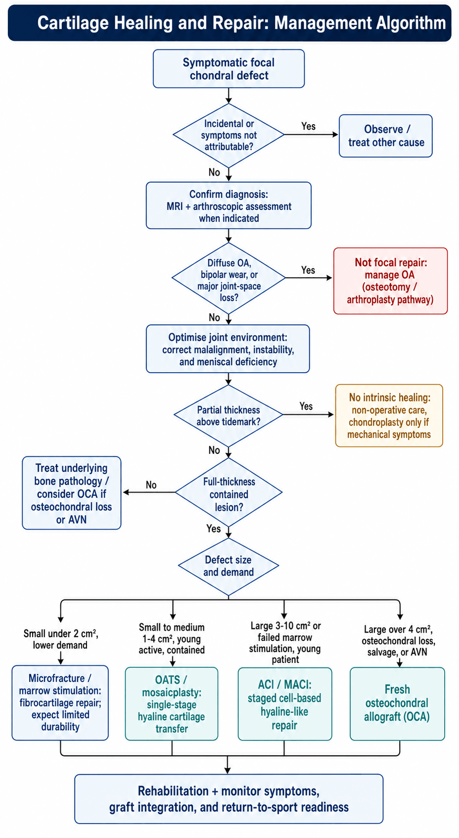
Management Algorithm
Why Cartilage Cannot Heal (AAAA)
- Avascular - no blood supply
- Aneural - no nerve supply
- Alymphatic - no lymphatics
- Amitotic - minimal cell division
Composition (WATER)
- Water 65-80%
- Aggrecan (proteoglycan)
- Type II collagen
- ECM 95% of volume
- Rare cells (chondrocytes 5%)
Repair Tissue Comparison
- Hyaline = Type II collagen (native)
- Fibrocartilage = Type I collagen (repair)
- Fibrocartilage biomechanically inferior
- Deteriorates under load over time
Treatment Options (MOCHA)
- Microfracture - marrow stim, fibrocartilage
- OATS - autograft, hyaline, single stage
- Cell-based (ACI/MACI) - hyaline-like
- Hyaluronic scaffolds - matrix-assisted
- Allograft (OCA) - large defects