Craniocervical and Subaxial Instability
- ADI (Atlantodental Interval): Normal is less than 3mm adults, less than 5mm children.
- PADI (Posterior ADI): Less than 14mm indicates cord compromise risk.
- Powers Ratio: Ratio greater than 1 indicates anterior OC-C1 dislocation.
- White-Panjabi Criteria: Score of 5 or greater indicates subaxial instability.
- RA patients: Must screen for AAI before anesthesia/intubation.
- “ADI greater than 3mm in adults is abnormal
- “PADI less than 14mm predicts poor neurological outcome
- “Powers ratio: BC/OA (greater than 1 = anterior dislocation)
- “Always screen RA and Down syndrome patients
- “Harms technique: C1 lateral mass + C2 pedicle screws
Clinical Imaging
Imaging Atlas


Adult: less than 3mm is Normal. Child: less than 5mm is Normal. Reflects transverse ligament integrity.
less than 14mm: Critical threshold for cord compromise. PADI is more prognostic than ADI.
Powers Ratio greater than 1: Anterior dislocation. BDI greater than 12mm: Abnormal Basion-Dens Interval.
Translation: greater than 3.5mm. Angulation: greater than 11 degrees. (White-Panjabi Criteria)
At a Glance
Cervical instability refers to abnormal motion between vertebrae that may cause neurological compromise, occurring at the craniocervical junction (OC-C1-C2) or subaxially (C3-C7). Key measurements include the ADI (less than 3mm adults, less than 5mm children) reflecting transverse ligament integrity, PADI less than 14mm indicating cord compromise risk, and Powers ratio greater than 1 for anterior OC-C1 dislocation. Rheumatoid arthritis patients have 25-80% prevalence of atlantoaxial instability and require preoperative screening before anesthesia. White-Panjabi criteria (score ≥5) diagnose subaxial instability. Surgical stabilization (e.g., Harms technique C1 lateral mass + C2 pedicle screws) prevents neurological deterioration.
ADIADI Values - AC/DC
Hook:AC/DC - Adult 3, Child 5, Danger at 14 (Cord)
PADSWhite-Panjabi Criteria - PADS
Hook:PADS - 5 Points And you have instability (greater than 5 = unstable)
OCOC-C1 Measurements - POB
Hook:POB - Powers, Occipital, Basion: Key craniocervical measurements
Overview/Epidemiology
Cervical instability is defined as abnormal motion of the cervical spine that results in neurological compromise, incapacitating pain, or structural deformity under physiological loads.
Epidemiology:
- Trauma: Most common cause overall - MVA, falls, sports
- Rheumatoid arthritis: 25-80% develop AAI over disease course
- Down syndrome: 15-20% have AAI on screening radiographs
- Congenital: Os odontoideum, basilar invagination, occipitalization of C1
- Tumors/Infections: Destabilize through bone destruction
Clinical Significance:
- OC-C1 dislocation has 70% pre-hospital mortality
- Untreated AAI can progress to myelopathy and quadriplegia
- Early recognition and stabilization prevents neurological decline
Anatomy
Upper Cervical Anatomy (OC-C1-C2):
Bony Structures:
- Occiput: Occipital condyles articulate with C1 lateral masses
- Atlas (C1): Ring-shaped, no vertebral body, lateral mass articulations
- Axis (C2): Odontoid process (dens) is the pivot for rotation
- C1-C2 articulation: Allows 50% of cervical rotation
Ligamentous Stabilizers:
- Transverse ligament: Primary restraint to anterior C1 translation
- Alar ligaments: Limit rotation and lateral bending
- Apical ligament: Connects dens tip to basion
- Tectorial membrane: Extension of PLL, covers dens posteriorly
- Cruciate ligament: Transverse + vertical bands
Subaxial Anatomy (C3-C7):
Stabilizing Structures:
- Anterior: ALL, disc, vertebral body
- Middle: PLL, posterior disc, posterior body
- Posterior: Facet capsules, ligamentum flavum, interspinous ligaments
Denis Three-Column Model applies but White-Panjabi criteria more specific for cervical.
Pathophysiology
Traumatic Instability
Mechanism:
- High-energy trauma causes ligamentous disruption or fracture
- Flexion-distraction injuries disrupt posterior ligaments
- Extension injuries disrupt anterior structures
- Rotation and lateral bending cause facet injuries
Patterns:
- OC-C1 dislocation: Usually fatal; survivors have tectorial membrane disruption
- Odontoid fractures: Type II most common, highest nonunion rate
- Hangman's fracture: Bilateral C2 pars fracture
- Facet dislocations: Unilateral (25% subluxation) or bilateral (50% subluxation)
Neurological Compromise Mechanism:
The spinal cord at C1-C2 level is approximately 10mm in diameter. The canal normally provides approximately 20mm of space.
Steel's Rule of Thirds at C1:
- 1/3 dens
- 1/3 cord
- 1/3 space (buffer)
When PADI (space available for cord) drops below 14mm, cord compression becomes likely. Myelopathy results from:
- Direct mechanical compression
- Vascular compromise (anterior spinal artery)
- Dynamic cord impingement with flexion/extension
Classification
Upper Cervical Instability (OC-C1-C2)
Occipitoatlantal (OC-C1) Dislocation:
- Type I: Anterior dislocation (most common in survivors)
- Type II: Longitudinal distraction
- Type III: Posterior dislocation
Atlantoaxial Instability (C1-C2):
- Anterior subluxation: ADI greater than 3mm (most common)
- Posterior subluxation: Rare, associated with dens fracture
- Rotatory subluxation: Fielding-Hawkins Types I-IV
- Lateral translation: Associated with Jefferson fracture
Fielding-Hawkins Rotatory Subluxation:
- Type I: Rotatory fixation without anterior shift
- Type II: Rotatory fixation with 3-5mm anterior shift
- Type III: Rotatory fixation with greater than 5mm shift
- Type IV: Rotatory fixation with posterior shift

Clinical Presentation
History:
- Mechanism of injury (trauma)
- Neck pain, especially with motion
- Myelopathic symptoms: weakness, numbness, gait disturbance
- Electric shock sensation with flexion (Lhermitte's sign)
- History of RA, Down syndrome, or connective tissue disorder
Neurological Assessment
Upper Motor Neuron Signs (Myelopathy):
- Hyperreflexia
- Hoffmann sign positive
- Ankle clonus
- Babinski sign positive
- Spastic gait
- Inverted radial reflex
Motor Examination:
- Test all myotomes C5-T1
- Assess grip strength
- Intrinsic hand muscle wasting
Sensory:
- Dermatomal assessment
- Proprioception (posterior columns)
- Cape-like sensory loss (central cord)
Gait:
- Spastic, wide-based
- Difficulty with tandem walking
Investigations
Plain Radiographs
Standard Views:
- AP, lateral, open mouth odontoid
- Flexion-extension laterals (supervised, if neurologically intact)
Key Measurements:
- Normal
- Less than 3mm
- Abnormal
- Greater than 3mm
- Normal
- Less than 5mm
- Abnormal
- Greater than 5mm
- Normal
- Greater than 14mm
- Abnormal
- Less than 14mm
- Normal
- Less than 1
- Abnormal
- Greater than 1
- Normal
- Less than 12mm
- Abnormal
- Greater than 12mm
- Normal
- Less than 12mm
- Abnormal
- Greater than 12mm
Powers Ratio (BC/OA):
- BC = basion to posterior C1 arch
- OA = opisthion to anterior C1 arch
- Greater than 1 = anterior OC-C1 dislocation
- Less than 0.7 = posterior dislocation
Flexion-extension views are essential but should only be done with supervision if patient is neurologically intact.

Management
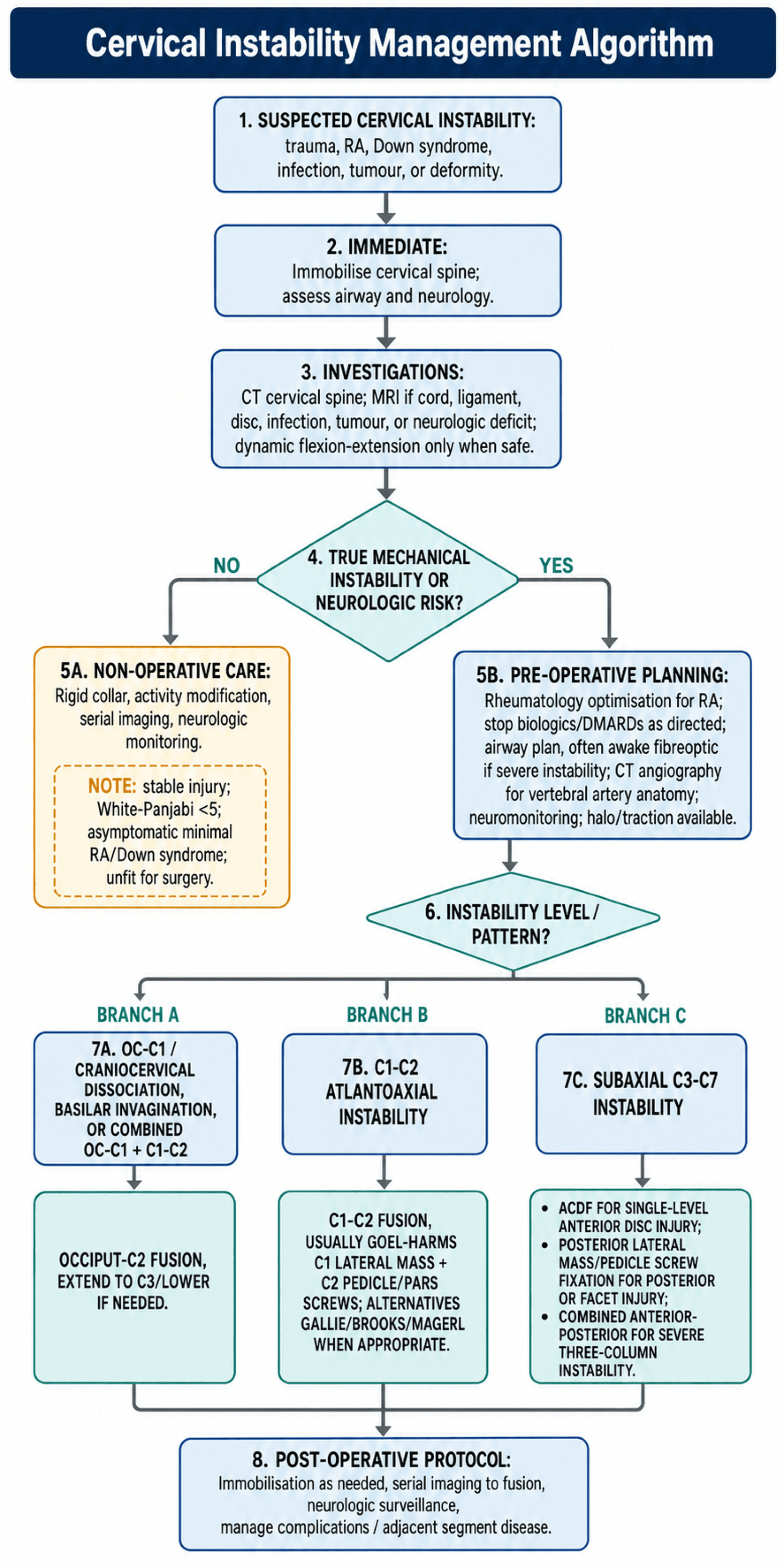
Pre-operative Planning:
- RA patients need pre-op rheumatology optimization
- Stop biologics/DMARDs as directed
- Airway plan with anesthesia (awake fiberoptic if severe instability)
- Prepare for prone vs lateral positioning
- Have halo available if needed for intra-op traction
Non-operative Management
Indications:
- Stable injuries (White-Panjabi less than 5)
- Asymptomatic RA with minimal ADI increase
- Asymptomatic Down syndrome with AAI on screening
- Patient unfit for surgery
Treatment:
- Rigid cervical collar (Miami J, Aspen)
- Activity modification
- Serial imaging to monitor progression
- Neurological monitoring
Duration:
- 6-12 weeks for stable injuries
- Indefinite monitoring for RA/Down syndrome
Limitations:
- True instability rarely heals without surgery
- High failure rate for type II odontoid in elderly

Complications
Perioperative Complications
Neurological:
- Cord injury (rare, less than 1%)
- Root injury (C2 - occipital numbness)
- Worsening myelopathy
Vascular:
- Vertebral artery injury (2-4% with C1-C2 screws)
- May be asymptomatic if contralateral dominant
- Cerebellar stroke if bilateral injury
Airway:
- Post-op swelling and airway compromise
- Delayed extubation often needed
- Consider ICU monitoring
Positioning:
- Pressure sores (prone positioning)
- Brachial plexus injury
- Peripheral nerve injury
Complication Prevention:
- Pre-operative CT angiography for vertebral artery dominance
- Neuromonitoring (SSEPs, MEPs) intraoperatively
- Meticulous technique with anatomical landmarks
- Appropriate collar immobilization post-op
Differential Diagnosis
The key task is to separate true mechanical instability from mimics that cause neck pain or myelopathic signs without abnormal segmental motion.
| 0 | 1 | 2 |
|---|---|---|
| True cervical instability | Abnormal segmental motion under physiological load | Dynamic flexion-extension imaging, ADI/PADI, White-Panjabi |
| Cervical spondylotic myelopathy | Static canal stenosis, no abnormal motion | MRI cord signal, static canal diameter |
| Acute disc herniation | Radiculopathy or myelopathy without translation | MRI disc, neutral alignment |
| Pseudosubluxation (children) | Physiological C2-C3 / C3-C4 step-off that reduces on extension | Swischuk spinolaminar line |
| Os odontoideum | Fixed ossicle, may mimic acute fracture | CT (corticated margins) vs fracture |
| Ankylosing spondylitis fracture | Rigid fused spine, highly unstable transverse fracture | CT/MRI whole spine, low threshold |
| Klippel-Feil with adjacent hypermobility | Congenital fusion shifting motion to open segments | Dynamic radiographs of mobile levels |
Controversies & Areas of Uncertainty
- Routine flexion-extension radiographs for Down syndrome sport clearance — historically mandated, now widely questioned because asymptomatic radiographic AAI poorly predicts catastrophic injury; many bodies emphasise clinical screening and symptom-directed imaging instead.
- Powers ratio vs modern measurements — the Powers ratio is insensitive for non-anterior and distraction injuries; CT-based BDI/BAI (Harris lines) and the condyle-C1 interval are more reliable for craniocervical dissociation.
- Type II odontoid fracture in the elderly — operative vs non-operative management remains debated, balancing high non-union rates against the morbidity of surgery and halo immobilisation; fibrous stable non-union may be an acceptable endpoint.
- C2 fixation choice — pedicle vs pars vs translaminar screws are selected by vertebral artery anatomy on pre-operative CT angiography; no single technique is universally superior.
- Closed reduction before MRI in facet dislocations — awake serial traction reduction is advocated by some in alert, cooperative patients, but many centres mandate pre-reduction MRI to exclude a herniated disc that could be driven into the cord.
- Defining instability — White-Panjabi and SLIC are checklists, not validated thresholds for every scenario; clinical judgement integrating dynamic imaging, neurology, and patient factors remains essential.
Evidence Base
- 73 RA patients; PADI correlated with paralysis whereas anterior ADI did not
- All Class III patients had PADI/subaxial canal under 14mm; PADI best predicted recovery
- C1 lateral mass + C2 pars/pedicle polyaxial screws with rods; 37 patients, solid fusion in all
- No neural or vascular injury; allows intra-operative reduction unlike transarticular screws
- 10 cadaveric spines; both screw constructs reduced motion more than Gallie wiring alone
- No significant stability difference between screw-rod and transarticular constructs
- 30 atlantoaxial dislocations fixed via lateral mass plate-and-screw; 100% union
- No morbidity, mortality, or implant failure; rigid direct C1-C2 segmental fixation
- 404 screened; 14.6% had AAI (13.1% asymptomatic, only 1.5% symptomatic needing surgery)
- ADI greatest in flexion; follow-up showed no significant progression
- Defined the Powers ratio (BC/OA); ratio greater than 1 indicates anterior OC-C1 dislocation
- Created for immediate radiographic recognition of an often-fatal injury
- SLIC scores morphology, discoligamentous complex, and neurology (ICC 0.49-0.90)
- Total score greater than 4 favours surgery; under 4 favours non-operative care
- Point-based subaxial checklist; translation over 3.5mm and angulation over 11 degrees each score 2
- Total score of 5 or greater defines clinical instability
Viva Scenarios
Practise clinical reasoning and management decisions out loud
“55-year-old female with 20-year history of rheumatoid arthritis presents with progressive neck pain, electric shock sensation down her spine when flexing her neck, and difficulty with fine motor tasks in her hands. She is on methotrexate and a TNF-inhibitor. How would you assess and manage this patient?”
This patient likely has **atlantoaxial instability (AAI)** from rheumatoid arthritis, given her Lhermitte's sign and hand dysfunction suggesting myelopathy.
Assessment:
- Full neurological exam - document myelopathic signs (hyperreflexia, Hoffmann's, gait)
- Lateral cervical flexion-extension X-rays - measure ADI (abnormal if greater than 3mm) and PADI (critical if less than 14mm)
- CT cervical spine - bony detail, rule out vertical settling
- MRI cervical spine - assess pannus, cord compression, cord signal change
Management:
- If PADI less than 14mm or symptomatic myelopathy, surgical stabilization is indicated
- Pre-op: Rheumatology consult to optimize disease control, stop biologics 2-4 weeks pre-op
- Anesthesia consult - plan for awake fiberoptic intubation given cervical instability
- Surgery: Posterior C1-C2 fusion using Harms technique (C1 lateral mass screws + C2 pedicle screws)
“Parents of a 12-year-old boy with Down syndrome bring him for clearance to participate in school gymnastics. They have heard about neck problems in Down syndrome. How do you approach this?”
This is a common clinical scenario. Children with Down syndrome have a 15-20% prevalence of atlantoaxial instability due to ligamentous laxity and dens hypoplasia.
Assessment:
- History: Any neck pain, torticollis, neurological symptoms (weakness, gait changes)
- Examination: Full neurological exam, check for myelopathic signs
- Imaging: Lateral cervical X-ray with flexion-extension views - measure ADI
- Normal ADI in children: less than 5mm
Management:
- If asymptomatic and ADI less than 5mm: Can participate in sports with activity modification
- Avoid high-risk activities: diving, gymnastics floor exercises, contact sports
- If ADI greater than 5mm but asymptomatic: Restrict from high-risk activities, annual monitoring
- If symptomatic or ADI greater than 10mm: Surgical stabilization indicated
“24-year-old restrained driver involved in high-speed head-on MVA. GCS 15, complaining of severe neck pain. Initial trauma CT shows 'possible craniocervical dissociation'. How do you assess and manage?”
Occipitoatlantal (OC-C1) dislocation is often fatal at scene. Survivors need urgent stabilization to prevent catastrophic neurological deterioration.
Immediate Management:
- Maintain cervical immobilization - rigid collar, sandbags
- Do NOT apply traction - can cause distraction injury
- Full ATLS assessment for associated injuries
Imaging Review - Key Measurements:
- Powers Ratio (BC/OA): greater than 1 = anterior OC-C1 dislocation
- Basion-Dens Interval (BDI): greater than 12mm = abnormal
- Basion-Axis Interval (BAI): greater than 12mm = abnormal
- Condyle-C1 Interval (CCI): greater than 2mm = abnormal
Management:
- MRI: Assess ligamentous injury, cord status
- If craniocervical dissociation confirmed: Surgical stabilization indicated
- Technique: Occiput to C2 posterior fusion (may extend to C3 if needed)
- Consider halo for temporary immobilization while optimizing patient
“32-year-old male dove into shallow water and now has severe neck pain and bilateral arm weakness. X-ray shows C6 anterolisthesis on C7. How do you manage this patient?”
This patient has a likely bilateral facet dislocation at C6-C7 with incomplete spinal cord injury (arm weakness suggests central cord component).
Initial Management:
- Immobilize in rigid collar
- Complete neurological assessment and document (ASIA score)
- CT cervical spine - confirm facet dislocation, assess for fractures
- MRI - assess cord compression, disc herniation, cord signal change
Key Decision - MRI First:
- Obtain MRI before reduction to rule out disc herniation
- If large disc herniation, may need anterior discectomy before posterior reduction
- If no significant disc, can proceed with closed or open reduction
Treatment:
- Closed reduction with Gardner-Wells tongs and sequential weights (if awake, cooperative)
- If closed reduction fails or disc herniation: Anterior discectomy and reduction, then anterior fusion
- Alternatively: Posterior open reduction and lateral mass screw fixation
- Consider combined approach for 3-column instability
MCQ Practice Points
Q: What is the normal atlantodental interval (ADI) in adults? A: Less than 3mm. ADI 3-5mm indicates transverse ligament incompetence; greater than 5mm indicates both transverse and alar ligament failure. In children, normal ADI can be up to 5mm due to ligamentous laxity.
Q: What PADI measurement indicates the spinal cord is at risk? A: Posterior Atlantodental Interval (PADI) less than 14mm predicts neurological deficit. PADI represents the space available for the cord (SAC) at C1-2 and is more predictive of myelopathy than ADI.
Q: How is the Powers Ratio calculated and what does a ratio greater than 1 indicate? A: Powers Ratio = BC/OA where B = basion, C = posterior arch C1, O = opisthion, A = anterior arch C1. Ratio greater than 1 indicates anterior occipito-atlantal dislocation. Normal ratio is 0.77 (range 0.55-1.0).
Q: Which patient populations require routine screening for atlantoaxial instability? A: Down syndrome (trisomy 21) and rheumatoid arthritis patients require screening. Down syndrome patients have 15-20% incidence of atlantoaxial instability due to ligamentous laxity. RA patients develop pannus erosion of the transverse ligament.
Guidelines, Registries & Global Practice
Global epidemiology:
- Trauma is the leading cause worldwide, with motor-vehicle and fall mechanisms predominating in high-income settings and a higher proportion of high-energy/road-traffic trauma in low- and middle-income countries.
- Rheumatoid arthritis historically caused cervical instability in 25-80% over the disease course; the incidence of severe craniocervical disease has fallen markedly in regions with early access to modern DMARDs and biologics.
- Down syndrome screening series report atlantoaxial instability in roughly 10-20%, the large majority asymptomatic (Pueschel and Scola: 14.6%).
Side-by-side guidance:
- Position
- Subaxial trauma stratified by morphology, discoligamentous complex, neurology; score over 4 favours surgery
- Position
- Major-trauma cervical clearance pathways; MRI before reduction where ligamentous or disc injury is suspected
- Position
- Emphasis on PADI and dynamic imaging for rheumatoid AAI; surgery before neurological decline
- Position
- Down syndrome: clinical screening emphasised; routine flexion-extension radiographs for sport no longer universally mandated
Registry and outcome notes:
- Craniocervical and upper-cervical fusion volumes are low, so high-quality evidence is dominated by single-centre series and biomechanical studies rather than large registries.
- Spine registries (e.g. national spine surgery and trauma databases in the UK, US, Scandinavia) increasingly track subaxial fusion outcomes, adjacent-segment disease, and revision rates.
High- vs limited-resource practice:
- Where CT angiography, intra-operative neuromonitoring, and navigation are available, screw-based constructs (Goel-Harms, lateral mass, pedicle) are standard.
- In limited-resource settings, wiring techniques, halo immobilisation, and traction-based reduction retain a larger role; awake intubation and meticulous landmark-based technique remain universally applicable.
KEY MEASUREMENTS
- ADI: less than 3mm (adult), less than 5mm (child) = normal
- PADI: less than 14mm = cord at risk
- Powers Ratio: greater than 1 = anterior OC-C1 dislocation
- BDI/BAI: greater than 12mm = craniocervical dissociation
- Translation greater than 3.5mm = subaxial instability
- Angulation greater than 11 degrees = subaxial instability
WHITE-PANJABI CRITERIA
- Score 5 or greater = clinically unstable
- Anterior elements destroyed = 2 points
- Posterior elements destroyed = 2 points
- Translation greater than 3.5mm = 2 points
- Angulation greater than 11 degrees = 2 points
- Cord damage = 2 points
SURGICAL TECHNIQUES
- Harms: C1 lateral mass + C2 pedicle screws (standard for AAI)
- Magerl: Transarticular C1-C2 screws (higher VA risk)
- Gallie: Wire + graft (supplements screws)
- OC-C2 fusion: For OC-C1 instability
- ACDF: Subaxial anterior approach
- Lateral mass screws: C3-C6 posterior
RA CERVICAL DISEASE
- AAS (atlantoaxial subluxation) = 65%
- SAS (subaxial subluxation) = 20%
- Basilar invagination = 15% (most dangerous)
- PADI less than 14mm = operate before neuro decline
- Stop biologics 2-4 weeks pre-op
- Awake fiberoptic intubation if severe