Progressive UMN Signs | Cord Compression | Surgical Decompression
- Progressive disease - natural history is stepwise or gradual decline without treatment
- MRI is gold standard - shows cord compression and T2 signal change (edema/gliosis)
- Upper motor neuron signs - hyperreflexia, Hoffman sign, Babinski, inverted radial reflex
- Surgery halts progression - improves or stabilizes symptoms in 70-80% of patients
- Surgical timing matters - early intervention (Nurick 1-2) has better outcomes than late (4-5)
- “Myelopathy = UMN signs; radiculopathy = LMN signs (important distinction)
- “Lhermitte sign (neck flexion causes electric shock down spine) suggests cord compression
- “Hoffman sign (flick middle finger, thumb flexes) is sensitive but not specific
- “T2 hyperintensity on MRI correlates with worse prognosis and poor recovery
- “AOSpine prospective studies: surgical decompression improves function across all severity grades
Clinical Imaging
Imaging Atlas

Myelopathy is UMN (hyperreflexia, Hoffman, Babinski, clonus, spasticity). Radiculopathy is LMN (hyporeflexia, weakness, dermatomal sensory loss). Can coexist as myeloradiculopathy. The level of cord compression determines which nerve roots are affected (LMN at level, UMN below).
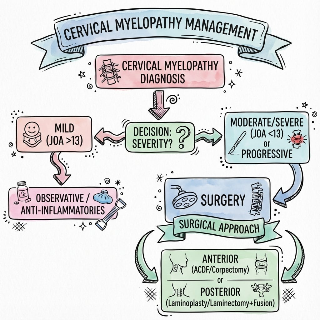
Natural history is progressive decline. Stepwise deterioration (20%) or gradual decline (60%). Only 20% remain stable without treatment. Surgery halts progression in most cases, with improvement in 50-70% if operated early.
Early surgery (Nurick 1-2) yields better outcomes than late surgery (Nurick 4-5). Severe myelopathy (Nurick 5, T2 hyperintensity, long symptom duration over 18 months) predicts poor recovery. Operate before irreversible cord damage occurs.
Anterior approach: 1-2 levels, anterior compression (disc, osteophyte). Posterior approach: multilevel (more than 3 levels), congenital stenosis, OPLL. Combined approach: severe multilevel with kyphosis. Comparative data show no consistent neurological advantage of one approach when each is matched to the appropriate compression pattern.
| Scenario | Compression Pattern | Approach | Key Pearl |
|---|---|---|---|
| Single level C5-6 disc herniation | Anterior compression | ACDF (anterior cervical discectomy fusion) | Gold standard for 1-2 level anterior pathology |
| Multilevel stenosis (C3-7), lordotic spine | Circumferential compression | Laminoplasty or laminectomy + fusion | Posterior approach preserves motion with laminoplasty |
| Severe OPLL (more than 60% canal), ossified | Anterior compression, multilevel | Posterior laminoplasty (avoid anterior if OPLL thick) | Anterior approach risks CSF leak with OPLL removal |
| Multilevel stenosis with kyphosis | Circumferential, deformity | Combined anterior + posterior or PCF alone | Correct deformity to prevent progression |
HOFFMANClinical Signs of Cervical Myelopathy
Hook:HOFFMAN is the classic myelopathy sign - remember it encompasses all UMN features of cord compression!
GRADESNurick Grading Scale for Myelopathy Severity
Hook:GRADES 0 to 5: remember the Nurick scale progresses from no cord signs to complete disability!
SIGNALMRI Findings in Cervical Myelopathy
Hook:SIGNAL changes on T2 MRI tell the story of cord compression and predict surgical outcome!
OPERATEIndications for Surgery in Cervical Myelopathy
Hook:OPERATE criteria guide surgical decision-making - do not wait for severe disability before intervening!
Handy Legs Walk BadlyProgression of Myelopathy Symptoms
Hook:Myelopathy progresses from hands to legs to gait to bladder!
Canal is Half the BodyStenosis Measurement (Pavlov Ratio)
Hook:Pavlov ratio = Canal / Body. Less than 0.8 = stenosis, less than 1.0 = borderline!
VITAMINSDifferential Diagnosis of Myelopathy
Hook:Rule out VITAMINS before diagnosing spondylotic myelopathy!
Overview and Epidemiology
Cervical myelopathy is the most common cause of spinal cord dysfunction in adults over 55 years. It results from chronic compression of the cervical spinal cord, most commonly due to degenerative changes (spondylosis, disc herniation, ligamentum flavum hypertrophy, OPLL). The natural history without surgery is progressive neurological decline in 80% of cases. Early surgical decompression halts progression and improves outcomes in 70-80% of patients.
- Peak age 50-60 years (degenerative), younger with trauma or congenital stenosis
- Men more than women (2:1 ratio) due to higher rates of cervical spondylosis
- Most common levels: C5-6 and C6-7 (maximum motion and degeneration)
- Risk factors: congenital stenosis (canal less than 13mm AP), occupation (repetitive neck flexion/extension)
- Progressive disability: 60% gradual decline, 20% stepwise deterioration, 20% stable
- Functional impairment: gait disturbance, hand clumsiness (buttoning, writing), falls
- Surgical outcomes: 50-70% improve, 20-30% stabilize, 5-10% worsen despite surgery
- Predictors of poor recovery: severe myelopathy (Nurick 4-5), T2 hyperintensity, long symptom duration (more than 18 months)
Pathophysiology and Anatomy
The cervical spinal cord occupies approximately 60-70% of the spinal canal cross-sectional area. Normal AP canal diameter is 14-17mm. When the canal narrows to less than 13mm (relative stenosis) or less than 10mm (absolute stenosis), the cord is at risk of compression. Compression can be static (anatomical narrowing) or dynamic (worsened with neck flexion/extension). The pathophysiology involves:
- Direct mechanical compression - squeezing of cord parenchyma
- Ischemia - compression of anterior spinal artery or intramedullary vessels
- Chronic repetitive trauma - microtrauma with neck motion leading to gliosis and myelomalacia

| Cause | Mechanism | Typical Age | Treatment Approach |
|---|---|---|---|
| Cervical spondylosis | Degenerative: disc bulging, osteophytes, facet hypertrophy | More than 50 years | Anterior (1-2 levels) or posterior (multilevel) |
| OPLL (ossification of PLL) | Heterotopic ossification of posterior longitudinal ligament | East Asian, more than 50 years | Posterior laminoplasty preferred (avoid anterior CSF leak risk) |
| Congenital stenosis | Developmental narrow canal (Pavlov ratio less than 0.8) | Younger age, trauma | Posterior decompression if symptomatic |
| Disc herniation (acute) | Posterior disc extrusion compressing cord | 40-60 years | Anterior discectomy and fusion |
| Ligamentum flavum hypertrophy | Posterior compression from ligament thickening | Elderly | Posterior laminectomy or laminoplasty |
The cervical cord is most vulnerable at C5-6 and C6-7 due to maximal motion and narrowest canal diameter at these levels. The cord's blood supply is watershed between anterior spinal artery (80% of cord) and posterior spinal arteries (20%). Compression can cause ischemia and infarction, leading to irreversible damage.
Flexion narrows the canal anteriorly (ligamentum flavum buckles posteriorly). Extension narrows posteriorly (disc and osteophytes protrude anteriorly). This pincer effect causes repetitive microtrauma with daily activities, explaining the progressive nature of myelopathy.
Classification Systems
Nurick Scale - Functional Impairment (Most Commonly Used)
| Grade | Clinical Presentation | Gait | Surgical Indication |
|---|---|---|---|
| 0 | Root signs only, no cord signs | Normal | Treat radiculopathy, observe for myelopathy |
| 1 | Cord signs present, no gait difficulty | Normal | Consider early surgery (controversial) |
| 2 | Mild gait difficulty, employment possible | Abnormal but independent | Surgery recommended |
| 3 | Gait difficulty prevents employment | Abnormal, independent in ADLs | Surgery indicated |
| 4 | Unable to walk without assistance | Requires aid (cane/walker) | Urgent surgery |
| 5 | Chair-bound or bedridden | Non-ambulatory | Urgent surgery, poor recovery expected |
The Nurick scale is functional and focuses on gait and employment. It is the most widely used grading system for myelopathy severity and correlates with surgical outcomes. Grades 2-3 are clear surgical indications. Grade 1 is controversial - some advocate early surgery, others observe. Grades 4-5 have poorer outcomes but surgery still halts progression.
Clinical Assessment
- Progressive symptoms: worsening over weeks to months (not acute unless trauma)
- Hand clumsiness: difficulty with fine motor tasks (buttoning, writing, picking up coins)
- Gait disturbance: wide-based gait, unsteadiness, frequent falls
- Neurogenic bladder: urgency, frequency, hesitancy (advanced disease)
- Lhermitte sign: electric shock sensation down spine with neck flexion
- Pain is NOT a prominent feature (unlike radiculopathy) - if severe neck/arm pain, think radiculopathy or mixed picture
- Upper motor neuron signs: hyperreflexia, Hoffman sign, Babinski sign, clonus (ankle/patella)
- Inverted radial reflex: tap brachioradialis, finger flexion occurs instead of forearm flexion (C5-6 level lesion)
- Gait assessment: tandem gait (balance), heel-toe walk, Romberg sign (posterior column dysfunction)
- Hand coordination: rapid alternating movements, finger tapping (assess speed and coordination)
- Sensory level: may have dermatomal level or glove-and-stocking neuropathy pattern
- Grip and release test: inability to rapidly open/close fist 20 times in 10 seconds suggests myelopathy
Elderly patients with diabetes or alcohol use may have superimposed myelopathy that is missed because their hand numbness is attributed to peripheral neuropathy. Key distinguishing features:
- Myelopathy: UMN signs (hyperreflexia, Hoffman, Babinski), gait disturbance, MRI shows cord compression
- Peripheral neuropathy: LMN signs (hyporeflexia), stocking-glove distribution, nerve conduction studies abnormal, MRI normal
Clinical pearl: If a patient with "peripheral neuropathy" has brisk reflexes and Hoffman sign, obtain a cervical spine MRI to rule out myelopathy!
| Feature | Myelopathy (UMN) | Radiculopathy (LMN) | Mixed (Myeloradiculopathy) |
|---|---|---|---|
| Reflexes | Hyperreflexia, clonus | Hyporeflexia or absent | Hyperreflexia below level, hyporeflexia at level |
| Hoffman sign | Positive (flick middle finger, thumb flexes) | Negative | Positive (indicates cord involvement) |
| Babinski sign | Positive (upgoing toe) | Negative (downgoing toe) | Positive (UMN tract involvement) |
| Sensory pattern | Sensory level, proprioception loss, gait ataxia | Dermatomal numbness | Both patterns present |
| Pain | Minimal or absent (dull neck ache) | Severe radicular pain (shooting down arm) | Radicular pain plus myelopathy signs |
| Gait | Wide-based, ataxic, spastic | Normal | Abnormal gait |
| Treatment urgency | Urgent if progressive | Elective (most resolve non-operatively) | Urgent (myelopathy component dictates) |
| Condition | Distinguishing Features | Key Investigation |
|---|---|---|
| Motor neuron disease (ALS) | Mixed UMN and LMN signs, fasciculations, NO sensory loss, bulbar involvement, normal cord on MRI | EMG/nerve conduction studies (denervation), clinical pattern |
| Multiple sclerosis / demyelination | Younger patient, relapsing-remitting course, optic neuritis, brain lesions, Lhermitte sign | MRI brain and whole spine, CSF oligoclonal bands |
| Subacute combined degeneration (B12 deficiency) | Dorsal column + corticospinal signs, peripheral neuropathy, macrocytic anaemia, glossitis | Serum B12, methylmalonic acid, homocysteine |
| Copper deficiency myelopathy | Mimics B12 picture; history of bariatric surgery, zinc excess, malabsorption | Serum copper and caeruloplasmin |
| Intramedullary or extramedullary tumour | Progressive, may have nocturnal pain, level-specific deficit; ependymoma/astrocytoma/meningioma/metastasis | MRI with contrast |
| Syringomyelia | Dissociated (cape) sensory loss, may follow Chiari malformation or trauma | MRI showing intramedullary syrinx |
| Spinal cord infarction (anterior spinal artery) | Acute onset, motor and spinothalamic loss with preserved proprioception | MRI with diffusion, vascular risk assessment |
| Infective / inflammatory myelitis (e.g. transverse myelitis, epidural abscess) | Subacute, fever or sepsis features (abscess), enhancing cord lesion | MRI with contrast, inflammatory markers, CSF |
Investigations

Diagnostic Imaging Protocol
Purpose: Assess alignment, instability, bony pathology
- Lateral: Measure Pavlov ratio (canal diameter / vertebral body diameter). Normal more than 1.0, stenotic less than 0.8
- Flexion/Extension: Assess for dynamic instability (more than 3mm subluxation or more than 11 degrees angulation)
- AP: Look for degenerative changes, osteophytes
- Limitations: Cannot visualize cord, disc, or soft tissue compression
Purpose: Visualize cord compression, cord signal changes, disc pathology
- T2 sagittal: Shows cord compression, canal stenosis, disc bulging
- T2 axial: Shows cord deformity and T2 hyperintensity (edema/gliosis - poor prognostic sign)
- T1 sagittal: Shows T1 hypointensity (myelomalacia - severe chronic compression)
- Interpretation: Cord compression + T2 signal change + clinical myelopathy = surgical indication
Purpose: Bony detail, OPLL assessment, preoperative planning
- When indicated: OPLL suspected (assess ossification thickness), preoperative planning for screw trajectories
- CT myelogram: If MRI contraindicated (pacemaker, claustrophobia), shows cord compression but not intrinsic signal
Purpose: Assess dynamic compression not visible on neutral MRI
- Rationale: Some patients have cord compression only in flexion or extension
- Limitations: Not widely available, increased cost
Purpose: Assess cord function, intraoperative monitoring
- Preoperative: Can confirm diagnosis if MRI equivocal (prolonged latency or reduced amplitude)
- Intraoperative: Monitor cord function during decompression (more than 50% amplitude drop = cord ischemia risk)
T2 hyperintensity (bright signal on T2 within the cord) indicates:
- Edema (early, reversible) or gliosis/myelomalacia (late, irreversible)
- Prognostic marker: T2 hyperintensity predicts poorer surgical outcomes
- Snake-eye sign: Bilateral symmetric T2 hyperintensity on axial MRI at grey matter, suggests severe chronic compression and very poor prognosis
T1 hypointensity (dark signal on T1) indicates:
- Myelomalacia (cord necrosis) - irreversible damage
- Very poor prognosis for neurological recovery
Clinical correlation: Severity of T2 changes correlates with Nurick grade and recovery potential. Early surgery (before T2 changes develop) yields best outcomes.
Management Algorithm
Conservative Management - Limited Role
80% of patients with cervical myelopathy will progress without surgery (60% gradual decline, 20% stepwise deterioration). Only 20% remain stable. Therefore, observation is only appropriate in very select cases:
- Nurick 0-1 (minimal symptoms, no gait disturbance)
- Patient unfit for surgery (severe medical comorbidities)
- Patient refuses surgery after informed consent about natural history
Conservative Measures (Temporizing or Palliative)
Soft collar for symptom relief during acute exacerbation. Avoid prolonged use (causes muscle atrophy and stiffness). Not a long-term solution for cord compression.
- Neuropathic pain: Gabapentin, pregabalin (for coexisting radiculopathy)
- Spasticity: Baclofen, tizanidine (for UMN spasticity)
- No role for NSAIDs or steroids in chronic degenerative myelopathy (unlike acute cord injury)
Goal: Maintain ROM, strengthen supporting musculature, gait training
- Does NOT halt progression of cord compression
- Avoid manipulation or traction - risk of worsening cord compression
Clinical assessment: Nurick grade, mJOA score, gait, UMN signs Repeat MRI if worsening: Assess progression of cord compression Threshold for surgery: If symptoms progress, convert to surgical management
Observation is reasonable for Nurick 0-1 IF:
- MRI shows mild compression without T2 signal change
- Patient is high surgical risk (severe cardiac/pulmonary disease)
- Informed consent given about natural history (80% progression risk)
Surgery is indicated even in Nurick 1 IF:
- T2 signal change present (suggests cord edema/gliosis)
- Progression documented (worsening symptoms over months)
- Young patient with long life expectancy
Surgical Techniques - Anterior Approaches
Anterior Cervical Discectomy and Fusion (ACDF)
Indications:
- 1-2 level anterior compression (disc herniation, osteophyte)
- Focal cord compression at specific levels
- Most common surgical treatment for cervical myelopathy
ACDF Steps
Supine, head neutral or slight extension, shoulder roll to extend neck Smith-Robinson approach: Transverse or oblique incision along anterior border SCM, typically left side (less risk to RLN) Dissection: Medial to carotid sheath, lateral to trachea/esophagus, retract medially
Localization: Fluoroscopy to confirm level (C6 has prominent anterior tubercle) Discectomy: Remove disc completely, including posterior annulus and posterior osteophytes Decompress cord: Remove PLL if ossified, ensure adequate canal decompression Foraminotomy: If coexisting radiculopathy, decompress nerve root laterally
Options:
- PEEK cage + autograft/allograft (most common)
- Structural allograft (femoral ring, fibula)
- Cage size: Trial to ensure adequate height restoration and lordosis
Plate fixation: Anterior cervical plate with screws into vertebral bodies above and below Rationale: Increases fusion rate (95% vs 85% without plate), prevents cage subsidence
Closure: Layer closure, drain optional (controversial) Collar: Soft collar for comfort, not structural (plate provides stability) Diet: NPO until swallow assessment (dysphagia risk), advance as tolerated
- Complete PLL removal: Essential for adequate posterior decompression
- Avoid over-distraction: Causes facet joint distraction and postoperative pain
- Check fluoroscopy: Ensure plate does not cross adjacent disc space
- Fusion rate: 95% with plate at 1 year
- Inadequate decompression: Leaving posterior osteophyte causes persistent compression
- RLN injury: Use left-sided approach, gentle retraction (1-2% risk)
- Dysphagia: 10-20% temporary, 2-5% persistent (worse with multilevel)
- Adjacent segment disease: 5-10% per decade (natural history debate)
Surgical Techniques - Posterior Approaches
Cervical Laminoplasty - Motion-Preserving Decompression
Indications:
- Multilevel stenosis (3 or more levels, typically C3-7)
- Circumferential compression (anterior and posterior)
- Congenital stenosis with acquired spondylosis
- OPLL (preferred over anterior approach due to CSF leak risk)
Key principle: Open-door laminoplasty - hinge on one side, open on contralateral side, prop open with spacer. Cord drifts posteriorly away from anterior compression.
Laminoplasty Steps
Prone, head in Mayfield clamp, neutral neck position (avoid excessive flexion/extension) Midline incision C2-T1, subperiosteal dissection, preserve C2 and C7 spinous processes Expose lateral masses to lateral edge of facet joints bilaterally
Hinge side (usually left): Thin lamina with high-speed burr to create greenstick fracture (do not penetrate dura) Open side (usually right): Complete trough at lamina-lateral mass junction, detach ligamentum flavum Lift door: Elevate lamina at hinge, opens like a door, increases canal diameter by 4-5mm
Spacer placement: Titanium miniplate with spacer, hydroxyapatite block, or suture (older technique) Goal: Maintain 5-7mm opening, prevent door closure Check decompression: Visualize dural pulsation, ensure adequate space for cord drift
Muscle reattachment: Reattach extensor musculature to C2 and C7 spinous processes (preserve to reduce axial pain) Drain: Subfascial drain for 24-48 hours No collar needed: Laminoplasty inherently stable
- Motion preservation: Maintains neck ROM (vs fusion)
- Multilevel decompression with single posterior approach
- No pseudarthrosis risk (no fusion)
- Lower infection risk than anterior approach
- Preferred for OPLL (avoids CSF leak risk of anterior approach)
- Axial neck pain: 20-30% (aching posterior neck pain)
- C5 palsy: 5-10% (deltoid weakness, usually recovers)
- Loss of ROM: 30-50% loss of flexion/extension despite "motion preservation"
- Cannot correct kyphosis: Requires lordotic or neutral alignment preoperatively
C5 palsy occurs in 5-10% of laminoplasty/laminectomy cases. Presents as deltoid and biceps weakness (C5 myotome) within 24-72 hours postoperatively.
Mechanism: Cord drifts posteriorly after decompression, tethering of C5 nerve root causes traction injury. More common with greater posterior drift (wider decompression, severe preoperative compression).
Management: Observation (70-80% recover fully within 6-12 months). Rule out epidural hematoma with urgent MRI if sudden severe weakness.
Prevention: Avoid excessive posterior drift by limiting decompression width, perform foraminotomy to release tethering.
Complications
| Complication | Incidence | Risk Factors | Management |
|---|---|---|---|
| C5 palsy (deltoid/biceps weakness) | 5-10% (posterior approach) | Greater posterior cord drift, severe preoperative stenosis | Observation (70-80% recover in 6-12 months), MRI to rule out hematoma |
| Dysphagia (swallowing difficulty) | 10-20% temporary, 2-5% persistent (anterior approach) | Multilevel ACDF, excessive retraction, revision surgery | Speech therapy, diet modification, usually resolves in 6-12 weeks |
| Recurrent laryngeal nerve injury (hoarseness) | 1-2% (anterior approach) | Right-sided approach (RLN non-recurrent variant), excessive retraction | Voice rest, speech therapy, most recover in 3-6 months, vocal cord injection if persistent |
| Dural tear/CSF leak | 1-3% (higher with OPLL) | OPLL adherent to dura, revision surgery | Primary repair with suture or sealant, bed rest 48 hours, rarely requires reoperation |
| Neurological deterioration | 1-2% (new or worsened deficit) | Excessive manipulation, cord ischemia, epidural hematoma | Urgent MRI, return to OR if hematoma, observation if ischemia (usually improves) |
| Pseudarthrosis (non-union) | 5-10% ACDF, 15-20% corpectomy | Smoking, multilevel fusion, inadequate fixation | Revision fusion if symptomatic (pain, instability), observe if asymptomatic |
| Adjacent segment disease | 5-10% per decade after fusion | Fusion (vs laminoplasty), preexisting degeneration | Surgical decompression if symptomatic stenosis develops |
| Axial neck pain (posterior approach) | 20-30% (laminoplasty or fusion) | Muscle dissection, loss of ROM | NSAIDs, physical therapy, usually improves over 6-12 months |
| Infection (deep wound or epidural abscess) | 1-3% | Diabetes, obesity, immunosuppression, revision surgery | Antibiotics (if early), return to OR for washout (if deep), drain epidural abscess urgently |
| Postoperative kyphosis (laminectomy without fusion) | 30-50% if laminectomy alone | Multilevel laminectomy, preexisting kyphosis | Prevention: Always fuse after multilevel laminectomy. Treatment: Revision fusion if symptomatic |
Clinical presentation: Sudden neurological deterioration within 24-48 hours postoperatively (severe weakness, sensory loss, bladder dysfunction).
Diagnosis: Urgent MRI - shows epidural fluid collection compressing cord.
Management: Immediate return to operating room for hematoma evacuation. Every hour of delay worsens prognosis. This is a surgical emergency.
Prevention: Meticulous hemostasis, drain placement, avoid anticoagulation in immediate postoperative period.
Postoperative Care and Rehabilitation
ACDF Postoperative Protocol
Monitoring: Neurological checks Q2H (motor/sensory), swallow assessment before PO Collar: Soft collar for comfort (not structural - plate provides stability) Mobilization: Out of bed Day 1, early ambulation reduces DVT risk Diet: NPO until swallow cleared, advance as tolerated (soft diet if dysphagia) Pain control: Multimodal analgesia (acetaminophen, gabapentin, PRN opioids)
Activity: Avoid heavy lifting (more than 5kg), no driving while in collar or on opioids Collar: Wean at 2-4 weeks (earlier if single-level, longer if multilevel) Physical therapy: Gentle ROM exercises, posture training Imaging: X-rays at 6 weeks to assess alignment, hardware position
Activity advancement: Gradual return to activities, progressive strengthening Fusion assessment: X-rays or CT at 12 weeks if concern for pseudarthrosis Functional goals: Most patients independent in ADLs by 3 months
Surveillance: Annual X-rays for 2 years, then PRN if symptomatic Adjacent segment disease: Monitor for new symptoms (5-10% per decade) Return to full activity: 3-6 months depending on occupation
Outcomes and Prognosis
| Factor | Good Prognosis | Poor Prognosis |
|---|---|---|
| Preoperative Nurick grade | Nurick 1-2 (mild disease) | Nurick 4-5 (severe disease, especially more than 18 months duration) |
| MRI signal changes | No T2 hyperintensity or T1 hypointensity | T2 hyperintensity (gliosis) or T1 hypointensity (myelomalacia) |
| Symptom duration | Less than 12 months | More than 18 months (chronic irreversible cord damage) |
| Age | Younger (under 60 years) | Elderly (over 75 years, but age alone not contraindication) |
| Comorbidities | Healthy, no diabetes | Diabetes, vascular disease (impairs cord perfusion and healing) |
| Surgical timing | Early intervention (before severe disability) | Delayed surgery (after prolonged severe compression) |
Three factors predict poor recovery after surgery for cervical myelopathy:
- Severe preoperative myelopathy: Nurick 4-5, especially if duration more than 18 months
- T2 hyperintensity or T1 hypointensity on MRI: Indicates gliosis/myelomalacia (irreversible cord damage)
- "Snake-eye sign" on axial MRI: Bilateral symmetric T2 hyperintensity in anterior grey matter - very poor prognosis (most do not improve)
Clinical pearl: These patients should still undergo surgery to halt progression, but counsel realistic expectations (stabilization rather than improvement).
Evidence Base and Key Trials
AOSpine North America Prospective Study: Efficacy and Safety of Surgical Decompression
- Prospective multicentre study at 12 North American centres: 278 patients with symptomatic cervical spondylotic myelopathy and MRI cord compression
- Severity at baseline: 30.6% mild (mJOA 15 or more), 39.6% moderate (mJOA 12-14), 29.9% severe (mJOA less than 12)
- Significant improvement at 1 year in mJOA, Nurick grade, Neck Disability Index, and SF-36v2 across all severity categories
- Overall complication rate 18.7% (52 of 278), with no significant difference between severity groups
Surgery for Mild Degenerative Cervical Myelopathy (AOSpine NA and International Cohorts)
- 193 patients with mild DCM (mJOA 15-17) pooled from the AOSpine CSM-NA and CSM-International trials
- Even mild DCM caused marked baseline impairment in all SF-36v2 domains versus population norms
- Significant improvement to 2 years in mJOA, Nurick grade, NDI, and SF-36v2 physical and mental component summaries
- Complication rate was low in this mild-severity group
Kadanka RCT: Conservative versus Surgical Treatment of Mild/Moderate CSM (3-year)
- Prospective randomised trial: 68 patients with mild to moderate, non-progressive or slowly progressive CSM (35 conservative, 33 surgical)
- No significant difference in mean mJOA score between groups over 3-year follow-up
- Timed 10-metre walk favoured the conservative group; self-evaluation slightly favoured surgery at 6 months only
- Surgery was not shown to be superior to conservative treatment on average for mild/moderate non-progressive disease
Kadanka 10-Year Follow-up RCT: Conservative versus Surgical CSM
- Ten-year prospective randomised study: 64 patients randomised 1:1 to conservative versus surgical treatment of mild/moderate CSM
- No statistically significant difference in mJOA, blinded video assessment of daily activities, or timed 10-metre walk at 10 years
- No difference between groups in the proportion losing the ability to walk
- Authors emphasise the study is underpowered and treat the finding as a hypothesis needing confirmation
Tetreault Systematic Review: Predictors of Surgical Outcome in CSM
- Systematic review of 91 graded studies (16 excellent, 38 good, 37 poor quality) on predictors of surgical outcome in CSM
- Longer symptom duration was consistently associated with poorer mJOA/JOA and Nurick outcomes
- More severe baseline mJOA/JOA score predicted worse postoperative mJOA/JOA outcome
- Age was NOT a significant independent predictor of functional outcome across the higher-quality studies
AOSpine / Global Spine Journal Clinical Practice Guidelines for DCM
- International GRADE-based guidelines for degenerative cervical myelopathy developed by a multidisciplinary group (AOSpine, CSRS)
- Surgery recommended for moderate and severe DCM
- For mild DCM, recommend offering surgery OR a supervised trial of structured rehabilitation, with operative management if neurological decline occurs or rehabilitation fails
- Non-myelopathic patients with cord compression but no radiculopathy do not warrant prophylactic surgery and should be counselled on red-flag symptoms and followed
Clinical Decision Scenarios
Practise clinical reasoning and management decisions out loud
“A 58-year-old man presents with 6 months of progressive hand clumsiness and difficulty walking. He drops objects frequently and feels unsteady going downstairs. On examination, you find hyperreflexia in all four limbs, positive Hoffman sign bilaterally, and upgoing plantars. His gait is wide-based and spastic. MRI shows multilevel cervical stenosis C3-7 with cord compression and T2 hyperintensity at C5-6. What is your diagnosis and management?”
“You are performing an ACDF for C5-6 myelopathy. After discectomy, you encounter a large posterior osteophyte that extends behind the vertebral body. The posterior longitudinal ligament appears ossified. How do you proceed? What are the risks, and how would you mitigate them?”
“You performed a C3-7 laminoplasty yesterday for cervical myelopathy. On postoperative day 1, the nurse calls you because the patient has suddenly developed severe bilateral arm and leg weakness (MRC grade 2/5) that was not present immediately after surgery. What is your differential diagnosis, and how do you manage this?”
“On examination of a patient with suspected cervical myelopathy, you elicit a positive Hoffman sign and inverted radial reflex. What do these signs indicate, and what other examination findings would you look for to confirm the diagnosis?”
MCQ Practice Points
Q: The cervical spinal cord is most vulnerable to compression at which levels? A: C5-6 and C6-7 due to the combination of maximal motion at these levels and the narrowest canal diameter. These levels also have watershed blood supply between the anterior spinal artery territory (80% of cord) and posterior spinal arteries (20%), making them vulnerable to ischemia with compression.
Q: A patient with cervical myelopathy has mild gait difficulty but is still able to work full-time. What is their Nurick grade? A: Nurick grade 2 - mild gait abnormality but employment not affected. This is a clear surgical indication, as Nurick grades 2-3 benefit most from surgery. Grade 1 has cord signs but normal gait (controversial whether to operate). Grade 3 has gait difficulty preventing employment.
Q: What does T2 hyperintensity within the cervical cord on MRI represent, and what is its prognostic significance? A: T2 hyperintensity can represent edema (reversible) or gliosis/myelomalacia (irreversible). It is a poor prognostic sign - patients with T2 signal changes have worse surgical outcomes and less neurological recovery compared to those without signal changes. The "snake-eye sign" (bilateral symmetric T2 hyperintensity in anterior grey matter on axial MRI) predicts very poor prognosis.
Q: What is the primary indication for choosing a posterior approach (laminoplasty) over an anterior approach (ACDF) for cervical myelopathy? A: Multilevel compression (3 or more levels, typically C3-7), circumferential compression, congenital stenosis, or OPLL. Posterior laminoplasty decompresses multiple levels through a single approach, allows the cord to drift posteriorly away from anterior compression, and avoids the CSF leak risk of removing adherent OPLL anteriorly. Anterior ACDF is preferred for 1-2 level focal anterior compression (disc, osteophyte).
Q: What is C5 palsy, and how is it managed? A: C5 palsy is isolated deltoid and biceps weakness (C5 myotome) occurring in 5-10% of patients after posterior cervical decompression (laminoplasty or laminectomy). It typically presents 24-72 hours postoperatively. The mechanism is cord drift posteriorly after decompression, causing tethering and traction injury to the C5 nerve root. Management is observation - 70-80% recover fully within 6-12 months. Urgent MRI is needed if sudden severe weakness to rule out epidural hematoma. Prevention includes avoiding excessive posterior drift and performing C5 foraminotomy to release tethering.
Q: According to the AOSpine CSM studies, which surgical approach (anterior vs posterior) has better outcomes for cervical myelopathy? A: The large AOSpine North America prospective study (Fehlings et al, JBJS Am 2013) showed that surgical decompression significantly improved mJOA, Nurick grade, NDI, and SF-36 across all severity grades, with an overall complication rate of 18.7%. It did not directly compare anterior versus posterior approaches. Comparative and systematic-review data (Tetreault et al, Eur Spine J 2013) indicate that surgical approach is not a consistent independent predictor of neurological outcome - the decision should be based on anatomical pathology (number of levels, compression pattern, sagittal alignment, OPLL). Approach-specific complication profiles differ: anterior approaches carry more dysphagia and recurrent laryngeal nerve injury, posterior approaches more C5 palsy and axial neck pain.
Guidelines, Registries & Global Practice
Degenerative cervical myelopathy (DCM) is the commonest cause of non-traumatic spinal cord dysfunction in adults worldwide. North American estimates place incidence at a minimum of 41 per million and prevalence at a minimum of 605 per million, with CSM-related hospitalisations around 4.04 per 100,000 person-years and rising surgical rates (Nouri et al, Spine 2015). OPLL as a cause is markedly more common in East Asian populations than in Western cohorts (Tetreault et al, Neurosurgery 2015).
| Body / Source | Mild DCM (mJOA 15-17) | Moderate-Severe DCM (mJOA less than 15) | Asymptomatic Cord Compression | Evidence Basis |
|---|---|---|---|---|
| AOSpine / Global Spine Journal (Fehlings 2017) | Offer surgery OR supervised structured rehabilitation; operate if decline or rehab fails | Surgical decompression recommended | No prophylactic surgery; counsel on red flags and follow up | GRADE methodology, international multidisciplinary |
| WFNS Spine Committee recommendations | Surgery or close monitoring acceptable; shared decision-making | Surgery recommended to halt progression | Surveillance, patient education on symptom onset | Consensus + systematic review |
| General regional practice variation | More observation in some European/UK settings; earlier surgery in some North American/Asian centres | Universal surgical recommendation where access permits | Variable surveillance intervals; no consensus screening protocol | Practice-pattern, not trial-based |
- Moderate and severe DCM: surgical decompression is recommended across guidelines to halt neurological decline
- Asymptomatic cord compression without myelopathy: no prophylactic surgery; educate and monitor for red-flag symptoms
- Diagnosis: MRI is the key investigation; T2 hyperintensity carries prognostic weight
- Prognosis: longer symptom duration and worse baseline severity predict poorer recovery (Tetreault et al, Eur Spine J 2013)
- Mild DCM: AOSpine/GSJ permit either surgery or a supervised rehabilitation trial - the main area of genuine equipoise (Kadanka RCTs)
- Surveillance intervals for non-operative and asymptomatic patients are not standardised
- Approach selection (anterior vs posterior vs combined) is guided by levels, compression pattern, alignment and OPLL rather than a single recommended technique
- OPLL management weighting differs by region given higher East Asian prevalence
Consistent across health systems for cervical myelopathy surgery:
-
Informed consent:
- Natural history of established myelopathy is progression in the majority without treatment
- Realistic expectations: surgery aims to halt decline and stabilise or improve function, not cure
- Major risks to document: C5 palsy (5-10%, posterior), dysphagia (10-20%, anterior), recurrent laryngeal nerve injury (1-2%, anterior), neurological deterioration (1-2%), infection (1-3%)
- Document T2 signal change if present (poor prognostic sign)
-
Avoiding delayed diagnosis (a recurrent litigation theme globally):
- Document the UMN examination (reflexes, Hoffman, Babinski, gait) and obtain MRI when myelopathy is suspected
- Do not delay surgery in progressive myelopathy
-
Perioperative care: surgical antibiotic prophylaxis per local guidelines (e.g. a single preoperative cephalosporin dose), and VTE prophylaxis individualised to bleeding/cord-haematoma risk.
Key Anatomy
- Cervical cord occupies 60-70% of canal cross-sectional area
- Normal canal AP diameter = 14-17mm; stenotic = less than 10mm (absolute) or less than 13mm (relative)
- Most vulnerable levels = C5-6 and C6-7 (maximal motion, narrowest canal, watershed blood supply)
- Anterior spinal artery supplies 80% of cord; posterior spinal arteries supply 20%
- Compression mechanism = static (anatomical stenosis) + dynamic (flexion/extension pincer effect)
Classification
- Nurick 0 = root signs only (no cord involvement)
- Nurick 1 = cord signs, normal gait (controversial surgical indication)
- Nurick 2 = mild gait abnormality, employment not affected (clear surgical indication)
- Nurick 3 = gait prevents employment (surgical indication)
- Nurick 4 = requires assistance to walk (urgent surgery, guarded prognosis)
- Nurick 5 = chair-bound/bedridden (urgent surgery, poor prognosis)
- mJOA score = 18 points total (motor UE/LE, sensory UE/LE/trunk, sphincter)
Treatment Algorithm
- Anterior ACDF = 1-2 level anterior compression (disc, osteophyte)
- Posterior laminoplasty = multilevel (more than 3 levels), circumferential compression, OPLL
- Laminectomy + fusion = multilevel with instability or kyphosis (always fuse after multilevel laminectomy)
- Combined approach = severe multilevel with kyphotic deformity
- Surgical timing = early (Nurick 1-2) has better outcomes than late (Nurick 4-5)
- Natural history = 80% progressive decline without surgery (60% gradual, 20% stepwise)
Surgical Pearls
- ACDF: Complete PLL removal for adequate posterior decompression; avoid over-distraction
- Laminoplasty: Create hinge on one side, open door on other, maintain 5-7mm opening with spacer
- OPLL: Posterior laminoplasty preferred (avoids anterior CSF leak risk); thin OPLL = leave, thick = remove carefully
- Always fuse after multilevel laminectomy (prevents postoperative kyphosis)
- C5 palsy prevention: Avoid excessive posterior drift, perform C5 foraminotomy
Complications
- C5 palsy = 5-10% (deltoid/biceps weakness, observe, 70-80% recover in 6-12 months)
- Dysphagia = 10-20% temporary, 2-5% persistent (anterior approach)
- RLN injury = 1-2% (hoarseness, use left-sided approach, most recover)
- Epidural hematoma = less than 1% (acute quadriparesis, urgent MRI and OR for evacuation)
- Pseudarthrosis = 5-10% ACDF, 15-20% corpectomy (smoking, multilevel)
- Adjacent segment disease = 5-10% per decade (fusion patients)
- Axial neck pain = 20-30% (posterior approach, NSAIDs + PT, usually improves)