Progressive UMN Signs | Cord Compression | Surgical Decompression
- Progressive disease - natural history is stepwise or gradual decline without treatment
- MRI is gold standard - shows cord compression and T2 signal change (oedema/gliosis)
- Upper motor neuron signs - hyperreflexia, Hoffman sign, Babinski, inverted radial reflex
- Surgery halts progression - improves or stabilises symptoms in 70-80% of patients
- Surgical timing matters - early intervention (Nurick 1-2) has better outcomes than late (4-5)
- “Myelopathy = UMN signs; radiculopathy = LMN signs (important distinction)
- “Lhermitte sign (neck flexion causes electric shock down spine) suggests cord compression
- “Hoffman sign (flick middle finger, thumb flexes) is sensitive but not specific
- “T2 hyperintensity on MRI correlates with worse prognosis and poor recovery
- “AOSpine prospective studies: surgical decompression improves function across all severity grades
Overview and Epidemiology
Cervical myelopathy is the most common cause of spinal cord dysfunction in adults over 55 years, and it is the clinical result of chronic compression of the cervical spinal cord. Degenerative change accounts for most of it: spondylosis, disc herniation, ligamentum flavum hypertrophy and ossification of the posterior longitudinal ligament (OPLL).
Who presents. Symptoms peak between 50 and 60 years in degenerative disease, and earlier where trauma or congenital stenosis is the substrate. Men outnumber women 2:1, following the higher rates of cervical spondylosis, and the levels involved are usually C5-6 and C6-7, where motion and therefore degeneration are greatest. Two risk factors are worth asking after: a congenitally narrow canal, under 13mm in AP diameter, and an occupation of repetitive neck flexion and extension.
The natural history is the argument for treating. Left alone, about 80% of patients deteriorate, 60% declining gradually and 20% stepwise, and only 20% remain stable. Early surgical decompression halts progression and improves outcomes in 70-80%: broken down, 50-70% improve, 20-30% stabilise, and 5-10% worsen despite surgery.
What it costs the patient. The disability is gait disturbance, hand clumsiness with buttons and handwriting, and falls, which is why the grading scales are built around walking and employment.
Pathophysiology and Anatomy
How much room the cord has. The cervical spinal cord occupies approximately 60-70% of the cross-sectional area of the spinal canal, whose normal AP diameter is 14-17mm. Narrowing to under 13mm is relative stenosis and under 10mm absolute stenosis, and at those diameters the cord is at risk of compression. Compression may be static, from the anatomical narrowing itself, or dynamic, worsened by neck flexion and extension.
The cord is injured in three ways:
- Direct mechanical compression of the cord parenchyma
- Ischaemia, from compression of the anterior spinal artery or the intramedullary vessels
- Chronic repetitive trauma, microtrauma with neck motion leading to gliosis and myelomalacia
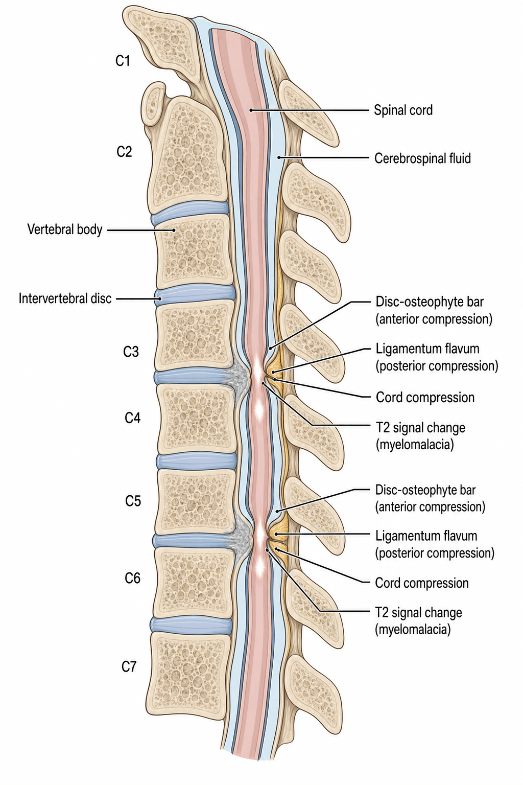
The pincer. The cord is squeezed between an anterior disc-osteophyte bar and a posteriorly buckled, hypertrophied ligamentum flavum, and the pincer tightens dynamically with neck extension, as disc and osteophyte protrude anteriorly. Ordinary daily movement therefore delivers repetitive microtrauma to a cord that has no reserve space, which is what makes the disease progressive rather than static.
Why C5-6 and C6-7. These are the levels of maximal motion and the narrowest canal diameter, so they are where the cord is most vulnerable. The cord's blood supply is a watershed between the anterior spinal artery, which supplies 80% of the cord, and the posterior spinal arteries, which supply 20%; compression can cause ischaemia and infarction, and that damage is irreversible.
- Mechanism
- Degenerative: disc bulging, osteophytes, facet hypertrophy
- Typical Age
- More than 50 years
- Treatment Approach
- Anterior (1-2 levels) or posterior (multilevel)
- Mechanism
- Heterotopic ossification of posterior longitudinal ligament
- Typical Age
- East Asian, more than 50 years
- Treatment Approach
- Posterior laminoplasty preferred (avoid anterior CSF leak risk)
- Mechanism
- Developmental narrow canal (Pavlov ratio less than 0.8)
- Typical Age
- Younger age, trauma
- Treatment Approach
- Posterior decompression if symptomatic
- Mechanism
- Posterior disc extrusion compressing cord
- Typical Age
- 40-60 years
- Treatment Approach
- Anterior discectomy and fusion
- Mechanism
- Posterior compression from ligament thickening
- Typical Age
- Elderly
- Treatment Approach
- Posterior laminectomy or laminoplasty
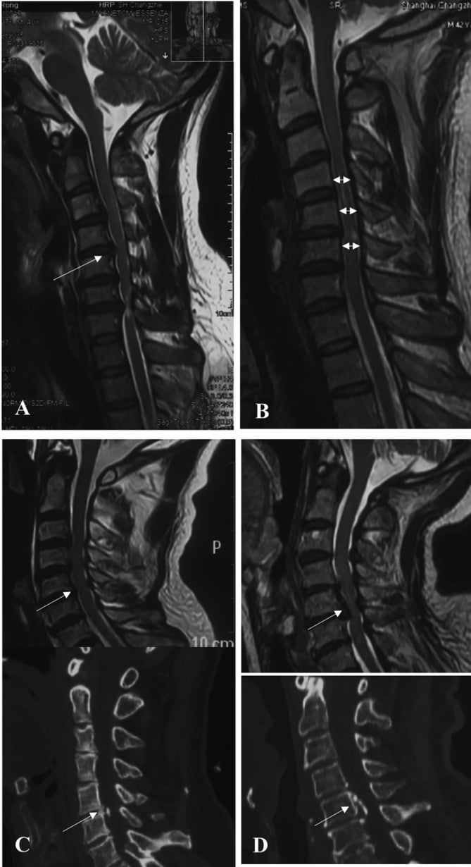
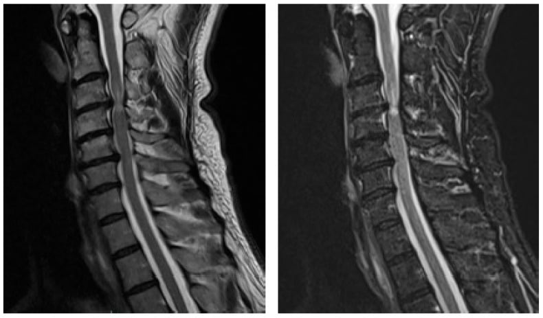
Congenital / Developmental Cervical Canal Stenosis
A constitutionally narrow canal lowers the threshold at which any later degenerative change produces cord compression, so these patients present younger and a relatively minor spondylotic insult can precipitate myelopathy. It is a risk factor, a cause and a radiographic measurement all at once.
Developmental against acquired. A developmentally narrow canal results from short pedicles present from skeletal maturity, reducing the cord's reserve space before any degeneration. Acquired (spondylotic) stenosis is superimposed degenerative narrowing. The two commonly coexist: the small congenital canal is the substrate, and age-related disc bulging, osteophytes and ligamentum flavum buckling tip it into symptomatic compression.
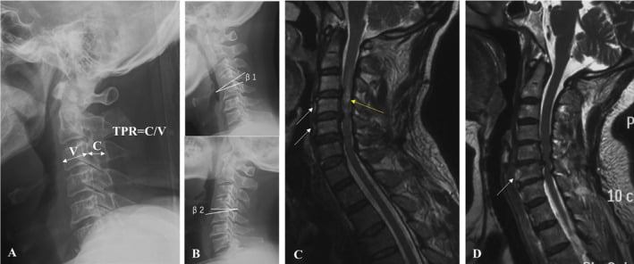
Measuring it, and the trap in the measurement. Developmental stenosis is suggested by an absolute AP canal diameter below about 13mm on the lateral radiograph or MRI, and is critical below 10mm. The Torg-Pavlov ratio divides canal AP diameter by vertebral body AP diameter, and a ratio less than 0.8 suggests stenosis; it is a useful screen with poor specificity and low positive predictive value, especially in athletes and large individuals, because a large vertebral body lowers the ratio without any true canal narrowing. MRI is more reliable than the ratio because it shows the true canal diameter and the cord-CSF reserve, so the diagnosis of clinically meaningful stenosis should rest on MRI rather than the plain-film ratio alone.
What it means clinically. A narrow canal needs less degeneration to become symptomatic, so spondylotic myelopathy arrives earlier. In contact-sport athletes, a hyperflexion or hyperextension load on a stenotic canal can cause cervical cord neurapraxia, transient bilateral sensorimotor symptoms that fully resolve, also called transient quadriparesis. Return to play is individualised and controversial: decisions rest on the true canal and cord reserve on MRI rather than the Torg-Pavlov ratio, and recurrent episodes, or persistent cord compression with T2 signal change, argue against return to collision sport.
Approach implication. Developmental stenosis is a globally narrow canal across multiple levels rather than a focal anterior lesion, so it typically favours a posterior canal-expanding procedure, laminoplasty or laminectomy with fusion, that lets the cord drift back.
Classification Systems
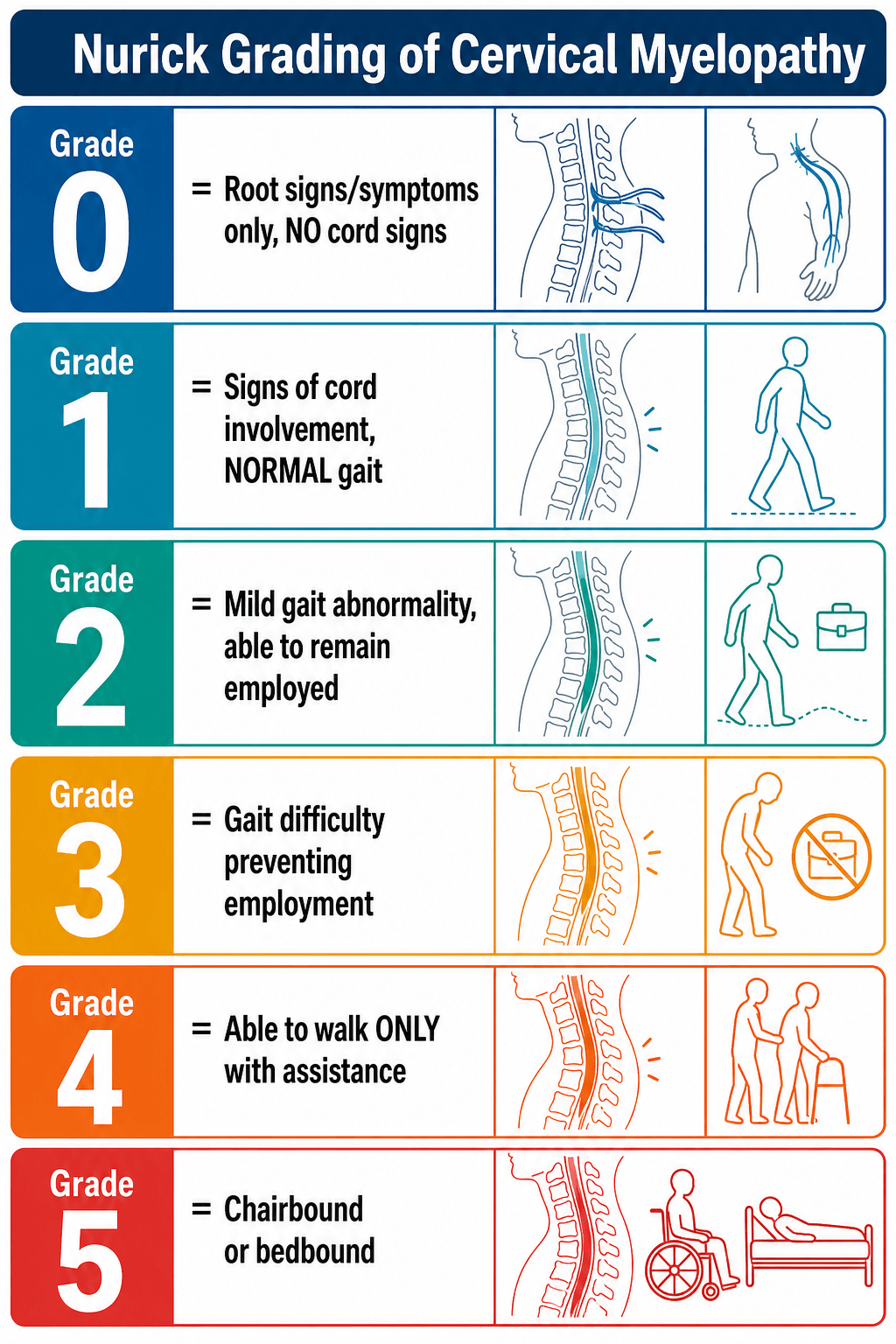
Nurick Scale - Functional Impairment (Most Commonly Used)
Nurick grades the legs. The scale is functional and focuses on gait and employment. It is the most widely used grading system for myelopathic severity and it correlates with surgical outcomes. Grades 2 and 3 are clear surgical indications. Grade 1, cord signs with a normal gait, is the controversial one: some advocate early surgery, others observe. Grades 4 and 5 have poorer outcomes, but surgery still halts progression.
- Clinical Presentation
- Root signs only, no cord signs
- Gait
- Normal
- Surgical Indication
- Treat radiculopathy, observe for myelopathy
- Clinical Presentation
- Cord signs present, no gait difficulty
- Gait
- Normal
- Surgical Indication
- Consider early surgery (controversial)
- Clinical Presentation
- Mild gait difficulty, employment possible
- Gait
- Abnormal but independent
- Surgical Indication
- Surgery recommended
- Clinical Presentation
- Gait difficulty prevents employment
- Gait
- Abnormal, independent in ADLs
- Surgical Indication
- Surgery indicated
- Clinical Presentation
- Unable to walk without assistance
- Gait
- Requires aid (cane/walker)
- Surgical Indication
- Urgent surgery
- Clinical Presentation
- Chair-bound or bedridden
- Gait
- Non-ambulatory
- Surgical Indication
- Urgent surgery, poor recovery expected
Clinical Assessment
The history is of a slow decline. Symptoms worsen over weeks to months and are not acute unless there has been trauma. What patients describe is clumsy hands, trouble with buttons, handwriting and picking up coins, a wide-based unsteady gait with frequent falls, and in advanced disease a neurogenic bladder with urgency, frequency, hesitancy and retention.
The order matters. Hand clumsiness is often the first symptom, lower limb weakness follows, gait disturbance develops, and bladder symptoms indicate severe disease.
Pain is not a prominent feature, unlike radiculopathy; severe neck or arm pain points to a radiculopathy or a mixed picture. Lhermitte sign, an electric shock down the spine on neck flexion, suggests cord compression.
The examination hunts upper motor neuron signs. Hyperreflexia, Hoffman sign, Babinski sign and clonus at the ankle or patella. The inverted radial reflex is worth eliciting: tapping the brachioradialis produces finger flexion instead of forearm flexion, and localises to a C5-6 level lesion.
Then quantify the hands and the gait:
- Grip and release test: inability to open and close the fist 20 times in 10 seconds suggests myelopathy
- Finger escape sign: ulnar drift of the ring and little finger when the hand is held extended, from intrinsic weakness
- Rapid alternating movements and finger tapping, assessing speed and coordination
- Tandem gait for balance, heel-toe walking, and Romberg sign for posterior column dysfunction
- Sensory testing: numb clumsy hands, loss of proprioception, a sensory level, or a glove-and-stocking pattern
An elderly patient with diabetes or a high alcohol intake has a ready explanation for numb hands, and a superimposed myelopathy is missed behind it. Myelopathy gives UMN signs, gait disturbance and cord compression on MRI; peripheral neuropathy gives hyporeflexia, a stocking-glove distribution, abnormal nerve conduction studies and a normal MRI.
Brisk reflexes and a Hoffman sign in a patient already labelled with peripheral neuropathy are reason enough to obtain a cervical spine MRI.
Myelopathy is an upper motor neuron syndrome and radiculopathy a lower motor neuron one, and the two coexist as myeloradiculopathy: the compressed level gives lower motor neuron signs and everything below it upper motor neuron signs.
- Myelopathy (UMN)
- Hyperreflexia, clonus
- Radiculopathy (LMN)
- Hyporeflexia or absent
- Mixed (Myeloradiculopathy)
- Hyperreflexia below level, hyporeflexia at level
- Myelopathy (UMN)
- Positive (flick middle finger, thumb flexes)
- Radiculopathy (LMN)
- Negative
- Mixed (Myeloradiculopathy)
- Positive (indicates cord involvement)
- Myelopathy (UMN)
- Positive (upgoing toe)
- Radiculopathy (LMN)
- Negative (downgoing toe)
- Mixed (Myeloradiculopathy)
- Positive (UMN tract involvement)
- Myelopathy (UMN)
- Sensory level, proprioception loss, gait ataxia
- Radiculopathy (LMN)
- Dermatomal numbness
- Mixed (Myeloradiculopathy)
- Both patterns present
- Myelopathy (UMN)
- Minimal or absent (dull neck ache)
- Radiculopathy (LMN)
- Severe radicular pain (shooting down arm)
- Mixed (Myeloradiculopathy)
- Radicular pain plus myelopathy signs
- Myelopathy (UMN)
- Wide-based, ataxic, spastic
- Radiculopathy (LMN)
- Normal
- Mixed (Myeloradiculopathy)
- Abnormal gait
- Myelopathy (UMN)
- Urgent if progressive
- Radiculopathy (LMN)
- Elective (most resolve non-operatively)
- Mixed (Myeloradiculopathy)
- Urgent (myelopathy component dictates)
Before settling on spondylosis, exclude the mimics. Anything that damages the cord or the corticospinal tracts produces the same upper motor neuron picture, so each alternative is separated by its distinguishing features and the one investigation that settles it.
- Distinguishing Features
- Mixed UMN and LMN signs, fasciculations, NO sensory loss, bulbar involvement, normal cord on MRI
- Key Investigation
- EMG/nerve conduction studies (denervation), clinical pattern
- Distinguishing Features
- Younger patient, relapsing-remitting course, optic neuritis, brain lesions, Lhermitte sign
- Key Investigation
- MRI brain and whole spine, CSF oligoclonal bands
- Distinguishing Features
- Dorsal column + corticospinal signs, peripheral neuropathy, macrocytic anaemia, glossitis
- Key Investigation
- Serum B12, methylmalonic acid, homocysteine
- Distinguishing Features
- Mimics B12 picture; history of bariatric surgery, zinc excess, malabsorption
- Key Investigation
- Serum copper and caeruloplasmin
- Distinguishing Features
- Progressive, may have nocturnal pain, level-specific deficit; ependymoma/astrocytoma/meningioma/metastasis
- Key Investigation
- MRI with contrast
- Distinguishing Features
- Dissociated (cape) sensory loss, may follow Chiari malformation or trauma
- Key Investigation
- MRI showing intramedullary syrinx
- Distinguishing Features
- Acute onset, motor and spinothalamic loss with preserved proprioception
- Key Investigation
- MRI with diffusion, vascular risk assessment
- Distinguishing Features
- Subacute, fever or sepsis features (abscess), enhancing cord lesion
- Key Investigation
- MRI with contrast, inflammatory markers, CSF
VITAMINSDifferential Diagnosis of Myelopathy
Hook:Rule out VITAMINS before diagnosing spondylotic myelopathy!
Asymptomatic Cord Compression and the Non-Myelopathic Patient
Cord compression on MRI without myelopathy is a common real-world dilemma, because incidental cervical cord contact or compression is found in an appreciable proportion of asymptomatic adults and increases with age. Finding compression on a scan does not, by itself, mandate surgery. Nurick grade 0, root signs only, describes the same pre-myelopathic patient seen from the clinical side.
The AO Spine guideline separates two non-myelopathic patients (Fehlings et al, Global Spine J 2017, PMID 29164035):
- Cord compression without myelopathy and without radiculopathy: do not offer prophylactic surgery. Counsel the patient about the risk of progression, educate them on the red-flag symptoms of myelopathy (hand clumsiness, deteriorating handwriting or buttoning, gait unsteadiness, falls, bladder change) and follow clinically, with a low threshold to image and treat if myelopathy develops.
- Cord compression without myelopathy but with radiculopathy, clinical or electrophysiological: these patients are at higher risk of developing myelopathy and should be counselled accordingly. Offer either surgery or non-operative management with close serial follow-up or a supervised rehabilitation trial, converting to surgery if myelopathy emerges.
Why radiculopathy and electrophysiology change the plan. In the natural-history evidence synthesised by the guideline, reported progression from asymptomatic compression to clinical myelopathy is on the order of 8% at 1 year and roughly a fifth to a quarter by about 4 years in the higher-risk subgroup - those with clinical radiculopathy or abnormal electrophysiology, meaning EMG radiculopathy or prolonged SSEPs or MEPs - and considerably lower in patients without these features. That is the rationale for stratifying the intensity of surveillance rather than operating on every compressed cord.
Asymptomatic cervical cord compression without myelopathy does not warrant prophylactic surgery. The moment objective myelopathy appears, the standard surgical algorithm applies.
Investigations
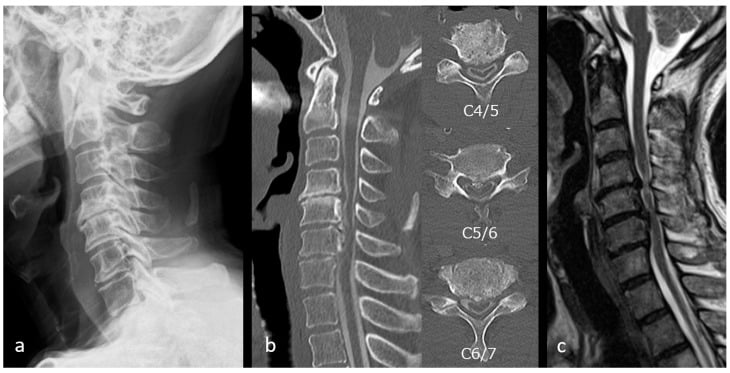
Plain films first, then the MRI that makes the diagnosis. Radiographs show alignment, instability and bony pathology; MRI shows the cord, and cord compression with clinical myelopathy is what decides treatment.
Diagnostic Imaging Protocol
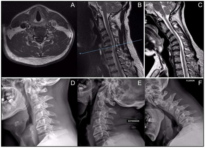
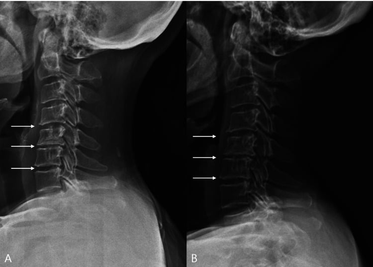
Alignment, instability and bony pathology. The lateral film gives the Pavlov ratio, canal diameter divided by vertebral body diameter, normal over 1.0, stenotic under 0.8 and borderline in between. Flexion and extension views look for dynamic instability, subluxation of more than 3mm or angulation of more than 11 degrees. The AP shows degenerative change and osteophytes.
Radiographs cannot visualise the cord, the disc or any soft-tissue compression.
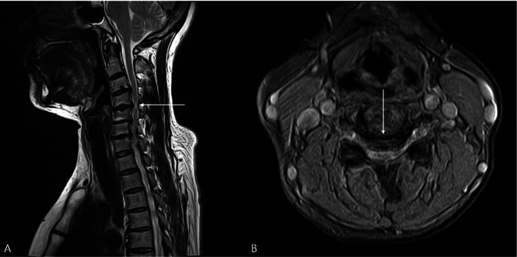
Cord compression, cord signal and disc pathology. T2 sagittal shows the compression, the canal stenosis, the disc bulging and the obliteration of the CSF in front of and behind the cord. T2 axial shows cord deformity and indentation at each compressed level, and intramedullary T2 hyperintensity. T1 sagittal shows T1 hypointensity.
Cord compression plus T2 signal change plus clinical myelopathy is a surgical indication; coexisting root compression on the same study is what makes a patient myeloradiculopathic.
Bone detail, OPLL and planning. CT measures the thickness of an ossified posterior longitudinal ligament and plans screw trajectories. CT myelography is the substitute where MRI is contraindicated by a pacemaker or claustrophobia: it shows cord compression but not intrinsic cord signal.
Dynamic compression invisible on a neutral scan. Some patients have cord compression only in flexion, or only in extension. It is not widely available and costs more.
Cord function, before and during surgery. Preoperatively, prolonged latency or reduced amplitude can confirm the diagnosis when the MRI is equivocal. Intraoperatively they monitor the cord during decompression, where an amplitude drop of more than 50% signals a risk of cord ischaemia.
T2 hyperintensity, bright signal within the cord on T2, is either oedema, which is early and reversible, or gliosis and myelomalacia, which is late and irreversible. Either way it predicts poorer surgical outcomes, its severity tracks the Nurick grade and the recovery available, and the best results follow surgery done before it appears.
The snake-eye sign - bilateral symmetric T2 hyperintensity in the anterior grey matter on axial MRI, with cord atrophy - means severe chronic compression and a very poor prognosis: most of these patients do not improve. T1 hypointensity, dark signal on T1, is established myelomalacia, cord necrosis, and recovery from it is very poor.
Management Algorithm
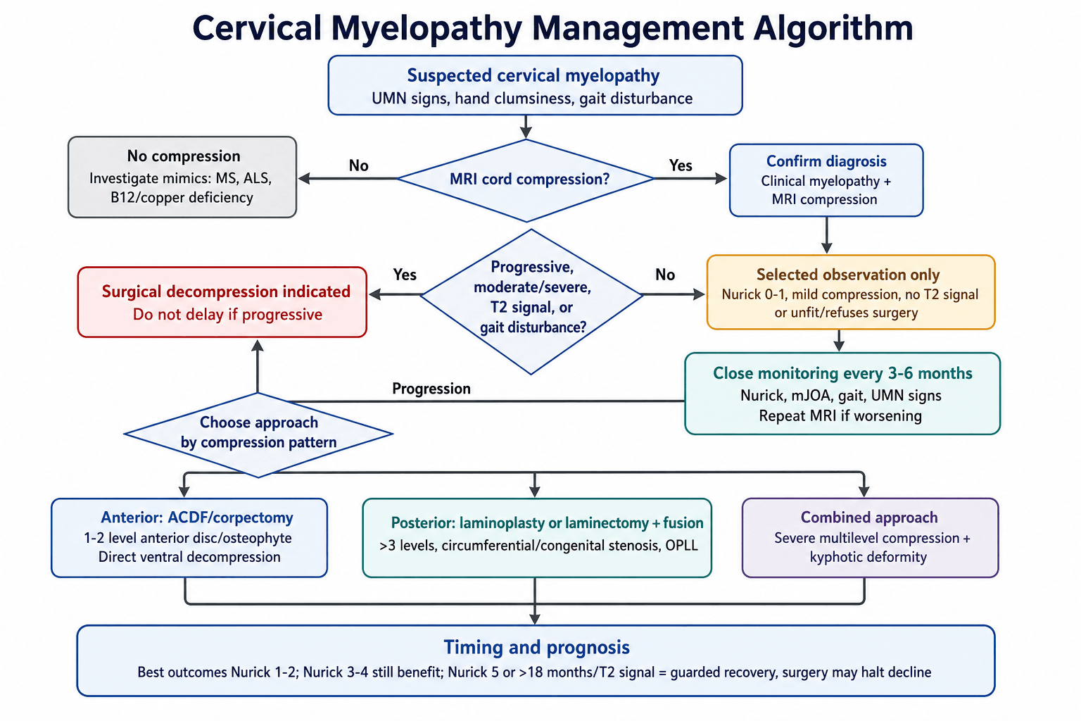
The indication is clinical and radiological together. Myelopathic signs on examination with cord compression on MRI is what justifies decompression; neither the scan nor the symptoms decide it alone, and what sets the urgency is progression.
Conservative Management - Limited Role
Because established myelopathy progresses in most patients, observation is only appropriate in three situations: Nurick 0-1, with minimal symptoms and no gait disturbance; the patient unfit for surgery through severe medical comorbidity; and the patient who refuses surgery after informed consent about the natural history.
What conservative care can and cannot do. These measures run in parallel and are aimed at symptoms, not at the disease: myelopathy is caused by mechanical compression of the spinal cord, and nothing in this list decompresses anything. The realistic goals are comfort, function and surveillance that detects deterioration early enough to still operate usefully, because cord recovery depends on how long it has been compressed, and the damage that accrues while a patient is managed conservatively is not recovered by later surgery.
Neck immobilisation is short-term only. A soft collar relieves symptoms during an acute exacerbation. Avoid prolonged use: it causes muscle atrophy and stiffness, and the deconditioned neck is then worse at protecting itself.
Medication treats symptoms only. Gabapentin or pregabalin for the neuropathic pain of a coexisting radiculopathy, and baclofen or tizanidine for upper motor neuron spasticity. There is no role for NSAIDs or steroids in chronic degenerative myelopathy: unlike acute cord injury there is no inflammatory oedema to suppress, only chronic mechanical compression.
Physiotherapy maintains function without touching the cord. Range of motion, strengthening of the supporting musculature and gait training are all worth having, and none of them halts progression of cord compression. Avoid manipulation and traction outright - forcing movement in a stenotic canal risks worsening the compression, and this is the one active harm on the list.
Monitoring is the part that actually matters. Assess clinically every 3-6 months: Nurick grade, mJOA score, gait and upper motor neurone signs, so that deterioration is measured rather than remembered, and repeat the MRI if the examination worsens. Converting to surgery as soon as symptoms progress is the plan rather than a fallback, and the monitoring exists solely to catch the moment it applies.
Where the line sits. Observation is reasonable at Nurick 0-1 when the MRI shows mild compression without T2 signal change, when the patient is a high surgical risk with severe cardiac or pulmonary disease, and when consent has covered the natural history. Operate even at Nurick 1 when T2 signal change is present, when progression is documented over months, or when the patient is young with a long life expectancy.
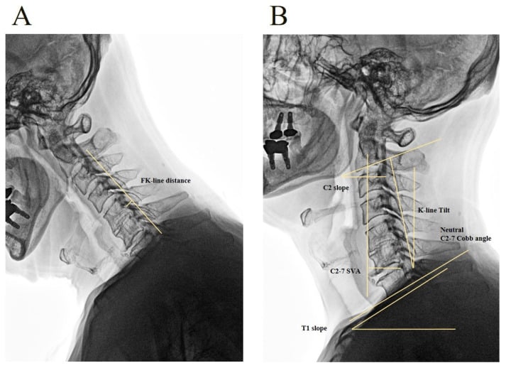
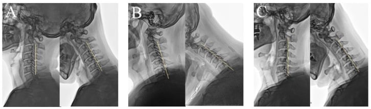
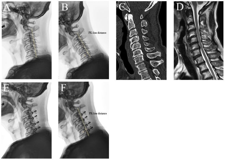
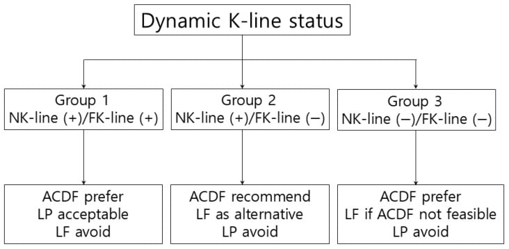
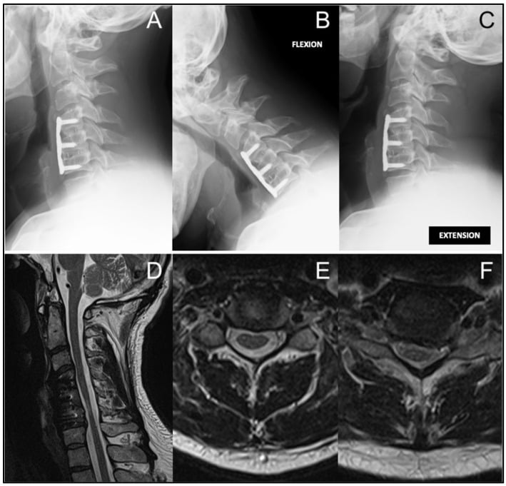
In multilevel OPLL the approach is also judged dynamically, using the K-line. What matters is whether the line stays positive once the neck flexes, so the assessment needs the neutral and flexion films together.
Surgical Techniques - Anterior Approaches
Anterior Cervical Discectomy and Fusion (ACDF)
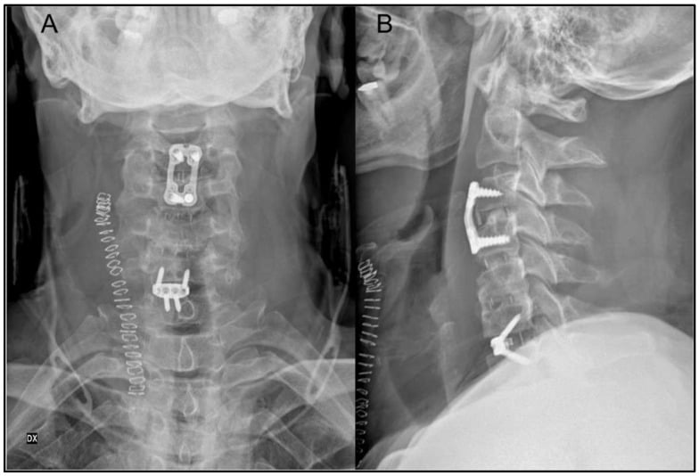
When. One or two levels of anterior compression from disc herniation or osteophyte, with focal cord compression at those levels. It is the most common surgical treatment for cervical myelopathy.
ACDF Steps
Supine, head neutral or slightly extended, with a shoulder roll to extend the neck. The Smith-Robinson approach uses a transverse or oblique incision along the anterior border of the sternocleidomastoid, typically on the left, where the recurrent laryngeal nerve is less at risk. Dissect medial to the carotid sheath and lateral to the trachea and oesophagus, retracting them medially.
Confirm the level on fluoroscopy; the C6 anterior tubercle is prominent and helps. Remove the disc completely, including the posterior annulus and the posterior osteophytes, and take the PLL if it is ossified, until the canal is adequately decompressed. Add a foraminotomy to decompress the root laterally if there is a coexisting radiculopathy.
A PEEK cage with autograft or allograft is the usual reconstruction; structural allograft (femoral ring, fibula) is the alternative. Trial the cage to restore height and lordosis. An anterior plate with screws into the bodies above and below raises the fusion rate at one year to 95% against 85% without a plate, and prevents the cage subsiding.
Layered closure, with a drain optional and still argued over. The soft collar is for comfort rather than structure, since the plate provides the stability, and the patient stays nil by mouth until swallowing is assessed, then advances as tolerated.
What makes the decompression adequate. Complete PLL removal is the step that decompresses the cord posteriorly, and leaving a posterior osteophyte behind causes persistent compression. Avoid over-distraction, which distracts the facet joints and causes postoperative pain, and check on fluoroscopy that the plate does not cross an adjacent disc space.
What goes wrong. Recurrent laryngeal nerve injury is minimised by the left-sided approach and gentle retraction, dysphagia is worse after multilevel surgery, and adjacent segment disease follows the fusion, with a live debate over how much of that is simply natural history.
Surgical Techniques - Posterior Approaches
Cervical Laminoplasty - Motion-Preserving Decompression
When. Multilevel stenosis of 3 or more levels, typically C3-7; circumferential compression from front and back; congenital stenosis with acquired spondylosis; and OPLL, where it is preferred to an anterior approach because of the CSF leak risk.
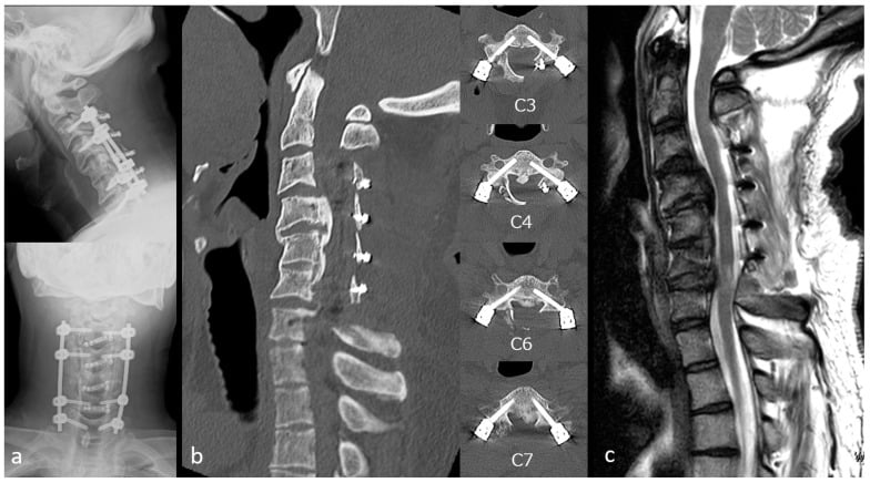
The principle. An open-door laminoplasty hinges the laminae on one side, opens them on the other, and props the door with a spacer. The canal enlarges and the cord drifts posteriorly, away from anterior compression, which is how a posterior operation treats a ventral problem.
Laminoplasty Steps
Prone, head in a Mayfield clamp, neck neutral and neither excessively flexed nor extended. Midline incision from C2 to T1 with subperiosteal dissection, preserving the C2 and C7 spinous processes, and exposure of the lateral masses out to the lateral edge of the facet joints on both sides.
On the hinge side, usually the left, thin the lamina with a high-speed burr to create a greenstick fracture without penetrating the dura. On the open side, usually the right, complete the trough at the lamina-lateral mass junction and detach the ligamentum flavum. Elevating the lamina at the hinge opens the door and increases canal diameter by 4-5mm.
Hold the door with a titanium miniplate and spacer, a hydroxyapatite block, or suture in the older technique, aiming for a 5-7mm opening that will not close. Confirm the decompression by seeing dural pulsation and enough space for the cord to drift.
Reattach the extensor musculature to the C2 and C7 spinous processes, which were preserved to reduce axial pain. A subfascial drain stays 24-48 hours. No collar is needed: a laminoplasty is inherently stable.
What it offers. Motion is preserved rather than fused, multiple levels are decompressed through a single posterior approach, there is no pseudarthrosis to worry about because nothing is fused, and the infection risk is lower than with the anterior approach.
What it costs. Aching axial neck pain, C5 palsy, and a 30-50% loss of flexion and extension despite the label of motion preservation. It cannot correct kyphosis, so it needs a lordotic or neutral alignment to begin with.
C5 palsy. Deltoid and biceps weakness in the C5 myotome, often with shoulder pain and patchy deltoid numbness, in a patient who woke from surgery intact. Onset is delayed, at a pooled mean of 3 days (95% CI 2.6-3.6) across 748 cases, which is why it is missed on the day-zero check.
The incidence depends on the operation, and the spread is threefold. ACDF 3.3%, laminoplasty alone 5.1%, laminoplasty plus another posterior procedure 6.5%, anterior corpectomy and fusion 7.5%, and laminectomy with fusion 11.0%, against a pooled 5.3% across 13,621 patients.
The mechanism is contested, and it is worth knowing what the evidence shows. The traditional explanation is tethering: the cord drifts posteriorly after decompression and drags on a short C5 root. Two observations sit badly with it. First, when 107 studies are stratified by approach and diagnosis, anterior decompression for radiculopathy produces essentially no C5 palsy (95% CI 0.00-0.01), while for myelopathy the rates are 4% anterior and 7% posterior with no significant difference between the approaches - yet anterior surgery causes no posterior drift. Second, the risk factors that survive multivariate analysis are duration of symptoms over 12 months and preoperative T2 cord signal change at C4-5, which are properties of the cord rather than of the foramen. Together these favour reperfusion injury of the cord grey matter, with tethering at most contributory.
Prophylactic foraminotomy is widely performed and is not established. In 70 laminoplasties all done with bilateral C4-5 foraminotomy, 5.7% still developed a C5 palsy, no better than the pooled rate, and the authors concluded plainly that prophylactic foraminotomy does not eliminate it. In a prospective series using maximal expansion the foraminotomy subgroups fared worse, not better: 33% versus 20% after laminoplasty and 50% versus 20% after posterior decompression and fusion. What is defensible is avoiding unnecessarily wide decompression and accepting that a chronically compressed, signal-changed cord carries irreducible risk.
Management, and honest consent. Exclude a compressive cause - haematoma or a malpositioned construct - with urgent imaging if the weakness is sudden or severe, then treat expectantly with physiotherapy and shoulder protection. At one year the pooled recovery rates were 100% after ACDF but only 52.9% after laminoplasty and 50% after posterior decompression and fusion, so after the posterior operations described here roughly half of affected patients are still not fully recovered at twelve months, and consent should not promise resolution.
Complications
- Incidence
- 5.1% laminoplasty, 11.0% laminectomy with fusion (5.3% pooled; 3.3% ACDF)
- Risk Factors
- Symptoms over 12 months and preoperative C4-5 T2 cord signal change are the risk factors that survive multivariate analysis
- Management
- Urgent MRI if sudden or severe, to exclude haematoma or malpositioned construct; then observation with physiotherapy
- Incidence
- 10-20% temporary, 2-5% persistent (anterior approach)
- Risk Factors
- Multilevel ACDF, excessive retraction, revision surgery
- Management
- Speech therapy, diet modification, usually resolves in 6-12 weeks
- Incidence
- 1-2% (anterior approach)
- Risk Factors
- Right-sided approach (RLN non-recurrent variant), excessive retraction
- Management
- Voice rest, speech therapy, most recover in 3-6 months, vocal cord injection if persistent
- Incidence
- 1-3% (higher with OPLL)
- Risk Factors
- OPLL adherent to dura, revision surgery
- Management
- Primary repair with suture or sealant, bed rest 48 hours, rarely requires reoperation
- Incidence
- 1-2% (new or worsened deficit)
- Risk Factors
- Excessive manipulation, cord ischaemia, epidural haematoma
- Management
- Urgent MRI, return to theatre if haematoma, observation if ischaemia (usually improves)
- Incidence
- 5-10% ACDF, 15-20% corpectomy
- Risk Factors
- Smoking, multilevel fusion, inadequate fixation
- Management
- Revision fusion if symptomatic (pain, instability), observe if asymptomatic
- Incidence
- 5-10% per decade after fusion
- Risk Factors
- Fusion (vs laminoplasty), preexisting degeneration
- Management
- Surgical decompression if symptomatic stenosis develops
- Incidence
- 20-30% (laminoplasty or fusion)
- Risk Factors
- Muscle dissection, loss of ROM
- Management
- NSAIDs, physical therapy, usually improves over 6-12 months
- Incidence
- 1-3%
- Risk Factors
- Diabetes, obesity, immunosuppression, revision surgery
- Management
- Antibiotics (if early), return to OR for washout (if deep), drain epidural abscess urgently
- Incidence
- 30-50% if laminectomy alone
- Risk Factors
- Multilevel laminectomy, preexisting kyphosis
- Management
- Prevention: Always fuse after multilevel laminectomy. Treatment: Revision fusion if symptomatic
Sudden neurological deterioration within 24-48 hours of surgery - severe weakness, sensory loss, bladder dysfunction - is an epidural haematoma until proved otherwise. Obtain an urgent MRI, which shows an epidural fluid collection compressing the cord.
Return to the operating theatre immediately to evacuate it: every hour of delay worsens the prognosis, and this is a surgical emergency. Prevention is meticulous haemostasis, a drain, and avoiding anticoagulation in the immediate postoperative period.
Postoperative Care and Rehabilitation
ACDF Postoperative Protocol
Neurological checks every 2 hours, motor and sensory, and a swallow assessment before anything by mouth. The soft collar is for comfort, not structure, since the plate provides the stability. Out of bed on day 1, because early ambulation reduces the risk of DVT. Nil by mouth until swallowing is cleared, then a soft diet if there is dysphagia, with multimodal analgesia of paracetamol, gabapentin and opioids as required.
No lifting over 5kg, and no driving while in a collar or taking opioids. Wean the collar at 2-4 weeks, earlier after a single-level fusion and later after a multilevel one. Gentle range-of-motion exercises and posture training. Radiographs at 6 weeks to assess alignment and hardware position.
Gradual return to activity with progressive strengthening. Radiographs or CT at 12 weeks if there is any concern about pseudarthrosis. Most patients are independent in daily activities by 3 months.
Annual radiographs for 2 years, then as symptoms dictate. Monitor for the new symptoms of adjacent segment disease. Return to full activity takes 3-6 months, depending on the occupation.
Outcomes and Prognosis
Prognosis is set before the operation starts. Recovery tracks the severity of the myelopathy, the length of time the patient has had it, and the presence of signal change within the cord. Early intervention, before severe disability, outperforms surgery delayed until the cord has been compressed for years.
Counsel accordingly. Patients carrying those features should still be decompressed, to halt progression, but the honest expectation is stabilisation rather than improvement.
Age is not the deciding factor. Advanced age carries a poorer prognosis, but age alone is neither a contraindication nor an independent predictor of functional outcome, so it should not exclude a patient from surgery.
- Good Prognosis
- Nurick 1-2 (mild disease)
- Poor Prognosis
- Nurick 4-5 (severe disease, especially more than 18 months duration)
- Good Prognosis
- No T2 hyperintensity or T1 hypointensity
- Poor Prognosis
- T2 hyperintensity (gliosis) or T1 hypointensity (myelomalacia)
- Good Prognosis
- Less than 12 months
- Poor Prognosis
- More than 18 months (chronic irreversible cord damage)
- Good Prognosis
- Younger (under 60 years)
- Poor Prognosis
- Elderly (over 75 years, but age alone not contraindication)
- Good Prognosis
- Healthy, no diabetes
- Poor Prognosis
- Diabetes, vascular disease (impairs cord perfusion and healing)
- Good Prognosis
- Early intervention (before severe disability)
- Poor Prognosis
- Delayed surgery (after prolonged severe compression)
Guidelines, Registries & Global Practice
Degenerative cervical myelopathy (DCM) is the commonest cause of non-traumatic spinal cord dysfunction in adults worldwide. North American estimates place incidence at a minimum of 41 per million and prevalence at a minimum of 605 per million, with CSM-related hospitalisations around 4.04 per 100,000 person-years and rising surgical rates (Nouri et al, Spine 2015). OPLL as a cause is markedly more common in East Asian populations than in Western cohorts (Tetreault et al, Neurosurgery 2015).
- Mild DCM (mJOA 15-17)
- Offer surgery OR supervised structured rehabilitation; operate if decline or rehab fails
- Moderate-Severe DCM (mJOA less than 15)
- Surgical decompression recommended
- Asymptomatic Cord Compression
- No prophylactic surgery; counsel on red flags and follow up
- Evidence Basis
- GRADE methodology, international multidisciplinary
- Mild DCM (mJOA 15-17)
- Surgery or close monitoring acceptable; shared decision-making
- Moderate-Severe DCM (mJOA less than 15)
- Surgery recommended to halt progression
- Asymptomatic Cord Compression
- Surveillance, patient education on symptom onset
- Evidence Basis
- Consensus + systematic review
- Mild DCM (mJOA 15-17)
- More observation in some European/UK settings; earlier surgery in some North American/Asian centres
- Moderate-Severe DCM (mJOA less than 15)
- Universal surgical recommendation where access permits
- Asymptomatic Cord Compression
- Variable surveillance intervals; no consensus screening protocol
- Evidence Basis
- Practice-pattern, not trial-based
- Moderate and severe DCM: surgical decompression is recommended across guidelines to halt neurological decline
- Asymptomatic cord compression without myelopathy: no prophylactic surgery; educate and monitor for red-flag symptoms
- Diagnosis: MRI is the key investigation; T2 hyperintensity carries prognostic weight
- Prognosis: longer symptom duration and worse baseline severity predict poorer recovery (Tetreault et al, Eur Spine J 2015)
- Mild DCM: AOSpine/GSJ permit either surgery or a supervised rehabilitation trial - the main area of genuine equipoise (Kadanka RCTs)
- Surveillance intervals for non-operative and asymptomatic patients are not standardised
- Approach selection (anterior vs posterior vs combined) is guided by levels, compression pattern, alignment and OPLL rather than a single recommended technique
- OPLL management weighting differs by region given higher East Asian prevalence
Consistent across health systems for cervical myelopathy surgery:
-
Informed consent:
- Natural history of established myelopathy is progression in the majority without treatment
- Realistic expectations: surgery aims to halt decline and stabilise or improve function, not cure
- Major risks to document: C5 palsy (5.1% laminoplasty, 11.0% laminectomy with fusion), dysphagia (10-20%, anterior), recurrent laryngeal nerve injury (1-2%, anterior), neurological deterioration (1-2%), infection (1-3%)
- Document T2 signal change if present (poor prognostic sign)
-
Avoiding delayed diagnosis (a recurrent litigation theme globally):
- Document the UMN examination (reflexes, Hoffman, Babinski, gait) and obtain MRI when myelopathy is suspected
- Do not delay surgery in progressive myelopathy
-
Perioperative care: surgical antibiotic prophylaxis per local guidelines (e.g. a single preoperative cephalosporin dose), and VTE prophylaxis individualised to bleeding/cord-haematoma risk.
MCQ Practice Points
Q: The cervical spinal cord is most vulnerable to compression at which levels? A: C5-6 and C6-7 due to the combination of maximal motion at these levels and the narrowest canal diameter. These levels also have watershed blood supply between the anterior spinal artery territory (80% of cord) and posterior spinal arteries (20%), making them vulnerable to ischemia with compression.
Q: A patient with cervical myelopathy has mild gait difficulty but is still able to work full-time. What is their Nurick grade? A: Nurick grade 2 - mild gait abnormality but employment not affected. This is a clear surgical indication, as Nurick grades 2-3 benefit most from surgery. Grade 1 has cord signs but normal gait (controversial whether to operate). Grade 3 has gait difficulty preventing employment.
Q: What does T2 hyperintensity within the cervical cord on MRI represent, and what is its prognostic significance? A: T2 hyperintensity can represent edema (reversible) or gliosis/myelomalacia (irreversible). It is a poor prognostic sign - patients with T2 signal changes have worse surgical outcomes and less neurological recovery compared to those without signal changes. The "snake-eye sign" (bilateral symmetric T2 hyperintensity in anterior grey matter on axial MRI) predicts very poor prognosis.
Q: What is the primary indication for choosing a posterior approach (laminoplasty) over an anterior approach (ACDF) for cervical myelopathy? A: Multilevel compression (3 or more levels, typically C3-7), circumferential compression, congenital stenosis, or OPLL. Posterior laminoplasty decompresses multiple levels through a single approach, allows the cord to drift posteriorly away from anterior compression, and avoids the CSF leak risk of removing adherent OPLL anteriorly. Anterior ACDF is preferred for 1-2 level focal anterior compression (disc, osteophyte).
Q: What is C5 palsy, and how is it managed? A: C5 palsy is isolated deltoid and biceps weakness (C5 myotome), typically appearing at a mean of 3 days postoperatively in a patient who woke intact. Incidence tracks the operation: 3.3% after ACDF, 5.1% laminoplasty, 7.5% corpectomy and fusion, 11.0% laminectomy with fusion (5.3% pooled). The traditional mechanism is posterior cord drift tethering the C5 root, but that is contested: anterior decompression for myelopathy carries a comparable rate (4% versus 7% posterior, p=0.999) despite producing no drift, while anterior surgery for radiculopathy produces essentially none - so what predicts the palsy is a compressed cord, favouring reperfusion injury. The risk factors on multivariate analysis are symptom duration over 12 months and preoperative C4-5 T2 cord signal change. Management is observation after urgent MRI has excluded a haematoma or malpositioned construct when weakness is sudden or severe. Be accurate about prognosis: recovery at one year is 100% after ACDF but only 52.9% after laminoplasty and 50% after posterior decompression and fusion. Prophylactic C4-5 foraminotomy is not established prevention - 5.7% still developed palsy in a series where every patient had it, and a prospective series found higher rates in the foraminotomy subgroups.
Q: According to the AOSpine CSM studies, which surgical approach (anterior vs posterior) has better outcomes for cervical myelopathy? A: The large AOSpine North America prospective study (Fehlings et al, JBJS Am 2013) showed that surgical decompression significantly improved mJOA, Nurick grade, NDI, and SF-36 across all severity grades, with an overall complication rate of 18.7%. It did not directly compare anterior versus posterior approaches. Comparative and systematic-review data (Tetreault et al, Eur Spine J 2015) indicate that surgical approach is not a consistent independent predictor of neurological outcome - the decision should be based on anatomical pathology (number of levels, compression pattern, sagittal alignment, OPLL). Approach-specific complication profiles differ: anterior approaches carry more dysphagia and recurrent laryngeal nerve injury, posterior approaches more axial neck pain. Note that C5 palsy is not a clean discriminator between the approaches as is often assumed - once the cord is myelopathic, pooled rates are 4% anteriorly and 7% posteriorly with no significant difference (p=0.999). Where the approaches genuinely separate is the specific operation: 3.3% for ACDF against 11.0% for laminectomy with fusion.
Clinical Decision Scenarios
Practise clinical reasoning and management decisions out loud
“A 58-year-old man presents with 6 months of progressive hand clumsiness and difficulty walking. He drops objects frequently and feels unsteady going downstairs. On examination, you find hyperreflexia in all four limbs, positive Hoffman sign bilaterally, and upgoing plantars. His gait is wide-based and spastic. MRI shows multilevel cervical stenosis C3-7 with cord compression and T2 hyperintensity at C5-6. What is your diagnosis and management?”
“You are performing an ACDF for C5-6 myelopathy. After discectomy, you encounter a large posterior osteophyte that extends behind the vertebral body. The posterior longitudinal ligament appears ossified. How do you proceed? What are the risks, and how would you mitigate them?”
“You performed a C3-7 laminoplasty yesterday for cervical myelopathy. On postoperative day 1, the nurse calls you because the patient has suddenly developed severe bilateral arm and leg weakness (MRC grade 2/5) that was not present immediately after surgery. What is your differential diagnosis, and how do you manage this?”
“On examination of a patient with suspected cervical myelopathy, you elicit a positive Hoffman sign and inverted radial reflex. What do these signs indicate, and what other examination findings would you look for to confirm the diagnosis?”
Key Anatomy
- Cervical cord occupies 60-70% of canal cross-sectional area
- Normal canal AP diameter = 14-17mm; stenotic = less than 10mm (absolute) or less than 13mm (relative)
- Most vulnerable levels = C5-6 and C6-7 (maximal motion, narrowest canal, watershed blood supply)
- Anterior spinal artery supplies 80% of cord; posterior spinal arteries supply 20%
- Compression mechanism = static (anatomical stenosis) + dynamic (flexion/extension pincer effect)
Classification
- Nurick 0 = root signs only (no cord involvement)
- Nurick 1 = cord signs, normal gait (controversial surgical indication)
- Nurick 2 = mild gait abnormality, employment not affected (clear surgical indication)
- Nurick 3 = gait prevents employment (surgical indication)
- Nurick 4 = requires assistance to walk (urgent surgery, guarded prognosis)
- Nurick 5 = chair-bound/bedridden (urgent surgery, poor prognosis)
- mJOA score = 18 points total (motor UE/LE, sensory UE/LE/trunk, sphincter)
Treatment Algorithm
- Anterior ACDF = 1-2 level anterior compression (disc, osteophyte)
- Posterior laminoplasty = multilevel (more than 3 levels), circumferential compression, OPLL
- Laminectomy + fusion = multilevel with instability or kyphosis (always fuse after multilevel laminectomy)
- Combined approach = severe multilevel with kyphotic deformity
- Surgical timing = early (Nurick 1-2) has better outcomes than late (Nurick 4-5)
- Natural history = 80% progressive decline without surgery (60% gradual, 20% stepwise)
Surgical Pearls
- ACDF: Complete PLL removal for adequate posterior decompression; avoid over-distraction
- Laminoplasty: Create hinge on one side, open door on other, maintain 5-7mm opening with spacer
- OPLL: Posterior laminoplasty preferred (avoids anterior CSF leak risk); thin OPLL = leave, thick = remove carefully
- Always fuse after multilevel laminectomy (prevents postoperative kyphosis)
- C5 palsy: no prevention is proven. Prophylactic C4-5 foraminotomy does NOT eliminate it (5.7% despite it) and fared worse in a prospective series - avoid unnecessarily wide decompression and accept irreducible risk in a chronically signal-changed cord
Complications
- C5 palsy = 3.3% ACDF, 5.1% laminoplasty, 11.0% laminectomy+fusion (5.3% pooled). Mean onset 3 days. Recovery at 1 year: 100% ACDF but only ~50% after posterior surgery
- Dysphagia = 10-20% temporary, 2-5% persistent (anterior approach)
- RLN injury = 1-2% (hoarseness, use left-sided approach, most recover)
- Epidural hematoma = less than 1% (acute quadriparesis, urgent MRI and OR for evacuation)
- Pseudarthrosis = 5-10% ACDF, 15-20% corpectomy (smoking, multilevel)
- Adjacent segment disease = 5-10% per decade (fusion patients)
- Axial neck pain = 20-30% (posterior approach, NSAIDs + PT, usually improves)
Evidence Base and Key Trials
AOSpine North America Prospective Study: Efficacy and Safety of Surgical Decompression
- Prospective multicentre study at 12 North American centres: 278 patients with symptomatic cervical spondylotic myelopathy and MRI cord compression
- Severity at baseline: 30.6% mild (mJOA 15 or more), 39.6% moderate (mJOA 12-14), 29.9% severe (mJOA less than 12)
- Significant improvement at 1 year in mJOA, Nurick grade, Neck Disability Index, and SF-36v2 across all severity categories
- Overall complication rate 18.7% (52 of 278), with no significant difference between severity groups
Surgery for Mild Degenerative Cervical Myelopathy (AOSpine NA and International Cohorts)
- 193 patients with mild DCM (mJOA 15-17) pooled from the AOSpine CSM-NA and CSM-International trials
- Even mild DCM caused marked baseline impairment in all SF-36v2 domains versus population norms
- Significant improvement to 2 years in mJOA, Nurick grade, NDI, and SF-36v2 physical and mental component summaries
- Complication rate was low in this mild-severity group
Kadanka RCT: Conservative versus Surgical Treatment of Mild/Moderate CSM (3-year)
- Prospective randomised trial: 68 patients with mild to moderate, non-progressive or slowly progressive CSM (35 conservative, 33 surgical)
- No significant difference in mean mJOA score between groups over 3-year follow-up
- Timed 10-metre walk favoured the conservative group; self-evaluation slightly favoured surgery at 6 months only
- Surgery was not shown to be superior to conservative treatment on average for mild/moderate non-progressive disease
Kadanka 10-Year Follow-up RCT: Conservative versus Surgical CSM
- Ten-year prospective randomised study: 64 patients randomised 1:1 to conservative versus surgical treatment of mild/moderate CSM
- No statistically significant difference in mJOA, blinded video assessment of daily activities, or timed 10-metre walk at 10 years
- No difference between groups in the proportion losing the ability to walk
- Authors emphasise the study is underpowered and treat the finding as a hypothesis needing confirmation
Tetreault Systematic Review: Predictors of Surgical Outcome in CSM
- Systematic review of 91 graded studies (16 excellent, 38 good, 37 poor quality) on predictors of surgical outcome in CSM
- Longer symptom duration was consistently associated with poorer mJOA/JOA and Nurick outcomes
- More severe baseline mJOA/JOA score predicted worse postoperative mJOA/JOA outcome
- Age was NOT a significant independent predictor of functional outcome across the higher-quality studies
AOSpine / Global Spine Journal Clinical Practice Guidelines for DCM
- International GRADE-based guidelines for degenerative cervical myelopathy developed by a multidisciplinary group (AOSpine, CSRS)
- Surgery recommended for moderate and severe DCM
- For mild DCM, recommend offering surgery OR a supervised trial of structured rehabilitation, with operative management if neurological decline occurs or rehabilitation fails
- Non-myelopathic patients with cord compression but no radiculopathy do not warrant prophylactic surgery and should be counselled on red-flag symptoms and followed
C5 palsy: incidence by operation
- 79 studies pooled: 704 C5 palsy cases among 13,621 patients, overall prevalence 5.3% (95% CI 4.6-6.0%)
- By operation: ACDF 3.3% (lowest), laminoplasty alone 5.1%, laminoplasty plus another posterior procedure 6.5%, anterior corpectomy and fusion 7.5%, laminectomy with fusion 11.0% (highest)
- Posterior 5.8% versus anterior 5.2% overall; OPLL 5.8% versus cervical spondylotic myelopathy 4.5%; men 5.2% versus women 2.2%
- Most cases were unilateral, transient, and diagnosed within 3 days
C5 palsy: the meta-analysis that tests the mechanism
- 107 studies stratified by BOTH approach and underlying diagnosis - the design choice that turns an incidence review into a test of mechanism
- Anterior decompression for RADICULOPATHY: 0.00 (95% CI 0.00-0.01) - C5 palsy essentially does not occur
- For MYELOPATHY: 0.04 anterior and 0.07 posterior, with no significant difference between approaches (p=0.999)
- Radiculopathy versus myelopathy within anterior surgery differed highly significantly (p<0.001)
C5 palsy: onset and how incompletely it recovers
- 30 studies, 8,116 patients undergoing surgery for degenerative cervical myelopathy, 748 reported C5 palsy cases
- Onset is delayed at a pooled mean of 3 days (95% CI 2.56-3.60) - earliest after ACDF at 2 days, then laminoplasty 3.2 days and posterior decompression and fusion 3.6 days
- Recovery at ONE YEAR was 100% after ACDF but only 52.9% after laminoplasty and 50.0% after posterior decompression and fusion
- Recovery improved progressively with time, but posterior decompression and fusion showed the poorest recovery of the three