Cervical Lateral Mass Fracture-Separation
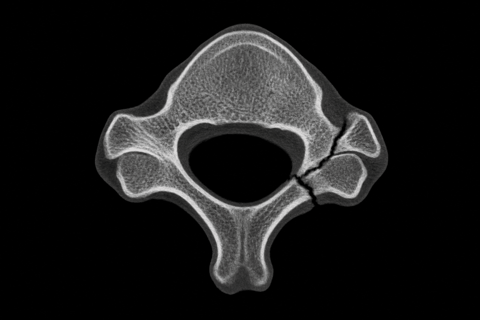
- A floating lateral mass (FLM) fracture-separation of the subaxial cervical spine is defined by a fracture through BOTH the ipsilateral LAMINA and the PEDICLE, which separates the lateral mass from the rest of the vertebra and thereby DISCONNECTS the superior and inferior articular processes (the facet joints above and below that level) - the lateral mass is left 'floating'.
- Because the lateral mass is the structural link between consecutive facet joints, isolating it removes the bony continuity of the posterolateral column and makes the segment HIGHLY UNSTABLE, prone to rotational malalignment, kyphosis and facet subluxation; the injured level often shows the lateral mass rotated into a horizontal ('reverse hamburger') orientation.
- FLM fractures result from HIGH-ENERGY trauma and are frequently associated with NERVE ROOT injury (the exiting root at that level is at risk) and with other cervical fractures or ligamentous injury, so a full assessment of the neurology and the rest of the cervical spine is essential.
- CT is the key investigation to DEFINE the pattern (lamina + pedicle fractures isolating the lateral mass, with assessment of facet alignment); because of the proximity of the lateral mass and pedicle to the TRANSVERSE FORAMEN, and because fracture patterns with DISLOCATION/SUBLUXATION carry the highest risk of BLUNT CEREBROVASCULAR INJURY (BCVI), CT ANGIOGRAPHY should be used to screen for VERTEBRAL ARTERY injury.
- Because the injury is unstable, OPERATIVE stabilisation (fusion) is commonly required and can be performed by an ANTERIOR, POSTERIOR or COMBINED approach; in reported series complications clustered in the posterior (hardware failure) and combined (respiratory) groups, while the anterior approach had fewest - so approach selection should weigh the injury pattern, alignment and associated injuries.
- NON-OPERATIVE management (rigid immobilisation) can be satisfactory for APPROPRIATELY SELECTED FLM fractures - in a reported cohort none of the carefully selected non-operatively treated patients required later surgery for subluxation - so management is individualised to stability, alignment, neurology and the patient, rather than mandating surgery for every FLM.
- “Floating lateral mass = fracture through BOTH lamina AND pedicle -> isolates the lateral mass and DISCONNECTS the facet joints above and below (highly unstable).
- “High-energy; watch for nerve root injury; CT defines it and CTA screens for vertebral artery (BCVI) injury - dislocation/subluxation patterns carry the highest BCVI risk.
- “Often operative (anterior/posterior/combined); selected cases manage non-operatively with rigid immobilisation.
Fracture through both the lamina and the pedicle isolates the lateral mass, so the facet joints above and below are disconnected - the mass 'floats'. Highly unstable.
Nerve root injury at that level, and vertebral artery injury - CTA screen (dislocation/ subluxation patterns carry the highest BCVI risk).
Definition, Instability & Workup
A floating lateral mass fracture is defined by a fracture through both the lamina and the pedicle on one side, which separates the lateral mass from the vertebra and disconnects the superior and inferior articular processes - the facet joints above and below. Because the lateral mass is the bony link between consecutive facets, isolating it removes the posterolateral column's continuity and leaves the segment highly unstable, prone to rotation, kyphosis and facet subluxation (the lateral mass may rotate into a horizontal orientation). These are high-energy injuries, often with nerve root involvement and associated cervical injuries. CT defines the pattern, and because the lateral mass/pedicle lie next to the transverse foramen - and fracture-dislocation/subluxation patterns carry the highest risk - CTA should screen for vertebral artery (BCVI) injury.
Management
- Assess fully. Document neurology (the exiting nerve root is at risk), assess alignment and the rest of the cervical spine, and CTA to screen for vertebral artery injury.
- Operative stabilisation is commonly required for this unstable injury - anterior, posterior or combined fusion. In reported series complications clustered in the posterior (hardware failure) and combined (respiratory) groups, with fewest in the anterior approach - so weigh injury pattern, alignment and associated injuries when choosing.
- Non-operative management (rigid immobilisation) is satisfactory for appropriately selected FLM fractures - in a reported cohort no carefully selected non-operative patient needed later surgery for subluxation.
- Individualise. Base the decision on stability, alignment, neurology and the whole injury picture rather than treating every FLM the same way.
Two associated injuries must not be overlooked in a floating lateral mass fracture. First, the VERTEBRAL ARTERY: the lateral mass and pedicle lie immediately adjacent to the transverse foramen, and fracture patterns with dislocation or subluxation carry the highest risk of blunt cerebrovascular injury, so CT angiography should be used to screen - a missed vertebral artery injury risks a posterior circulation stroke. Second, the NERVE ROOT: the exiting root at the injured level is frequently involved, so the neurological examination must be carefully documented. Recognise the injury as highly unstable because both the facet joints above and below have been disconnected, but individualise the operative-versus-non-operative decision, since carefully selected FLM fractures can be managed non-operatively.
Evidence & Key Studies
Assessing treatment of floating lateral mass (FLM) fractures of the subaxial cervical spine
- An FLM fracture involves separation of the lateral mass from the vertebra via disruption of BOTH the lamina and the pedicle, disconnecting the superior and inferior articular processes - a highly unstable injury.
- In a 10-year cohort of 45 patients (25 non-operative, 20 operative: 6 anterior, 12 posterior, 2 combined), no non-operative patient crossed over to surgery for subluxation.
- Complications occurred in the posterior (hardware failure) and combined (respiratory) groups, with none in the anterior group - suggesting carefully selected non-operative treatment can be satisfactory.
Cervical fracture patterns associated with blunt cerebrovascular injury on CT angiography
- The only fracture pattern associated with increased risk of blunt cerebrovascular injury (BCVI) was a fracture associated with dislocation/subluxation (odds ratio 3.8).
- Combined upper-cervical (occiput-C3) fractures and multilevel fractures were also associated with increased BCVI risk; among screened patients, BCVI was found in 30% and all strokes occurred in BCVI patients.
- The authors recommend CT-angiography screening for any upper-cervical, multilevel, or fracture-with-dislocation/subluxation pattern.
According to PubMed, the definition of the floating lateral mass (lamina + pedicle disruption disconnecting the articular processes), its instability, and the operative-versus-non-operative outcomes (including satisfactory selected non-operative treatment and the approach-specific complications) come from the cited Prezelski cohort; the association of dislocation/subluxation and upper-cervical/multilevel patterns with blunt cerebrovascular (vertebral artery) injury and the recommendation to screen with CT angiography from the cited Du study. The high-energy mechanism, the nerve-root risk and the general principles of subaxial cervical stabilisation are standard, well-established teaching. (See also our Subaxial Cervical Spine Injuries and Facet Dislocation topics.)
Clinical Decision Scenarios
Practise clinical reasoning and management decisions out loud
“What is a floating lateral mass fracture, and why is it unstable?”
“How would you manage a floating lateral mass fracture?”
Mnemonics & Memory Aids
FLOAT
Hook:FLOAT: Fracture lamina+pedicle, Lateral mass floats, Off-line facets (unstable), Artery (CTA)/root, Treat by stabilising or selected non-op.
Definition
- Fracture through BOTH lamina and pedicle isolating the lateral mass
- Disconnects superior and inferior articular processes (facets above and below)
- The lateral mass 'floats' - may rotate horizontal
Why it matters
- Highly unstable (loss of posterolateral column continuity)
- High-energy; nerve root injury common at that level
- Associated cervical fractures/ligamentous injury
Workup
- CT defines the pattern and facet alignment
- CTA to screen for vertebral artery / BCVI
- Dislocation/subluxation and upper-cervical/multilevel patterns = highest BCVI risk
Management
- Often operative stabilisation (anterior/posterior/combined)
- Anterior had fewest complications in series; posterior (hardware), combined (respiratory)
- Selected cases non-operative (rigid immobilisation) - individualise