Arm pain from cervical nerve root compression - C7 most common, 75-90% respond to conservative treatment
- C7 root most commonly affected (C6-7 disc) - triceps weakness, middle finger numbness
- Arm pain greater than neck pain is hallmark - if neck pain predominates, consider other diagnosis
- Spurling test: axial compression + rotation reproduces radicular symptoms
- 75-90% improve with conservative management - trial 6-12 weeks before surgery
- ACDF gold standard surgical treatment with 90-95% success rate
- “Know dermatomal and myotomal patterns for each root cold
- “C5: deltoid/biceps, C6: wrist extensors/brachioradialis, C7: triceps/wrist flexors
- “Progressive motor weakness or myelopathy are surgical indications
- “MRI is gold standard imaging - correlate with clinical findings
Overview and Epidemiology
Cervical radiculopathy is dysfunction of a cervical nerve root from compression or inflammation, producing pain, sensory change or motor weakness in the distribution of that root.
Companion page. This page is the radiculopathy — the nerve-root syndrome, its examination and conservative management. The disc pathology that usually causes it (degeneration, herniation, the ACDF/arthroplasty decision, and the myelopathy that develops when the cord rather than the root is compressed) is set out in cervical disc disease; cervical myelopathy covers the cord syndrome in full.
How common. The Rochester, Minnesota population study gives an age-adjusted annual incidence of 83.2 per 100,000, higher in men (107.3 per 100,000) than in women (63.5 per 100,000). Incidence peaks at 202.9 per 100,000 in the 50-54 year band, with mean onset around 48 years.
Where. C7 is the most commonly affected root, followed by C6, from the C6-7 and C5-6 levels respectively.
What causes it. Disc, spondylosis or both accounted for about 68% of cases, but a confirmed disc protrusion was demonstrated in only about 22% — most cervical radiculopathy is spondylotic rather than a soft disc. Only about 15% reported any preceding trauma or exertion, so the patient who cannot name the moment it started is the rule rather than the exception. Recurrence ran at about 32% over a median 4.9-year follow-up.
Who is at risk. The recognised associations combine mechanical load on the neck with factors that impair the disc or the nerve:
- Heavy manual labour, vibration exposure and prolonged neck flexion
- Smoking, which accelerates disc degeneration
- Diabetes mellitus, through peripheral nerve vulnerability
- Genetic predisposition to disc degeneration
- Prior lumbar radiculopathy, present in about 41% of patients historically
Pathophysiology
The foramen. The cervical root leaves the canal through the intervertebral foramen, bounded anteriorly by the uncovertebral joint, posteriorly by the facet, and above and below by the pedicles. The root itself occupies roughly a third of that space and the remainder is fat and vessels.
The artery. The vertebral artery ascends through the transverse foramina, and it is the structure that sets the lateral limit of a safe decompression.
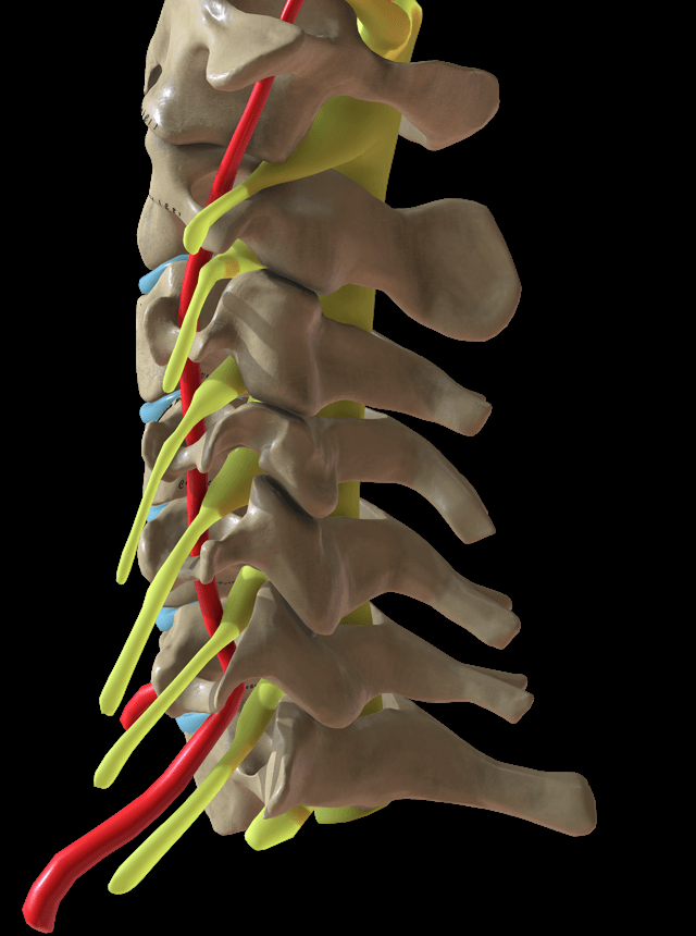
Which root. Cervical roots C1 to C7 exit above the pedicle of their numbered vertebra, and C8 exits between C7 and T1. The root that exits at the level of a disc is the root that disc compresses, so the C5-6 disc takes the C6 root and the C6-7 disc the C7 root. Name a disc by the two vertebrae it lies between; "the C6 disc" is ambiguous.
The lumbar convention runs the other way. A paracentral L4-5 herniation catches the traversing L5 root, while a far-lateral herniation at the same level catches the exiting L4 root instead.
Mechanical compression. A posterolateral herniation compresses the exiting root and is the most common pattern. A central herniation may instead compress the cord and cause myelopathy, and foraminal stenosis compresses the root chronically as uncovertebral and facet osteophytes encroach on the foramen.
Chemical radiculitis. Inflammatory mediators from the disc — phospholipase A2 and TNF-alpha among them — can produce radicular pain without major mechanical compression, which is why pain is sometimes disproportionate to what the scan shows.
Classification
The aetiological label is worth stating because each carries a typical age and tempo.
- Cause
- Acute nucleus pulposus herniation
- Age group
- 20-40 years
- Features
- Sudden onset, often central or centrolateral, may resolve spontaneously
- Cause
- Disc-osteophyte complex, spondylotic stenosis
- Age group
- Over 40 years
- Features
- Insidious and progressive, with cervical spondylosis
- Cause
- Soft and hard components together
- Age group
- —
- Features
- A common presentation
- Cause
- Chemical radiculitis
- Age group
- Any age
- Features
- Pain disproportionate to compression
- Cause
- Fracture, dislocation
- Age group
- Any age
- Features
- Associated with injury
Clinical Presentation
The story. Pain radiates from the neck into the shoulder, arm, forearm and hand in a dermatomal pattern, with paraesthesiae in specific digits and difficulty with specific motor tasks such as grip or lifting. Neck pain is usually the lesser complaint.
What to ask. The answers that change the diagnosis and the plan:
- Onset — sudden points to a soft disc, gradual to spondylosis
- Distribution — a specific dermatome, or vague and non-anatomic
- What makes it worse — extension and rotation, which narrow the foramen
- What makes it better — arm elevation, the shoulder abduction sign
- Red flags — myelopathic symptoms, trauma, weight loss, fever
Arm pain greater than neck pain is the hallmark. If neck pain predominates, question the diagnosis and think of a facet or muscular source instead.
Painless weakness points away from radiculopathy, which usually hurts. Think of a neurological cause such as ALS, or of tendon rupture. Radiculopathy hurts.
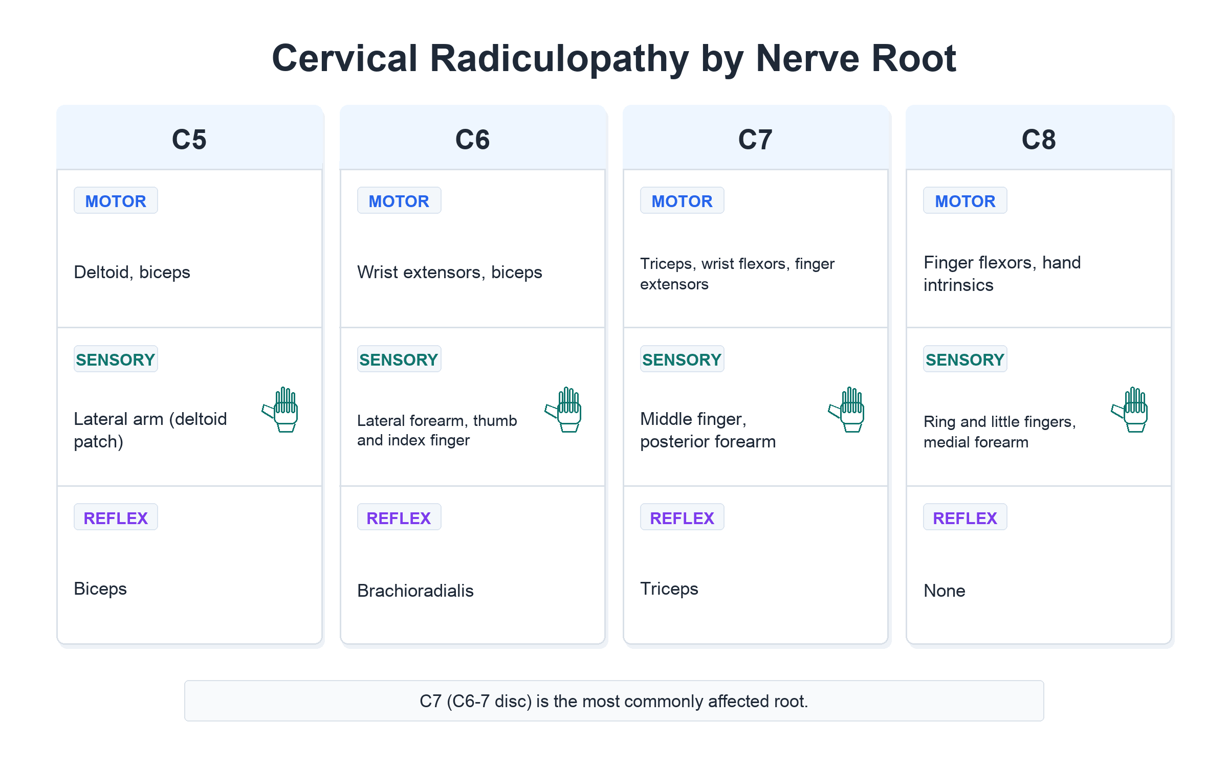
Localising the root. Three findings do it: where the numbness is, which movement is weak, and which reflex has gone.
- Sensory Distribution
- Lateral arm (deltoid patch)
- Motor Weakness
- Deltoid, biceps
- Reflex
- Biceps
- Sensory Distribution
- Lateral forearm, thumb, index finger
- Motor Weakness
- Wrist extensors, biceps
- Reflex
- Brachioradialis
- Sensory Distribution
- Middle finger, posterior forearm
- Motor Weakness
- Triceps, wrist flexors, finger extensors
- Reflex
- Triceps
- Sensory Distribution
- Ring and small fingers, medial forearm
- Motor Weakness
- Hand intrinsics, finger flexors
- Reflex
- None
- Sensory Distribution
- Medial arm
- Motor Weakness
- Hand intrinsics (abductor pollicis)
- Reflex
- None reliable
Provocative tests. Compression closes the foramen; relief on distraction suggests foraminal compression, and shoulder abduction opens the foramen. What counts as positive is reproduction or relief of the patient's own radicular symptoms.
- Technique
- Extend, rotate, axially compress neck
- Positive Finding
- Reproduction of radicular arm pain
- Interpretation
- High specificity, moderate sensitivity
- Technique
- Patient places hand on head
- Positive Finding
- Relief of arm pain
- Interpretation
- Suggests radiculopathy (opens foramen)
- Technique
- Axial traction on head
- Positive Finding
- Relief of symptoms
- Interpretation
- Suggests foraminal compression
- Technique
- Bear down/cough
- Positive Finding
- Increased radicular pain
- Interpretation
- Suggests disc herniation
Screen for myelopathy every time. Concurrent cord compression is the thing that must not be missed, and its signs are found only when they are looked for:
- Hoffman sign (finger flicking causes thumb flexion)
- Ankle clonus
- Lower limb hyperreflexia
- Babinski sign
- Gait disturbance (broad-based, spastic)
- Hand clumsiness (fine motor dysfunction)
Provocative Test Accuracy and the Wainner Cluster
No single provocative test is diagnostic; combining them is far more powerful. Individually, the Spurling test has high specificity (around 90 percent) but low-to-moderate sensitivity, so it rules radiculopathy IN but cannot rule it out, whereas the upper limb tension test (ULTT-A, median-biased) is highly sensitive but not specific, so a negative ULTT-A helps rule radiculopathy OUT.
Wainner described a four-test clinical prediction rule:
- Spurling test (A) — reproduction of radicular arm symptoms; rules in
- Cervical distraction — relief of radicular symptoms with traction; rules in when positive
- Ipsilateral cervical rotation under 60 degrees — reduced motion supports the diagnosis
- Upper limb tension test A (median) — symptom reproduction with a side-to-side difference; the sensitive one, and the useful rule-out
When 3 of the 4 tests are positive the likelihood ratio for cervical radiculopathy is roughly 6 (post-test probability about 65 percent); with all 4 positive the likelihood ratio rises to roughly 30 (post-test probability about 90 percent). Treat those as orders of magnitude rather than precise values: they come from a single derivation study of 82 patients in which the authors noted that the confidence intervals around every likelihood ratio were wide, and the cluster has never been prospectively validated.
The ULTT-A is the upper-limb analogue of the straight-leg raise: scapular depression, shoulder abduction, forearm supination with wrist and finger extension, shoulder external rotation and elbow extension, sensitised by contralateral neck side-bending.
Investigations
The sequence. Plain radiographs come first, to assess alignment and exclude instability. MRI follows when there are neurological symptoms or radiculopathy is suspected. CT is for osseous detail, for surgical planning, and when MRI is contraindicated; CT myelography for the patient who cannot have an MRI or when dynamic assessment is needed.
What the radiographs show. Disc-space narrowing, foraminal stenosis on the oblique views, uncovertebral joint hypertrophy, facet arthropathy, and loss of lordosis or frank kyphosis. Three measurements are worth carrying.
- Normal
- 9-10mm high, 4-6mm wide
- Abnormal
- Diameter under 4mm — high radiculopathy risk
- Normal
- 4-6mm
- Abnormal
- Under 3mm = degeneration
- Normal
- 17-18mm
- Abnormal
- Under 13mm = stenosis
What the MRI shows. The herniation is graded by its shape, and the shape predicts behaviour: a bulge is common and often means nothing, whereas an extruded fragment is usually the one causing the symptoms.
- Description
- Concentric expansion beyond margins
- Significance
- Common, often asymptomatic
- Description
- Focal bulge, base wider than apex
- Significance
- May cause compression
- Description
- Apex wider than base
- Significance
- Usually symptomatic
- Description
- Fragment separated from parent disc
- Significance
- May migrate, often needs surgery
- Description
- Loss of perineural fat on T1
- Significance
- Direct nerve compression
- Description
- T2 hyperintensity
- Significance
- Myelomalacia if present
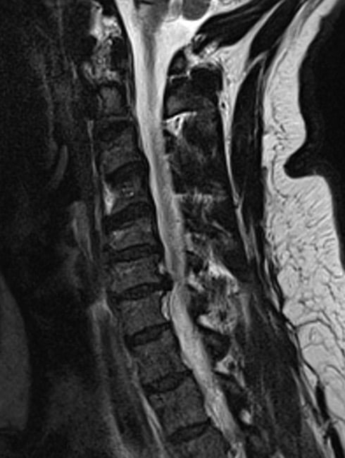
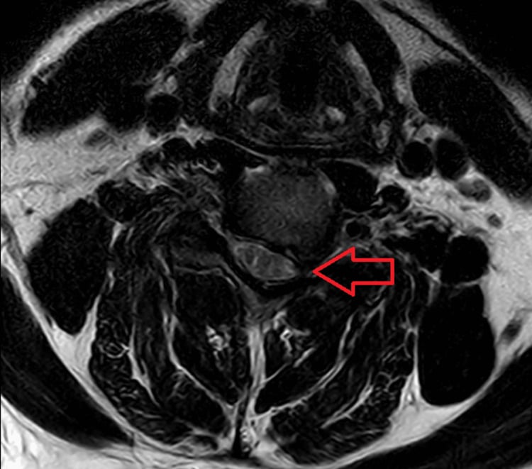
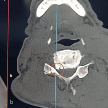
On T2-weighted sagittal images, look for "loss of CSF signal" around the cord (effacement). On axial images, assess foraminal fat signal loss on T1 and nerve root compression. Asymptomatic disc abnormalities are common, so always correlate imaging with the clinical level.
Electrodiagnostics. EMG and nerve conduction studies may help localise the affected root and separate radiculopathy from a peripheral neuropathy, but denervation changes take roughly 3 weeks or more from onset to appear. They are not routine, and earn their place in the atypical or multilevel presentation.
Differential Diagnosis
- Distinguishing Features
- Long tract signs, gait disturbance, bilateral symptoms (radiculopathy is typically unilateral)
- Investigation
- MRI shows cord compression/signal
- Distinguishing Features
- Multiple root pattern, may follow viral illness
- Investigation
- EMG/NCS, MRI of plexus
- Distinguishing Features
- Vascular symptoms, provocation with arm positions
- Investigation
- Doppler, nerve conduction studies
- Distinguishing Features
- Distal to root, Tinel positive at compression site
- Investigation
- EMG/NCS localizes to peripheral nerve
- Distinguishing Features
- Shoulder ROM limited, rotator cuff signs
- Investigation
- Shoulder X-ray, MRI, impingement tests
- Distinguishing Features
- Exertional, risk factors, associated symptoms
- Investigation
- ECG, cardiac enzymes
- Distinguishing Features
- Horner syndrome, severe pain, T1 involvement
- Investigation
- CXR, CT chest
- Distinguishing Features
- Diffuse pain, tender points, no neurological deficit
- Investigation
- Normal investigations, clinical diagnosis
Where each of these is worked up. The differential here is really a list of other diagnoses, each with its own page: cervical myelopathy when the picture is cord rather than root and the clock changes entirely; cervical disc disease for the disc itself and the operative thresholds; thoracic outlet syndrome for the positional lower-trunk mimic; cubital tunnel syndrome and carpal tunnel for the two peripheral compressions that most often masquerade as C8 and C6 roots; and rotator cuff tears for the shoulder pain that never was radicular. When the trial fails, the operation is ACDF.
The Double Crush Phenomenon
Cervical radiculopathy and a distal peripheral nerve entrapment can coexist and interact. The double crush phenomenon (Upton and McComas, 1973) proposes that a proximal compression of a nerve - for example a cervical root - impairs axoplasmic transport and renders the same axons more vulnerable to a second, more distal compression, such as carpal tunnel or cubital tunnel syndrome. Each lesion may be individually subclinical yet symptomatic in combination.
- Implication
- Symptoms may not fully fit a single root or single peripheral nerve
- Implication
- Consider an unaddressed second site
- Implication
- A distal lesion can sensitise the nerve to a proximal one
- Implication
- Careful examination plus EMG/NCS to localise both levels
Clinically, suspect double crush when the pattern is atypical, when EMG shows changes at more than one level, or when symptoms persist after treating only one site. Both sites may need to be addressed, and the more symptomatic or clearly compressive lesion is usually treated first.
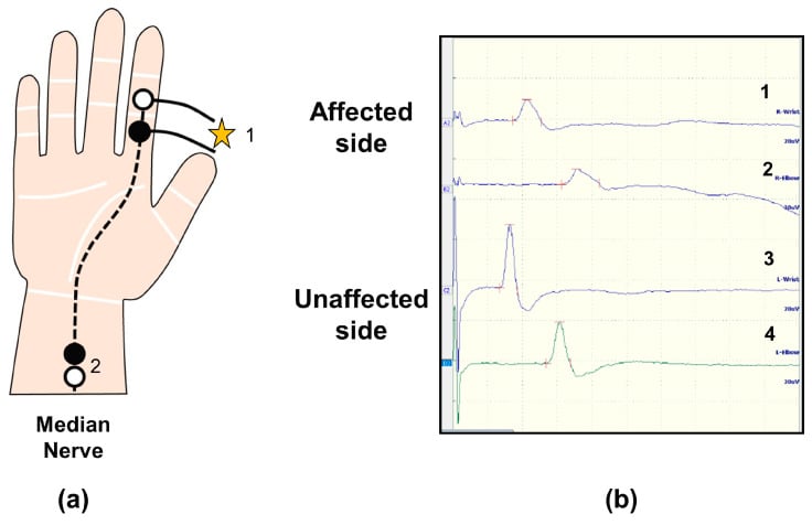
The double crush phenomenon explains why a cervical root compression and a distal entrapment (carpal or cubital tunnel) can coexist and reinforce each other through impaired axoplasmic flow. Persisting symptoms after decompressing one site should prompt a search for the second.
Management
Decision Sequence
- Exclude myelopathy, instability, infection, tumour and progressive motor deficit; these override a routine conservative trial.
- With stable neurology, begin activity modification, analgesia and structured physiotherapy, then reassess symptoms and strength over 6-12 weeks.
- If disabling concordant radicular pain persists, match the clinical root to MRI or CT pathology before considering injection or surgery.
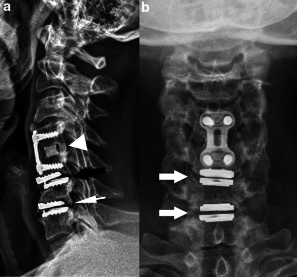
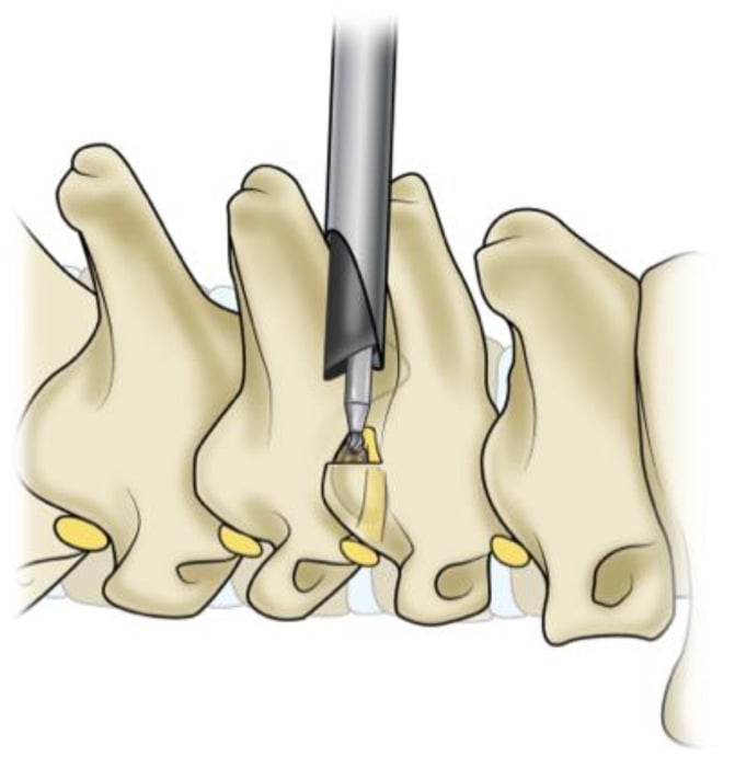
- Choose ACDF for central or multilevel pathology, arthroplasty for carefully selected motion-preservation candidates, and posterior foraminotomy for unilateral lateral pathology without instability or kyphosis.
Complications
Non-operative care has its own risks, which come from the drugs, from the injection, and from watching a deficit that is progressing.
- Cause
- Inadequate treatment
- Prevention/Management
- Multimodal approach
- Cause
- Prolonged use
- Prevention/Management
- PPI cover, limit duration
- Cause
- Natural history
- Prevention/Management
- Monitor closely, early surgery if progressive
- Cause
- ESI complication
- Prevention/Management
- Fluoroscopic guidance, interlaminar approach
After surgery, dysphagia is the most common complication and is usually transient; the rest are uncommon, and each carries a rate worth being able to quote.
- Rate
- 20-50% transient, 1-2% persistent
- Management
- Usually resolves over 2-4 weeks, soft diet
- Rate
- 1-2%
- Management
- Speech therapy, often recovers
- Rate
- Under 0.5%
- Management
- Primary repair if recognised, NPO
- Rate
- 0.5-1%
- Management
- Primary repair, fibrin glue, lumbar drain
- Rate
- Under 0.5%
- Management
- Avoid lateral dissection, careful burr technique
- Rate
- 3-10%
- Management
- CT assessment, revision if symptomatic
- Rate
- ~2.9% per year (cumulative)
- Management
- Surveillance, may require extension of fusion
- Rate
- 1-5% (higher posterior)
- Management
- Usually transient, recovers over ~6 months
ACDFACDF Complications
Hook:The procedure name lists the complications!
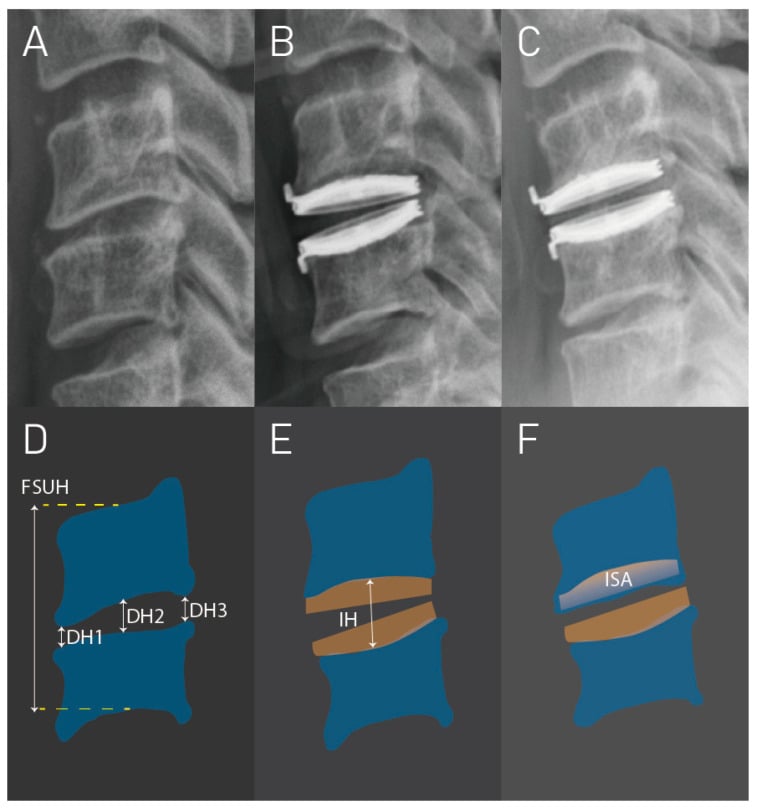
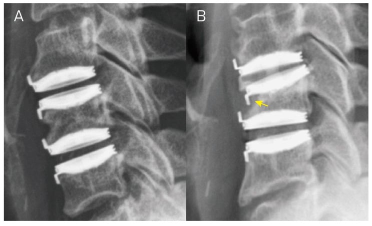
Subsidence is the arthroplasty and cage complication that a follow-up film is looking for, and it is graded by how much height the segment has lost.
Postoperative Care
Immediate (day 0-2). Monitor the airway, the risk being a haematoma, and assess the swallow before allowing diet. Mobilise early, with a soft collar if needed though it is not mandatory, and give DVT prophylaxis.
Early (weeks 1-6). Wound check at 2 weeks, a soft diet while there is dysphagia, cervical precautions with no heavy lifting, gentle range of motion from week 2, and sedentary work at 2-4 weeks.
Intermediate (weeks 6-12). Radiographs at 6 weeks, start physiotherapy, return to activities gradually, and manual work typically at 8-12 weeks.
Late (3-12 months). Assess the fusion, with CT if there is concern, full activity by 3-6 months, then watch for adjacent segment disease.
- Milestone
- Mobilizing, oral intake
- Milestone
- Return to light activities
- Milestone
- Driving resume
- Milestone
- Return to work (most occupations)
- Milestone
- Full recovery expected
Every source quotes a proportion who improve. Almost none quotes the TIMELINE, and the timeline is what the patient is actually asking about.
The best synthesis is a systematic review of the course and prognosis of symptomatic cervical disc herniation with radiculopathy (Wong et al, Spine J 2014, PMID 24614255), which found:
- Patients present with intense pain and moderate disability - the starting point is genuinely bad, and saying so protects your credibility when you then advise waiting.
- Substantial improvement occurs within the first 4 to 6 months. This is the part that matches the 6-to-12-week conservative trial: it is enough time to see the trajectory, not enough to see the endpoint.
- Complete recovery took 24 to 36 MONTHS in approximately 83%. Two to three years, not two to three months.
So "75-90% improve" and "83% recover" are not rival figures - they measure different things at different times. Improvement is early and common; complete resolution is late. A patient told "90% get better" who is still symptomatic at nine months believes the treatment has failed, when the published course says they are on it.
And be careful with the other number attached to this condition: the often-quoted 90% from the Rochester population study is a good-outcome figure at a median 4.9 years in a cohort where 26% had surgery and 31.7% recurred (PMID 8186959) - it is not a conservative success rate. See the evidence card above, which states it correctly.
Guidelines, Registries & Global Practice
Global Epidemiology
Population-based data (Rochester, Minnesota) give an age-adjusted annual incidence of ~83 per 100,000, peaking at ~203 per 100,000 in the 50-54 year age band, with C7 the most commonly affected root. The natural history is favourable: ~90% of patients are asymptomatic or only mildly affected at long-term follow-up.
Side-by-Side Society Guidance
- Key Recommendation
- MRI is the imaging modality of choice; a trial of conservative care is appropriate before surgery in patients without progressive deficit
- Key Recommendation
- Multimodal non-operative care first-line; surgery reserved for progressive deficit, myelopathy, or failed conservative management
- Key Recommendation
- Emphasise early reassurance, activity, and physiotherapy; image only when management would change or red flags are present
- Key Recommendation
- ACDF remains the benchmark; arthroplasty for selected single/two-level disc disease; posterior foraminotomy for lateral soft disc
Registry and Trial Evidence
- Randomized data (Engquist, Spine 2013) show surgery accelerates recovery but the advantage over structured physiotherapy narrows by 2 years - supporting an initial conservative trial in non-progressive cases.
- For lateral soft-disc pathology, motion-preserving posterior foraminotomy is equivalent to ACDF at 2 years (Ruetten, Spine 2008).
- Cervical disc arthroplasty preserves index-level motion with comparable or better outcomes than ACDF at 2 years in selected single-level disease (Heller/BRYAN, Spine 2009); arthroplasty registries and IDE data show lower reoperation for adjacent-level disease at longer follow-up.
High- vs Limited-Resource Variation
- Practice Pattern
- Early MRI access, arthroplasty and navigation available, faster pathway to elective surgery
- Practice Pattern
- Greater reliance on clinical diagnosis and plain radiographs; ACDF favoured over arthroplasty on cost grounds; longer waits for elective decompression
MCQ Practice Points
High-Yield Concepts
- C7 is the most commonly affected root, typically from the C6-7 disc level
- Cervical roots exit ABOVE their corresponding vertebra (unlike the lumbar spine)
- Spurling test: high specificity, moderate sensitivity
- Arm pain greater than neck pain is the classic radiculopathy pattern
- 75-90% improve with conservative management - trial for 6-12 weeks. But "improve" is not "recovered": complete recovery took 24-36 months in ~83% of patients in a systematic review (PMID 24614255)
- Progressive motor weakness is an indication for urgent surgery
- ACDF vs foraminotomy: ACDF for central disc, foraminotomy for lateral soft disc
- Dysphagia is the most common ACDF complication (usually transient)
Common Examination Pitfalls
- Missing myelopathy signs in a radiculopathy patient
- Attributing symptoms to the wrong root level
- Not checking reflexes systematically
- Forgetting that C8 exits between C7 and T1
- Overlooking progressive weakness as a surgical emergency
- Not differentiating from peripheral entrapment
Q: Which cervical nerve root is most commonly affected by radiculopathy and what is the typical disc level?
A: C7 root from a C6-7 disc herniation. Cervical roots exit ABOVE their corresponding vertebra (unlike the lumbar spine). The C7 root exits between C6 and C7, so a C6-7 disc herniation affects C7.
Q: How do you differentiate C6 vs C7 radiculopathy on clinical examination?
A: C6 radiculopathy: weakness of biceps and wrist extension, diminished biceps and brachioradialis reflexes, sensory loss in thumb and lateral forearm. C7 radiculopathy: weakness of triceps and wrist flexion, diminished triceps reflex, sensory loss in the middle finger.
Q: How should the Spurling test be interpreted?
A: It has high specificity but only moderate sensitivity. A positive test (reproduction of radicular symptoms with extension, lateral flexion, and axial compression) strongly supports radiculopathy, but a negative test does not rule it out.
Q: What are the surgical indications for cervical radiculopathy and which procedure is most appropriate?
A: Urgent surgery: progressive motor weakness or myelopathy signs. Elective surgery: failed 6-12 weeks of conservative management with persistent symptoms. Procedure choice: ACDF for central/paracentral disc or with axial neck pain; posterior foraminotomy for lateral soft disc without instability.
Exam Day Cheat Sheet
Key Diagnosis Facts
- Definition: Nerve root dysfunction from compression/inflammation
- Most common root: C7, then C6, from the C6-7 and C5-6 levels
- Classic pattern: Arm pain greater than neck pain in a dermatomal distribution
Level Specifics
- C6 pattern: Thumb/index numbness, wrist extension weakness, brachioradialis reflex
- C7 pattern: Middle finger numbness, triceps weakness, triceps reflex
Management & Outcome
- Key test: Spurling - extension + rotation + axial load, high specificity
- Conservative success: 75-90% improve in 6-12 weeks
- Surgery indication: Progressive weakness, refractory at 6-12 weeks, severe deficit
- ACDF success: 90-95% for radiculopathy
- Main complication: Dysphagia (20-50% transient)
Frequently asked questions
Which nerve root causes numbness in the thumb and index finger?
The C6 nerve root, usually from a C5-6 disc herniation or foraminal stenosis. C6 radiculopathy gives sensory change over the thumb and index finger, weakness of the wrist extensors and biceps (brachioradialis), and a reduced brachioradialis or biceps reflex.
Which nerve root causes middle finger numbness?
The C7 nerve root - the most commonly affected root in cervical radiculopathy, typically from the C6-7 disc. The pattern is middle finger sensory change with triceps weakness, weak wrist flexion, and a reduced triceps reflex.
How do you distinguish C6 from C7 radiculopathy at the bedside?
Localise by three findings. Sensory: thumb and index finger point to C6, the middle finger to C7. Motor: weak wrist extension and elbow flexion point to C6, weak elbow extension (triceps) and wrist flexion point to C7. Reflexes: brachioradialis and biceps are lost in C6, the triceps jerk in C7.
Why does a C6-7 disc prolapse affect the C7 nerve root?
In the cervical spine each root exits above the pedicle of its numbered vertebra, so the root traversing the C6-7 foramen is C7 - the opposite numbering rule to the lumbar spine, where a paracentral L4-5 disc hits the traversing L5 root. Cervical roots C1-7 exit above their vertebrae and C8 exits between C7 and T1.
Do most patients with cervical radiculopathy need surgery?
No - 75-90 percent improve with conservative treatment over 6-12 weeks (activity modification, analgesia, physiotherapy, with selective foraminal epidural steroid injection as an option). Surgery - anterior cervical discectomy and fusion or posterior foraminotomy - is reserved for progressive motor deficit, myelopathic signs, or persistent disabling pain despite adequate conservative care.
Clinical Decision Scenarios
Practise clinical reasoning and management decisions out loud
“A 45-year-old office worker presents with 4 weeks of right arm pain radiating from the neck to the thumb and index finger. He has numbness in these digits and weakness of wrist extension. Examination shows reduced brachioradialis reflex.”
“A 52-year-old presents with 3 weeks of left arm pain and progressive weakness. Initially had grip weakness but now cannot lift her arm against gravity. MRI shows large C6-7 disc extrusion with severe C7 root compression.”
“A 60-year-old man has bilateral arm symptoms with C5-6 and C6-7 disc disease on MRI. He has C6 and C7 dermatomal symptoms, mild weakness of right triceps, and subtle hyperreflexia in the legs.”
Evidence Base & Outcomes
Epidemiology of Cervical Radiculopathy (Landmark Population Study)
- Population-based survey, Rochester Minnesota 1976-1990, 561 patients
- Age-adjusted annual incidence 83.2 per 100,000; peak 202.9 per 100,000 at age 50-54
- C7 monoradiculopathy most frequent, followed by C6
- Disc/spondylosis cause in ~68%; confirmed disc protrusion in ~22%
- At final follow-up 90% asymptomatic or only mildly incapacitated; recurrence ~32%
Cervical Collar or Physiotherapy vs Wait-and-See (Landmark Conservative RCT)
- RCT of 205 patients with recent-onset cervical radiculopathy (under 1 month)
- Semi-hard collar with rest or physiotherapy vs wait-and-see
- Both active treatments gave significant additional arm and neck pain reduction at 6 weeks
- Wait-and-see group still improved (arm pain fell ~19 mm over 6 weeks)
- Supports a short course of collar or physiotherapy early in the disease
Surgery (ACDF) plus Physiotherapy vs Physiotherapy Alone (Landmark Surgical RCT)
- Prospective RCT, 63 patients with cervical radiculopathy, 2-year follow-up
- ACDF plus physiotherapy vs structured physiotherapy alone
- Surgery gave faster improvement and better neck pain/global outcome at 12 months
- At 24 months between-group differences largely disappeared
- Authors conclude structured physiotherapy should be tried before surgery
References
- Radhakrishnan K, Litchy WJ, O'Fallon WM, Kurland LT. Epidemiology of cervical radiculopathy: a population-based study from Rochester, Minnesota, 1976 through 1990. Brain 1994;117:325-35. PMID 8186959.
- Kuijper B, Tans JTJ, Beelen A, Nollet F, de Visser M. Cervical collar or physiotherapy versus wait and see policy for recent onset cervical radiculopathy: randomised trial. BMJ 2009;339:b3883. PMID 19812130.
- Engquist M, Lofgren H, Oberg B, et al. Surgery versus nonsurgical treatment of cervical radiculopathy: a prospective, randomized study. Spine 2013;38:1715-22. PMID 23778373.
- Ruetten S, Komp M, Merk H, Godolias G. Full-endoscopic cervical posterior foraminotomy for lateral disc herniations: a prospective, randomized, controlled study. Spine 2008;33:940-8. PMID 18427313.
- Heller JG, Sasso RC, Papadopoulos SM, et al. Comparison of BRYAN cervical disc arthroplasty with anterior cervical decompression and fusion: a randomized, controlled trial. Spine 2009;34:101-7. PMID 19112337.
- Diwan S, Manchikanti L, Benyamin RM, et al. Effectiveness of cervical epidural injections in the management of chronic neck and upper extremity pain. Pain Physician 2012;15:E405-34. PMID 22828692.