Radiculopathy | Dermatomal Patterns | ACDF Indications
- C5-C6 and C6-C7 are most common levels
- MRI is gold standard imaging
- The famous 90% is a 5-YEAR outcome from a cohort in which 26% had surgery - not a conservative resolution rate
- ACDF gold standard for surgical cases
- Progressive motor weakness is urgent surgical indication
- “C5 = deltoid and biceps, biceps reflex
- “C6 = brachioradialis reflex, wrist extension, thumb
- “C7 = triceps, middle finger (most common)
- “Spurling sign confirms radicular origin; Bakody relief points the same way
- “Red flags: myelopathy, progressive deficit
Overview and Epidemiology
Cervical disc disease covers both the acute soft disc herniation and the chronic spondylotic hard disc, and the same pathology can present as a nerve root syndrome, a cord syndrome, the two together, or axial neck pain with no signs of neural compression at all. C5-C6 and C6-C7 are the levels that fail most often, C6-C7 most commonly of all.
Risk factors. What to ask about in the history:
- Degenerative change with age
- Smoking
- Heavy manual labour
- Prior disc disease
- Genetic predisposition
Most cervical radiculopathy does settle without surgery, and a 6 to 12 week conservative trial is right. But the "90%" everyone attaches to that sentence does not say what it is used to say.
It comes from the Rochester population study of 561 patients (Radhakrishnan, Brain 1994, PMID 8186959), and the actual finding is: at last follow-up - median 4.9 YEARS - 90% were asymptomatic or only mildly incapacitated. Three things follow.
- It is not a conservative-management figure. 26% of that cohort underwent surgery. The 90% good outcome includes them. Quoting it as "90% resolve conservatively" inverts the study.
- It is not a 6-to-12-week figure. It is an outcome at nearly five years. Nothing in it describes how many settle within a conservative trial.
- "Asymptomatic or only mildly incapacitated" is not "resolved." And the same paper reports recurrence in 31.7% - a fact worth more to a patient than the 90% ever was.
What to say instead. Most patients improve enough with time and non-operative care that surgery is not required, a 6 to 12 week trial is reasonable in the absence of progressive deficit or myelopathy, roughly a quarter of a population-based cohort eventually had surgery, and about a third had a recurrence. Surgery is indicated for failed conservative treatment, progressive deficit, or myelopathy.
Companion pages. This page is the disc disease - the pathology, the ACDF and arthroplasty decision, and the myelopathy. If the picture is cord rather than root - gait, hands, hyperreflexia - the syndrome, its grading and its urgency belong to cervical myelopathy, where delay costs recovery rather than comfort. The nerve-root syndrome, with its dermatomal patterns, examination cluster and conservative management, is set out in cervical radiculopathy, and when the trial fails the operation is ACDF. Before accepting a root at all, note that two of the commonest mimics are peripheral rather than spinal - cubital tunnel syndrome for the C8 picture and thoracic outlet syndrome for the positional lower-trunk one.
Pathophysiology and Mechanisms
The disc. The nucleus pulposus is the central gelatinous material, with a high water content, and it does the cushioning; the annulus fibrosus is the outer fibrous ring that contains it and attaches to the vertebrae. The posterior longitudinal ligament is thin in the cervical spine, so there is less protection against a posterolateral herniation.
The uncovertebral joints of Luschka sit posterolaterally and are a common site of osteophyte formation - the same corner of the foramen a posterolateral herniation occupies. So hard disc and soft disc arrive at the root by the same route.
How compression happens. Disc degeneration costs height and provokes osteophyte formation, the neural foramen narrows, and the neural element is compressed; inflammatory mediators act alongside the mechanical compression.
The foramen and the cord. The foramen is bounded by the disc in front and the facet behind, and osteophyte from either narrows it, which compresses the exiting root. The cord sits centrally in the canal, where a central disc-osteophyte complex compresses it: myelopathy rather than radiculopathy.
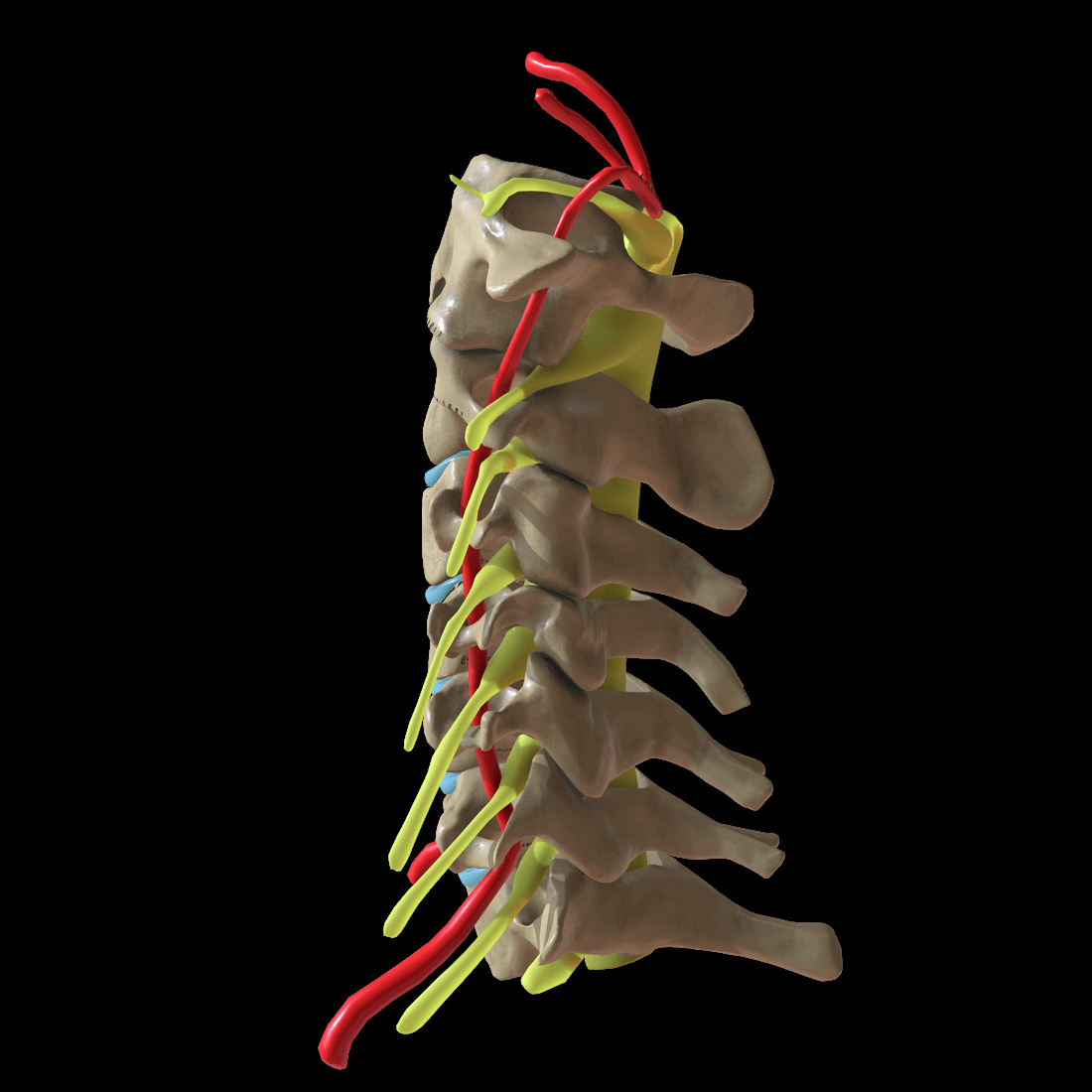
Why these levels. Flexion and extension are greatest at C5-C6, rotation at C1-C2 (atlantoaxial), and lateral bending is coupled with rotation. C5-C6 and C6-C7 carry the most motion and the most stress, which is why disc disease concentrates there - and why preserving motion is the argument for arthroplasty over fusion.
Cervical roots exit ABOVE their numbered vertebra, so a C5-C6 disc compresses the C6 root. The lumbar spine runs the other way: roots exit below, so an L4-L5 disc takes the L5 root, and the L5 root itself exits at L5-S1.
C8 is the exception, because there is no C8 vertebra: the C8 root exits at C7-T1. Matching the level on the scan to the root in the arm depends on getting this right.
Disc Herniation Nomenclature
The standard radiological lexicon separates a disc bulge from a true herniation and then grades herniations by shape and by continuity with the parent disc. This matters clinically because the morphology predicts the likelihood of spontaneous resorption and therefore supports the conservative-first approach.
- Definition
- Generalised extension of disc beyond the endplate margins over more than a quarter of the circumference
- Clinical Relevance
- Usually degenerative; rarely the sole cause of radiculopathy
- Definition
- Focal displacement where the base against the parent disc is wider than the displaced material
- Clinical Relevance
- Often contained; less likely to resorb spontaneously
- Definition
- Displaced material with a neck narrower than the fragment, or material extending above or below the disc space
- Clinical Relevance
- Often uncontained; more likely to resorb
- Definition
- Extruded fragment that has lost all continuity with the parent disc (free fragment)
- Clinical Relevance
- Uncontained; highest spontaneous resorption but can migrate
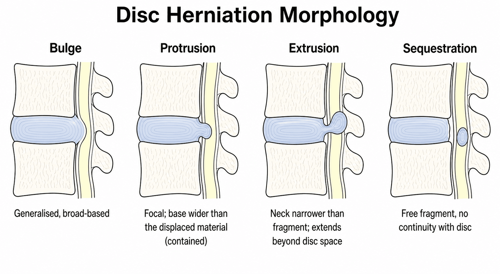
A herniation is additionally described as contained when the annulus or posterior longitudinal ligament still covers the displaced material, and uncontained when that barrier is breached.
Extruded and sequestered (uncontained) fragments provoke a macrophage-mediated inflammatory resorption and are more likely to shrink without surgery than a contained protrusion. This is the anatomical basis for the point that soft-disc herniations may resolve naturally, and it supports a conservative trial in the absence of progressive deficit or myelopathy.
Classification Systems
By pathology. The first question about a compressive lesion is whether it is soft or hard, because that decides both whether it can resolve and what the operation has to remove.
- Description
- Acute herniation
- Treatment Implication
- May resorb, good surgical outcome
- Description
- Osteophyte, chronic
- Treatment Implication
- May require osteophyte removal
- Description
- Disc plus osteophyte
- Treatment Implication
- Common, address both elements
A soft disc herniation may resolve naturally; a hard disc rarely does.
By clinical syndrome. The second question is which syndrome is in front of you, because that sets the urgency rather than the technique.
- Features
- Single root, sensory plus or minus motor
- Urgency
- Elective if no progression
- Features
- Long tract signs, gait, hands
- Urgency
- Urgent - early surgery better
- Features
- Combined features
- Urgency
- Urgent
- Features
- No neural compression signs
- Urgency
- Conservative, rarely surgical
Myelopathy requires urgent attention, and outcomes are worse with delay.
Myelopathy Severity Grading (mJOA and Nurick)
Once myelopathy is diagnosed it should be quantified, because severity guides the urgency of surgery and predicts recovery. Two scales are used.
The modified Japanese Orthopaedic Association (mJOA) score is an 18-point scale, where higher is better, summing four domains: upper-limb motor function (0 to 5), lower-limb motor function (0 to 7), upper-limb sensation (0 to 3) and bladder (sphincter) function (0 to 3). It defines the standard severity bands used in AO Spine pathways.
- mJOA Score
- 15 to 17
- Management Implication
- Surveillance versus surgery debated, but prospective data still show benefit from decompression
- mJOA Score
- 12 to 14
- Management Implication
- Surgical decompression recommended
- mJOA Score
- Under 12 (0 to 11)
- Management Implication
- Urgent decompression; recovery limited if longstanding or with cord signal change
The older Nurick grade (0 to 5) is a purely gait and ambulation-based scale: grade 0 root signs only without cord involvement; grade 1 cord signs without walking difficulty; grade 2 mild gait difficulty not preventing employment; grade 3 gait difficulty preventing employment; grade 4 walking only with assistance or a frame; grade 5 chairbound or bedridden.
Record the score before the operation is discussed: the debate about timing in mild disease turns on which band the patient is in.
Clinical Assessment
History. Take the pain first: where it sits in the neck and how far down the arm it travels, in a radicular pattern or not. Then the sensory symptoms, numbness and tingling, and the motor ones, weakness and clumsiness. Extension and rotation of the neck are the movements that aggravate it, and the answers that change the pathway are the red flags - gait disturbance, bladder symptoms, and bilateral arm symptoms.
Examination. The Spurling test reproduces the radicular pain and confirms that the origin is a root. Bakody's sign, the shoulder abduction relief sign, works in the opposite direction: pain eases with the arm lifted over the head, and that relief is a cervical radiculopathy finding. Then work through the motor groups (deltoid, biceps, triceps, grip), the dermatomal sensory pattern and the reflexes, and finish with the upper motor neurone signs that say the problem is the cord and not the root.
- Motor
- Deltoid, biceps
- Reflex
- Biceps
- Sensory
- Lateral arm
- Motor
- Biceps, wrist extensors
- Reflex
- Brachioradialis
- Sensory
- Thumb, lateral forearm
- Motor
- Triceps, wrist flexors
- Reflex
- Triceps
- Sensory
- Middle finger
- Motor
- Finger flexors, intrinsics
- Reflex
- None reliable
- Sensory
- Small finger, medial forearm
Do not miss myelopathy. Gait disturbance, hand clumsiness (buttons), hyperreflexia, Hoffmann sign, upgoing plantars (Babinski) and clonus are the findings that move a patient from a clinic problem to an urgent referral and imaging. A radiculopathy is root pain and weakness; myelopathy is balance, dexterity and upper motor neurone signs, and missing it fails the station.
Differential Diagnosis
Arm pain and hand symptoms have many causes outside the cervical disc. The examiner will reward a structured differential that distinguishes neural compression from peripheral entrapment, referred pain and non-musculoskeletal mimics.
- Distinguishing Features
- Dermatomal pain, positive Spurling, relief with Bakody (shoulder abduction)
- Confirmatory Test
- MRI plus clinical correlation, EMG/NCS
- Distinguishing Features
- Bilateral/gait symptoms, hand clumsiness, UMN signs (Hoffmann, hyperreflexia)
- Confirmatory Test
- MRI cord compression plus or minus signal change
- Distinguishing Features
- Median distribution, nocturnal, Tinel/Phalen positive, no neck pain
- Confirmatory Test
- Nerve conduction studies
- Distinguishing Features
- Ulnar distribution (small/ring finger), elbow flexion provokes
- Confirmatory Test
- NCS across elbow
- Distinguishing Features
- Positional symptoms, vascular features, lower trunk (C8-T1) pattern
- Confirmatory Test
- Provocative tests, vascular imaging
- Distinguishing Features
- Acute severe shoulder pain then patchy weakness, non-dermatomal
- Confirmatory Test
- EMG, clinical course
- Distinguishing Features
- Pain with shoulder movement, no neurology, painful arc
- Confirmatory Test
- Shoulder exam, ultrasound/MRI
- Distinguishing Features
- Lower trunk symptoms, Horner syndrome, weight loss, smoker
- Confirmatory Test
- Chest imaging, urgent CT
Sinister causes of neck and arm symptoms have to be excluded before the pain is treated as degenerative, because they change the pathway entirely. Pancoast tumour, with Horner syndrome, T1 weakness and a smoking history, is in the differential above. The other two are infection or discitis - fever, raised inflammatory markers, immunosuppression - and metastatic disease, with night pain or a known primary.
Investigations
MRI is the gold standard. Its soft tissue detail is what the diagnosis and the surgical decision rest on. Request it for suspected radiculopathy or myelopathy, for failed conservative treatment, for a progressive neurological deficit, and for preoperative planning.
What to assess on the scan:
- The level and side of the herniation
- Neuroforaminal stenosis
- Cord compression, and any signal change within the cord
- Whether the disease is multilevel
T2 signal change within the cord is myelomalacia, and it carries a poorer prognosis.

Radiographs. AP, lateral and flexion-extension views. They show what the bone is doing - disc space narrowing, osteophyte formation, foraminal narrowing, alignment including kyphosis, and instability on the dynamic views - and they are useful for that overall assessment, but they cannot see soft tissue.
CT gives the better bone detail, so it is the study for assessing an osteophyte, and CT myelography is the fallback when MRI is contraindicated.
EMG and nerve conduction studies confirm a radiculopathy, distinguish it from a peripheral neuropathy and show denervation changes; they earn their place when the clinical and radiological pictures do not match. The changes take 2 to 3 weeks to develop after the injury.
Management Algorithm
The decision. A pure radiculopathy with no myelopathy and no motor deficit gets a conservative trial of 6 to 12 weeks, and most settle in that window. Weakness with grade 4 power or better still earns a conservative trial, with close follow-up. A progressive motor deficit is an urgent surgical problem, decompressed by ACDF, and myelopathy is a surgical indication in its own right.
Conservative care runs in three phases, the last of which is a decision point.
Conservative Protocol
Activity modification. Analgesia (NSAIDs, neuropathic agents). Soft collar short-term if needed.
Physiotherapy. Postural education. Cervical exercises. Traction if helpful.
Reassess symptoms and function. If improving, continue. If not, consider intervention.
Injections. A cervical epidural steroid injection may give temporary relief and has diagnostic value; the risks are cord injury and infection. A selective nerve root block is both diagnostic and therapeutic, and it confirms which root is generating the pain. Evidence for long-term benefit is limited, but either can bridge a patient to recovery.
When to operate. Four situations turn a non-operative problem into an operative one, and myelopathy is the most urgent of them, because earlier surgery yields better outcomes.
FAILSurgical Indications
Hook:If conservative FAILS, consider surgery!
Surgical Technique
ACDF is the standard operation for single or two-level disease. The anterior Smith-Robinson exposure passes between the carotid sheath and the midline, and the disc is excised under the microscope or loupes, which removes the offending disc directly, decompresses the neural elements and restores disc height.
The key steps:
- Complete discectomy, including the posterior annulus
- Bilateral decompression of the neural foramina
- Endplate preparation that preserves the subchondral bone
- Cage placement, PEEK or titanium
- Plate fixation, optional at a single level
Fusion rates are over 95% with modern cage-and-plate technique.
Cervical disc arthroplasty preserves motion at the treated level, and the argument for it is a reduction in adjacent segment disease - reduced, not eliminated. It suits single-level disease with preserved disc height, no significant facet arthropathy and a young patient, and it is unsuitable for instability or multilevel disease. For radiculopathy the outcomes are similar to ACDF, and adjacent segment disease rates may be lower.
Posterior approaches avoid the anterior risks, dysphagia and the recurrent laryngeal nerve, and carry limitations of their own. A posterior foraminotomy deals with a lateral disc herniation and preserves motion, but does nothing for central stenosis. Laminoplasty or laminectomy is the answer to multilevel disease and myelopathy, preserving the posterior elements or sacrificing them respectively.
A left-sided approach is preferred for a primary ACDF, because the right recurrent laryngeal nerve has the more variable course. The side chosen does not excuse the rest of the care: protect the oesophagus and avoid excessive retraction.
Complications
- Incidence
- 5-30% early
- Notes
- Retraction-related
- Incidence
- 1-5%
- Notes
- Usually temporary, protect nerve
- Incidence
- 5.3% pooled overall; 3.3% ACDF, 7.5% corpectomy, 11.0% laminectomy with fusion
- Notes
- Delayed onset after an uneventful decompression
- Incidence
- Well under 1%
- Notes
- Rare but potentially fatal. Stay medial to the uncinate processes; know your preoperative anatomy, since a medially deviant artery is the usual setting
- Incidence
- 2-5%
- Notes
- May need revision; recurrent symptoms from pseudarthrosis or incomplete decompression
- Incidence
- 2.9% per year (Hilibrand)
- Notes
- 25.6% at 10 years; more than two-thirds of those affected needed a further operation
- Incidence
- Very rare
- Notes
- Devastating, meticulous technique
A patient who woke neurologically intact after a technically satisfactory decompression develops deltoid and biceps weakness a day or two later, sometimes with shoulder pain and patchy numbness over the badge area. This is C5 palsy, and it is not evidence that the operation went wrong.
It is a myelopathy phenomenon, not a nerve-stretch phenomenon. In pooled data across 107 studies, anterior decompression performed for radiculopathy carried an incidence of essentially zero (95% CI 0 to 1%), while for myelopathy it was 4% anteriorly and 7% posteriorly, with no significant difference between the two approaches. If tethering of a short C5 root by posterior cord drift were the mechanism, posterior surgery would be far worse than anterior; it is not. What separates the groups is the presence of a compressed cord, which points to reperfusion injury of the cord grey matter.
Practical consequences. Consent a myelopathic patient for it explicitly and a radiculopathy patient proportionately - the risks are genuinely different. If it occurs, first exclude a compressive cause (haematoma, graft or construct malposition) with urgent imaging, then treat expectantly: most cases are unilateral and most recover, though recovery can take many months and deltoid power may not return fully. Risk tracks the operation, highest after laminectomy with fusion and lowest after ACDF, and it is higher in men.
Postoperative Care
ACDF Recovery
Monitor swallowing. Watch for haematoma and airway compromise. Mobilise day 1.
Soft collar optional (surgeon preference). Avoid neck flexion or rotation. Light activity. Swallowing usually improves.
Gentle ROM exercises. Physiotherapy. Driving when comfortable and off opioid analgesia.
Return to most activities. Fusion consolidating. X-ray to confirm fusion.
Dysphagia after ACDF is common early and usually resolves, so the one worth chasing is the dysphagia that does not. Persistent dysphagia may indicate hardware prominence or an oesophageal injury.
Outcomes and Prognosis
Prognostic Factors
What predicts a better result. A short symptom duration, single-level disease, arm pain rather than neck pain as the dominant complaint, and no myelopathy.
What predicts a worse one. Long-standing symptoms, multilevel disease, myelopathy with cord signal change, and a workers' compensation claim, the last of these psychosocial rather than structural.
Controversies and Areas of Uncertainty
Arthroplasty versus ACDF. Arthroplasty reduces reoperation and superior-level degeneration in pooled RCTs, but most trials are industry-sponsored non-inferiority designs. Whether motion preservation truly prevents rather than delays adjacent-segment disease over 15-plus years remains unresolved. Note the awkward fact underneath the whole debate: in Hilibrand's original series, multilevel fusion carried a LOWER risk of adjacent-segment disease than single-level fusion, which is the opposite of what a stress-transfer mechanism predicts and points instead to progressive spondylosis in a susceptible spine.
Timing in mild myelopathy. Surgery clearly helps moderate-severe myelopathy. For mild myelopathy (mJOA 15 or above) the choice between early surgery and structured surveillance is debated, though prospective data show benefit even in mild disease. State both sides of that trade-off honestly: in the same AOSpine cohort the complication rate was 18.7 percent, and a patient at mJOA 16 has only two points of headroom, so the case for operating early rests on preventing decline and on the disability and quality-of-life scores rather than on a large mJOA gain.
Plate versus standalone cage. Anterior plating raises fusion rates and reduces subsidence but increases dysphagia and adjacent-level ossification. Standalone and zero-profile cages reduce dysphagia; the trade-off in multilevel constructs is unsettled.
Anterior versus posterior for myelopathy. For multilevel myelopathy with neutral or lordotic alignment, anterior and posterior approaches give comparable neurological recovery. Alignment, number of levels and ossified PLL drive selection more than outcome superiority.
Guidelines, Registries & Global Practice
Global Epidemiology
- Cervical radiculopathy has a reported annual incidence of approximately 80 per 100,000 population, peaking in the fifth and sixth decades.
- C7 (from C6-C7 disc) is the most commonly affected root, followed by C6 (from C5-C6).
- Cervical spondylotic myelopathy is the leading cause of acquired spinal cord dysfunction in adults worldwide and rises sharply with age.
- A minority of acute radiculopathy is true soft-disc herniation; most chronic presentations reflect spondylotic (hard-disc/osteophyte) compression.
Side-by-side Guideline Comparison
- Initial Management
- Conservative first for uncomplicated radiculopathy; MRI when surgery considered
- Surgical Trigger / Approach Emphasis
- Persistent disabling symptoms, progressive deficit, myelopathy; approach per pathology
- Initial Management
- Primary care led conservative care, physiotherapy, analgesia
- Surgical Trigger / Approach Emphasis
- Red flags or failed conservative care prompt spinal referral and MRI
- Initial Management
- Risk-stratified pathway; structured myelopathy assessment (mJOA)
- Surgical Trigger / Approach Emphasis
- Decompression for moderate-severe myelopathy; consider surgery in mild progressive disease
- Initial Management
- Evidence-based conservative trial before intervention
- Surgical Trigger / Approach Emphasis
- Approach matched to alignment, levels and ossified PLL
- Dedicated cervical-disc registries are less mature than hip/knee arthroplasty registries, but national spine registries (e.g. Swedish Swespine, UK British Spine Registry) capture ACDF and arthroplasty outcomes.
- Pooled RCT and registry data report fusion rates over 95% with modern cage-plate ACDF.
- Arthroplasty registries show low but real revision for heterotopic ossification and device migration.
- High-resource: ready MRI access, microscope/loupe ACDF, arthroplasty and navigation available; earlier myelopathy detection.
- Limited-resource: reliance on radiographs and CT myelography, longer conservative trials, autograft over costly implants, later myelopathy presentation with worse baseline mJOA.
- Core principles (conservative-first radiculopathy, timely myelopathy decompression) are universal.
Across all major societies the message is consistent: conservative management first for uncomplicated radiculopathy, MRI when surgery is contemplated, and timely surgical decompression for myelopathy or progressive deficit. Approach selection is driven by pathology, alignment and number of levels, not geography.
MCQ Practice Points
Q: Which root is affected by a C5-C6 disc herniation? A: C6 root. In the cervical spine, the root exits ABOVE the disc (C6 root exits at C5-C6).
Q: What dermatomal pattern characterizes C7 radiculopathy? A: Middle finger sensory changes, triceps weakness, triceps reflex diminished. C7 is most common radiculopathy.
Q: What is the most important surgical indication in cervical disc disease? A: Myelopathy. Long tract signs indicate cord compression and require urgent surgical decompression.
Q: Why is left-sided approach preferred for ACDF? A: Recurrent laryngeal nerve protection. Right RLN has more variable course around subclavian artery.
Q: How long should conservative treatment continue before considering surgery? A: 6-12 weeks for uncomplicated radiculopathy, absent progressive deficit or myelopathy. Resist the reflex "90% improve without surgery" - in the population study that figure comes from, 26% had surgery and 31.7% recurred (PMID 8186959).
Exam Viva Scenarios
Practise clinical reasoning and management decisions out loud
“A 45-year-old office worker presents with 6 weeks of right arm pain radiating to the middle finger with some weakness in triceps. MRI shows C6-C7 right posterolateral disc herniation. How do you manage?”
“A 62-year-old man presents with 3 months of gait unsteadiness, difficulty with buttons, and bilateral hand numbness. Examination shows hyperreflexia, positive Hoffmann sign, and broad-based gait. MRI shows multi-level stenosis C3-7 with cord signal change at C5-6.”
“You performed a single-level ACDF at C5-6 yesterday. The patient calls the ward complaining of difficulty breathing and voice change. What is your management?”
Root Levels
- C5: Deltoid, shoulder abduction, lateral arm
- C6: Biceps, wrist extension, thumb
- C7: Triceps, wrist flexion, middle finger
- C8: Intrinsics, finger flexion, small finger
Conservative Trial
- 6-12 weeks for uncomplicated
- Most settle; the quoted 90% is a 5-year outcome including 26% operated (PMID 8186959)
- NSAIDs, neuropathic agents, PT
- Red flags require urgent surgery
Surgical Indications (FAIL)
- Failed conservative (6-12 weeks)
- Advancing weakness (progressive)
- Intolerable pain (refractory)
- Long tract signs (myelopathy - urgent)
ACDF Pearls
- Left-sided approach (protect RLN)
- Complete discectomy and decompression
- Cage plus or minus plate
- Greater 95% fusion rate
Complications
- Hoarseness (RLN injury)
- Adjacent segment disease
- Recurrent symptoms (pseudarthrosis)
- Dysphagia (common, resolves)
- Spinal cord injury (rare)
Myelopathy Signs
- Gait disturbance
- Hand clumsiness (buttons)
- Hyperreflexia, Hoffmann, Babinski
- Do not delay surgery
Evidence Base and Key Studies
Persson RCT: Surgery vs Physiotherapy vs Collar
- 81 patients with long-lasting cervical radiculopathy randomised to surgery, physiotherapy or cervical collar
- At 4 months the surgery group had less pain, less sensory loss and greater muscle strength than both conservative groups
- By 16 months there were no differences between groups in pain, sensory loss or paraesthesia
- Slow conservative improvement and recurrent symptoms after surgery made one-year outcomes broadly equal
CASINO: the trial that was designed but never reported
- Designed as a multicentre RCT of early surgery (anterior discectomy or posterior foraminotomy) versus prolonged conservative care for MRI-proven cervical disc herniation with disabling radicular arm pain for at least 2 months
- Primary outcome was to be arm pain and paraesthesia VAS at one year, deliberately mirroring the Peul lumbar sciatica trial in which prolonged conservative care matched early surgery by one year
- NO RESULTS PAPER HAS EVER BEEN PUBLISHED - this design has not delivered an answer, and the cervical question therefore remains without the trial that was meant to settle it
- The successor is the MOVE-IT non-inferiority trial (126 patients, multimodal physiotherapy versus ACDF, NDI at 12 months with a 3-point non-inferiority margin), which began recruiting in 2024 with data collection due to complete in 2027
How small the physiotherapy-versus-surgery evidence base actually is (2024 systematic review)
- Searched five databases from inception and screened 2,109 records; after selection the entire world literature comparing personalised multimodal physiotherapy with surgery for cervical radiculopathy amounted to EIGHT PAPERS FROM ONLY TWO ORIGINAL TRIALS, totalling 117 PARTICIPANTS
- Low-certainty evidence of no significant difference in arm pain intensity or disability - except for heavy-work-related disability at 12 months and disability at 5 to 8 years
- Low-certainty evidence that physiotherapy improved SIGNIFICANTLY LESS than surgery on neck pain intensity, sensory loss and perceived recovery
- No significant differences in numbness, range of motion, medication use or quality of life; no adverse events reported; cost-effectiveness could not be assessed at all
Fehlings AOSpine North America CSM Study
- Prospective multicentre cohort of 278 patients undergoing surgical decompression for cervical spondylotic myelopathy
- Significant improvement at one year in mJOA, Nurick grade, NDI and most SF-36v2 domains across mild, moderate and severe disease
- Benefit was seen even in mild myelopathy, supporting earlier intervention
- Overall complication rate 18.7%, consistent with prior series