Dameron-Torg Classification of Proximal 5th Metatarsal Fractures
- Calling a Jones fracture a tuberosity avulsion. The Jones fracture is Zone 2 (metaphyseal-diaphyseal junction) — the tuberosity avulsion is Zone 1. This is the single most-tested distinction.
- Casting an athlete's Jones fracture. A Level I RCT showed ~44% cast failure vs ~5% with screw and union/return roughly twice as fast — athletes/high-demand get primary IM screw fixation.
- Ignoring sclerosis. Torg II/III (canal narrowing → obliteration) predicts cast failure — a Torg III will NOT heal in plaster; it needs a screw ± bone graft.
- The paediatric apophysis trap. The normal fifth-MT apophysis is longitudinal and bilateral (age ~9–17); a fracture is transverse — don't over-call.
- Undersizing the screw. Fill the canal (typically 5.5–6.5 mm) from a proximal-dorsal ('high and inside') entry; too small a screw and early return drive refracture.
- Over-treating a Zone 1 avulsion. Even displaced tuberosity fractures heal with a hard-soled shoe — ORIF only for intra-articular displacement over ~2 mm.
The Dameron zone classification
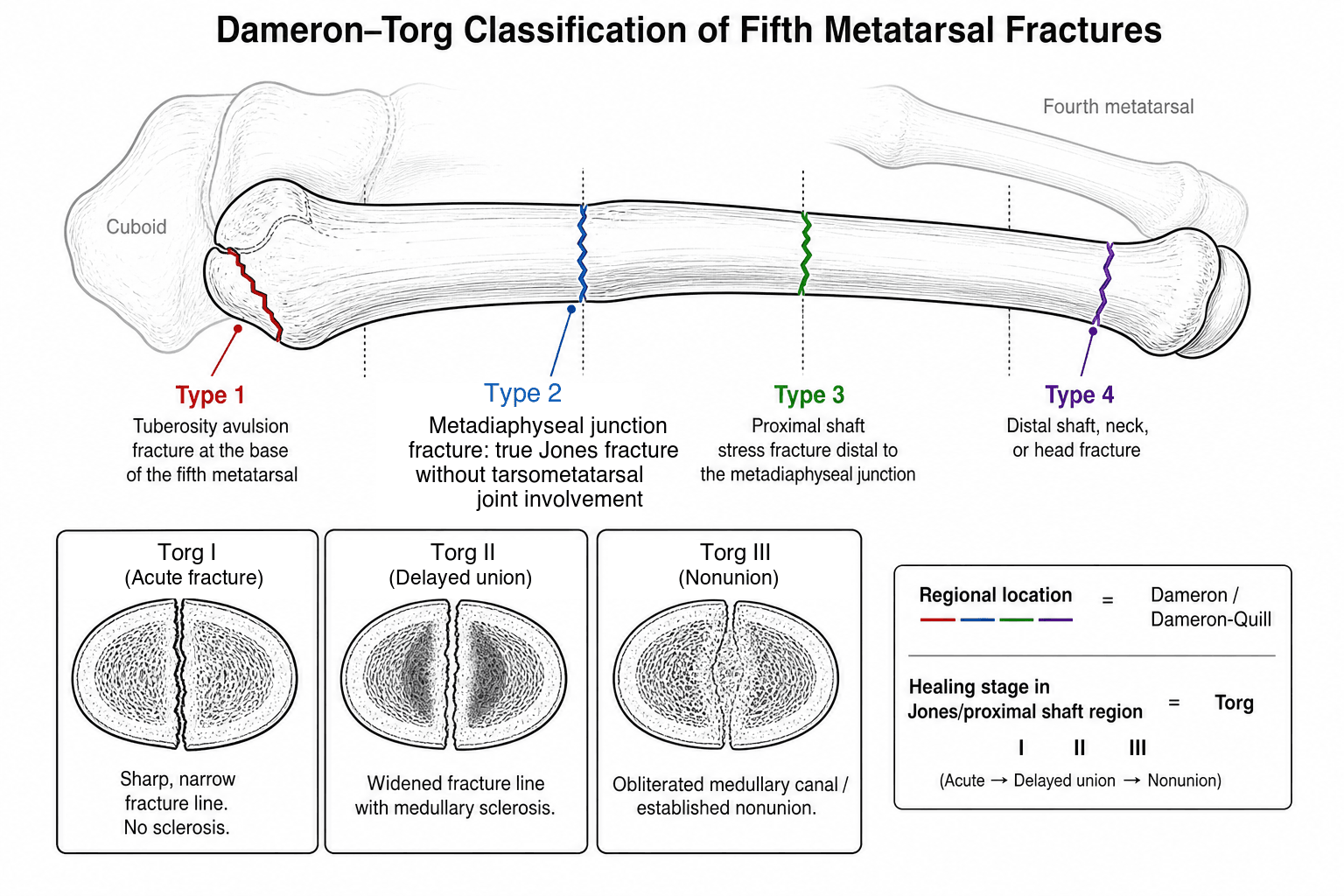
Proximal fifth metatarsal fractures are classified into three anatomical zones. The zone determines the vascular environment, healing potential, and treatment strategy.
- Location
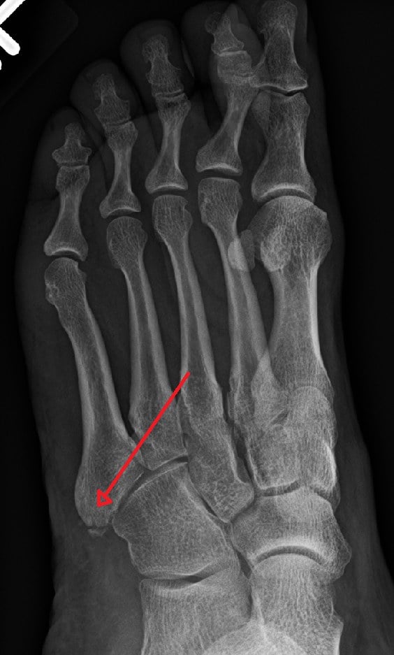
- Tuberosity (styloid process)
- Mechanism
- Inversion avulsion — lateral band of the plantar fascia (± peroneus brevis) pulls off the tuberosity
- Blood supply
- Excellent — rich cancellous metaphyseal supply
- Healing
- Reliable — near-universal union by 6–8 weeks
- Location
- Metaphyseal-diaphyseal junction (Jones fracture)
- Mechanism
- Acute inversion/load or repetitive stress; transverse fracture at the junction
- Blood supply
- Watershed — tenuous, between the metaphyseal and nutrient arteries
- Healing
- Poor — nonunion 15–30% non-operative; delayed union common
- Location
- Proximal diaphysis (stress-fracture zone)
- Mechanism
- Repetitive cyclic loading, typically in athletes; may present as a stress reaction
- Blood supply
- Marginal — diaphyseal nutrient artery alone
- Healing
- Variable — depends on chronicity and Torg type
TJSThe three zones
Hook:Moving distally from the Tuberosity to the Junction to the Shaft, the blood supply gets progressively more tenuous and healing gets worse.
The metaphyseal-diaphyseal junction receives a limited blood supply from two non-overlapping sources: a metaphyseal artery supplying the tuberosity region, and a nutrient artery entering the medial cortex more distally. The junction between these territories is a watershed with poor vascularity — which is why Zone 2 (Jones) fractures are prone to delayed union and nonunion.
Before calling a fracture, exclude the normal/accessory bones and assess the rest of the lateral foot:
- Apophysis (paediatric, age ~9 to 17): longitudinal, bilateral, smooth-corticated — a fracture is transverse (as above).
- Os vesalianum pedis: an accessory ossicle at the proximal tip of the 5th-MT base within the peroneus brevis tendon — smooth, corticated, usually bilateral; a fracture fragment is non-corticated/irregular, so compare the other foot.
- Os peroneum: a sesamoid within the peroneus longus tendon near the cuboid / calcaneocuboid joint (lateral foot, not the 5th-MT base itself); it can be bipartite or fractured (painful os peroneum syndrome).
- Don't tunnel-vision: acute lateral foot pain also includes a Lisfranc injury, a cuboid (nutcracker) fracture, an anterior calcaneal process avulsion, a lateral process of the talus fracture, and peroneal tendon pathology — examine and image accordingly.
Torg subclassification of zones 2 and 3
Torg et al. classified fractures of Zones 2 and 3 by radiographic appearance, which correlates with healing potential and guides management.
- Fracture line
- Sharp, narrow, well-defined margins
- Medullary canal
- No sclerosis — canal patent
- Periosteal reaction
- Minimal or none
- Management
- Non-weight-bearing cast 6–8 weeks; IM screw in athletes
- Fracture line
- Widened, with resorption at margins
- Medullary canal
- Partial sclerosis — some canal narrowing
- Periosteal reaction
- Present — attempted healing
- Management
- IM screw fixation; bone graft if sclerosis extensive
- Fracture line
- Wide, sclerotic margins
- Medullary canal
- Complete obliteration by sclerosis
- Periosteal reaction
- Absent — no healing response
- Management
- Surgical: IM screw with curettage/bone graft
ANSTorg types
Hook:A → N → S: as the canal progressively scleroses (Acute → Narrowing → Shut), conservative treatment progressively fails.
A Torg Type III fracture with complete medullary canal obliteration will NOT heal with cast immobilisation alone. Prolonged casting wastes the patient's time. Recognise the sclerosis pattern on radiographs — if the canal is sclerotic, the patient needs operative management.
Management by zone & type
- 1Zone it on the radiographZone 1 tuberosity, Zone 2 metaphyseal-diaphyseal junction (Jones), Zone 3 proximal diaphysis. In children, distinguish a transverse fracture from the normal longitudinal apophysis.
- 2Zone 1 → functional treatmentHard-soled shoe or walking boot, weight-bear as tolerated; even displaced tuberosity fractures heal. ORIF only for intra-articular displacement over 2 mm into the cubometatarsal joint.
- 3Zone 2/3 → grade by TorgTorg I acute → NWB cast for low-demand, IM screw for athletes; Torg II/III (sclerosis) → IM screw ± bone graft. Sclerosis predicts cast failure.
- 4Fix athletes primarilyPrimary intramedullary screw fixation for Jones fractures in athletes/high-demand patients minimises time lost and nonunion; fill the canal (5.5–6.5 mm) from a proximal-dorsal entry; graduated return to sport.
- Non-athlete
- Hard-soled shoe or boot, WBAT
- Athlete / high demand
- Same — early functional treatment
- Key technical point
- Even displaced tuberosity fractures heal; ORIF only for intra-articular displacement over 2 mm
- Non-athlete
- NWB short-leg cast 6–8 weeks
- Athlete / high demand
- Primary IM screw fixation
- Key technical point
- Screw 5.5 mm or larger, solid or cannulated, engaging the isthmus
- Non-athlete
- IM screw ± bone graft
- Athlete / high demand
- IM screw + bone graft
- Key technical point
- Refashion the sclerotic canal with a reamer before grafting
- Non-athlete
- NWB cast if Torg I; IM screw if Torg II/III
- Athlete / high demand
- Primary IM screw fixation
- Key technical point
- Higher refracture risk — address biomechanics, footwear, training errors
FASTWhen to fix a Jones fracture
Hook:If any one of FAST is positive, lean toward operative fixation.
Screw technique: entry at the proximal-dorsal tip of the fifth metatarsal, just medial to the peroneus brevis insertion ("high and inside"); guide wire down the canal under fluoroscopy; use the largest screw that fills the canal (typically 5.5–6.5 mm); avoid plantar cortex penetration and excessive countersinking (can fracture the dorsal cortex in hard young bone). Paediatric trap: the fifth metatarsal tuberosity has a normal apophysis (appears age 9–14, fuses 15–17) oriented longitudinally and is bilateral — a fracture line is transverse. Do not call an apophysis a fracture.
Even with surgery the Zone 2/3 fracture can fail; know the modes and address them:
- Screw-related: an undersized or too-short screw that does not engage the diaphyseal isthmus, and a straight screw forced down the naturally curved 5th MT (which can distract/gap the lateral fracture or perforate the lateral/plantar cortex) — use the largest screw that fits, respecting the bow, and consider a plantar/lateral plate or a tension construct in revision; a prominent screw head irritates the soft tissue.
- Biomechanical driver: a cavovarus foot / hindfoot varus / metatarsus adductus overloads the lateral column and is a recognised cause of nonunion and refracture — assess the hindfoot (Meary angle, hindfoot alignment, Coleman block) and consider a corrective osteotomy in recurrent cases.
- Metabolic: check and optimise vitamin D and calcium in nonunion/refracture.
- Return-to-sport: premature return is the leading cause of refracture — confirm union (radiograph ± CT), return gradually, and offload the lateral column with an orthotic.
Limitations & clinical nuances
- The original Jones description (1902) was not zone-specific. Sir Robert Jones described his own fracture sustained while dancing; the eponym now conventionally refers to Zone 2.
- Zone boundaries are not perfectly distinct on radiographs. A fracture can straddle Zones 2 and 3; the treatment implication is the same — poor healing potential, low threshold for fixation.
- Torg classification is radiographic, not histological. Sclerosis is a surrogate for biological activity; MRI identifies pre-radiographic stress reactions and may allow earlier intervention.
- Refracture after screw fixation occurs in roughly 5–10%, particularly in elite athletes returning too early — consider protected return and orthotic use.
- The avulsion mechanism of Zone 1: cadaveric work suggests the lateral band of the plantar aponeurosis is the primary avulsion force in inversion, rather than peroneus brevis contraction as traditionally taught.
Viva practice
Exam viva
Practise clinical reasoning and management decisions out loud
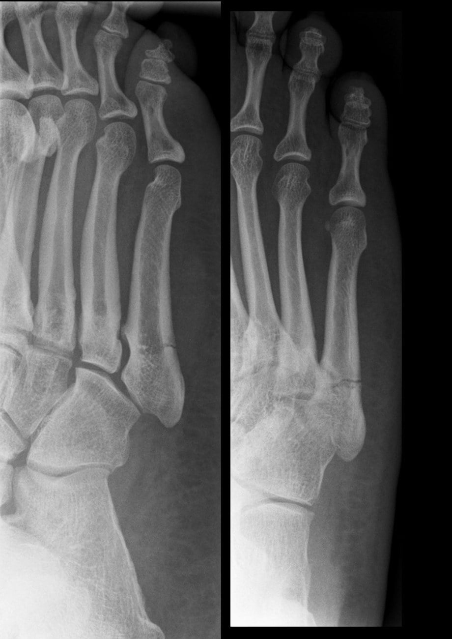
“A 22-year-old elite footballer sustains an inversion injury to his right foot. Weight-bearing radiographs show a transverse fracture at the metaphyseal-diaphyseal junction of the fifth metatarsal with no sclerosis and a narrow fracture line. How would you classify and manage this?”
“A 45-year-old recreational runner presents with lateral foot pain present for six weeks. Radiographs show a fracture line in the proximal diaphysis of the fifth metatarsal with sclerosis on both sides of the fracture and a narrowed medullary canal. How do you classify this and what is your management plan?”
Exam cheat sheet
- Zone 1 tuberosity avulsion (heals), Zone 2 Jones at the metaphyseal-diaphyseal junction (watershed, 15–30% nonunion), Zone 3 proximal diaphyseal stress.
- A Jones fracture is Zone 2 — NOT a tuberosity avulsion (the most-tested distinction).
- Torg I acute / II delayed / III nonunion by medullary sclerosis; sclerosis predicts cast failure.
- Athlete with a Jones fracture → primary IM screw (fill the canal, proximal-dorsal entry); a Level I RCT shows ~44% cast failure vs ~5% with screw and faster return.
- Watershed blood supply explains the poor healing of Zone 2.
- In children, the longitudinal bilateral apophysis is not a fracture (a fracture is transverse).
Three anatomical zones
- Zone 1: tuberosity avulsion — excellent healing, conservative treatment
- Zone 2: Jones fracture (metaphyseal-diaphyseal junction) — watershed, high nonunion
- Zone 3: proximal diaphyseal stress fracture — variable healing, assess Torg type
Torg subclassification (Zones 2 and 3)
- Type I (acute): sharp line, no sclerosis, canal patent — cast or screw (athletes get screw)
- Type II (delayed union): widened line, partial sclerosis — IM screw fixation
- Type III (nonunion): canal obliterated by sclerosis — IM screw with bone graft
Key clinical decision points
- Jones fracture is Zone 2 — NOT the tuberosity avulsion (examiners test this repeatedly)
- Athletes with a Jones fracture: primary IM screw fixation
- Screw size: fill the canal (5.5–6.5 mm); entry proximal-dorsal, just medial to the PB insertion
- In children: distinguish fracture (transverse) from the normal apophysis (longitudinal, bilateral, age 9–17)
Why Jones fractures fail to heal
- Watershed blood supply between the metaphyseal and nutrient artery territories
- Tension-side location and repetitive weight-bearing stress
- High mechanical demand on the lateral column of the foot
Evidence Base
Fracture of the Base of the Fifth Metatarsal Bone by Indirect Violence
- The original description of the fracture that bears his name — Jones sustained it himself while dancing.
- Distinguished this injury (caused by indirect violence through body weight on the inverted foot) from tuberosity avulsion fractures.
- Established the metaphyseal-diaphyseal-junction injury as a distinct entity.
Fractures of the Proximal Fifth Metatarsal: Selecting the Best Treatment Option
- Framed proximal fifth metatarsal fractures by three zones defined by their circulatory differences.
- Zone 1 (tuberosity) has good blood supply and fractures often extend into the metatarsocuboid joint; Zone 2 (Jones) heals more slowly and treatment must be individualised.
- Zone 3 (just distal to the metaphysis, the proximal ~1.5 cm of diaphysis) is often a stress injury in athletes and the most difficult to heal — without surgery, union may take 2 to 21 months.
According to PubMed, the historical entity comes from Jones 1902 (Ann Surg 1902;35(6):697-700, PMID 17861128), the zone-by-circulation framework from Dameron 1995 (DOI), the sclerosis-based Torg subclassification from Torg et al. 1984 (J Bone Joint Surg Am 1984;66(2):209-14, PMID 6693447), the three-fracture anatomical/vascular synthesis from Lawrence & Botte 1993 (DOI), and the Level I screw-vs-cast evidence from Mologne et al. 2005 (DOI).