Medial Ankle Stability | PER Mechanism | Rare Isolated Injury
- Deltoid complex 2-3x stronger than ATFL - isolated injury rare
- PER mechanism most common; a deltoid tear with a fibular fracture and an intact medial malleolus can be the SER-4 bimalleolar equivalent
- Medial clear space greater than 4mm indicates instability
- MRI gold standard for soft tissue assessment
- Conservative treatment 4-6 weeks for isolated injuries
- “Deep layer critical for rotational and translational control
- “Superficial layer for valgus and eversion restraint
- “Always suspect associated syndesmosis injury or fractures
- “Surgery for fracture/syndesmosis fixation or chronic instability
Deltoid Ligament Injuries
Anatomy and Biomechanics
The deltoid ligament is a multifascicular triangular structure running from the medial malleolus to the tarsal bones, and it provides primary medial ankle stability. It has a superficial and a deep layer, and they do different jobs.
The superficial layer. A fan-shaped sheet with a broad insertion covering the medial hindfoot, thinner than the deep layer at an average of 1-2mm. Its components act together as the primary restraint to hindfoot valgus and eversion, and the tibiocalcaneal is the strongest of them.
- Tibionavicular - anterior colliculus to navicular tuberosity
- Tibiocalcaneal (superficial) - anterior colliculus to sustentaculum tali
- Tibiospring - anterior colliculus to the superomedial calcaneonavicular (spring) ligament
- Posterior superficial tibiotalar - posterior colliculus to medial talus
The deep layer. Thick, cord-like bands, 3-5mm in thickness. The deep layer is the critical restraint to external rotation and prevents lateral translation of the talus, which is what holds the medial clear space. Its strongest component is the deep posterior tibiotalar ligament.
- Anterior tibiotalar - intercollicular groove to the anterior medial talus
- Posterior tibiotalar (deep) - posterior intercollicular region to the medial talar tubercle
- Tibiocalcaneal (deep) - intercollicular groove to the medial wall of the calcaneus
What a deep tear means. A deep-layer injury indicates severe force, a high-energy mechanism and a high risk of associated injuries, and it may require surgical intervention.
Strength and stability. The complex is 2-3 times stronger than the ATFL, with a tensile strength of 800-1200N against 300-400N, which explains why isolated injury is rare. With intact osseous anatomy it contributes 40-50% of total ankle stability, increasing to 70-80% when bony congruity is compromised.
Load with position. The complex also prevents valgus tilt of the talus and distributes medial ankle loads, and how the load is shared depends on the position of the ankle:
- Neutral - the deep posterior tibiotalar bears maximum load
- Dorsiflexion - the anterior tibiotalar and the superficial components engage
- Plantarflexion - the posterior components are maximally stressed
This position-dependent loading explains the injury patterns seen with each mechanism.
Neurovascular relations. These are the structures at risk during surgical approaches to the medial ankle.
- Posterior tibial neurovascular bundle - 1-1.5cm posterior to the medial malleolus, deep to the flexor retinaculum
- Medial plantar nerve branch - emerges at the level of the sustentaculum and innervates FHB
- Saphenous nerve - anterior to the deltoid, accompanying the great saphenous vein, whose system provides the medial venous drainage
- Arterial supply - medial malleolar branches of the posterior tibial artery
Mechanism and Classification
Pronation-external rotation (PER). The most common mechanism of deltoid injury. The medial structure fails first, and the injury then proceeds in sequence:
- Deltoid ligament rupture
- Anterior syndesmosis disruption (AITFL tear)
- Oblique or spiral fibular fracture above the level of the plafond (Weber C)
- Posterior syndesmosis disruption (PITFL) or posterior malleolus avulsion
A deltoid injury therefore indicates high energy and mandates assessment of the syndesmosis.
Supination-external rotation, stage 4 (SER-4). Here too the deltoid can be the medial injury: a fibular fracture with a deltoid rupture and an intact medial malleolus, the deltoid having failed instead of the malleolus. This is the bimalleolar-equivalent pattern shown in the figure under fibular reduction below.
Pronation-abduction (PAB). Less common, but a classic fracture pattern:
- Deltoid rupture or transverse medial malleolus fracture
- Anterior syndesmosis disruption (AITFL tear)
- Fibular fracture, transverse or comminuted, above the level of the plafond
Because the AITFL fails at stage 2, the syndesmosis must be assessed. A medial malleolus fracture may occur instead of a deltoid rupture depending on bone quality: osteoporotic bone fractures rather than the ligament tearing.
Direct valgus trauma. A pure valgus force without rotation causes a rare isolated deltoid injury, more common in sports with lateral impact such as football and rugby. It usually produces a partial tear of the superficial layer, with the deep layer intact.
Grading. Injuries are graded I-III by the extent of fibre disruption and functional impairment, assessed clinically and with advanced imaging.
- Clinical findings
- Mild medial ankle tenderness, minimal swelling, no instability on stress testing
- Imaging findings
- MRI: ligament intact with surrounding oedema, no fibre disruption. Radiographs normal
- Functional impact
- Able to weight-bear with discomfort, normal ROM with pain at extremes
- Treatment
- Conservative: protection 2-3 weeks, early ROM
- Clinical findings
- Moderate-severe tenderness, ecchymosis, mild instability on external rotation stress
- Imaging findings
- MRI: partial thickness tear with some fibre continuity. May show slight medial clear space widening (less than 4mm)
- Functional impact
- Difficulty weight-bearing, painful ROM, subjective instability
- Treatment
- Conservative: CAM boot immobilisation 4-6 weeks, protected weight-bearing
- Clinical findings
- Severe tenderness, extensive ecchymosis to hindfoot, positive instability tests, often associated injuries
- Imaging findings
- MRI: complete ligament discontinuity, fluid in ligament gap. Radiographs: medial clear space greater than 4mm, possible fracture
- Functional impact
- Unable to weight-bear, gross instability on examination, significant functional limitation
- Treatment
- Variable: conservative if isolated, surgical if associated syndesmosis/fracture
By associated injury. The same tear is managed differently according to what accompanies it:
- Isolated deltoid - rare, managed conservatively
- With a syndesmotic injury - requires fixation
- With a fracture (SER-4) - ORIF
Clinical Evaluation
History. The features of a medial-sided injury:
- Mechanism - pronation-external rotation, valgus stress or a twisting injury
- Ecchymosis extending to the hindfoot, not just the ankle
- Difficulty weight-bearing immediately after the injury
- A sensation of the medial ankle giving way
- Associated symptoms - syndesmotic pain, lateral ankle symptoms
- Pain directly over the medial malleolus and along the course of the deltoid
A high-energy mechanism and inability to weight-bear suggest a severe injury requiring imaging.
Asking about mechanism. Specific questions help predict the injury pattern and its associated pathology. "Did your foot twist outward while the ankle bent inward?" suggests a PER mechanism with a high risk of syndesmotic injury; "Was there a direct impact to the outside of the ankle?" suggests a PAB pattern. Previous ankle injuries and pre-existing instability are relevant in chronic presentations.
Inspection. Note whether the swelling is isolated medially or circumferential, which suggests syndesmotic injury. Extensive hindfoot ecchymosis indicates a grade 2-3 injury. Note whether the patient can weight-bear, and compare hindfoot alignment with the other side: valgus suggests chronic deltoid insufficiency or an acute deformity.
Palpation. In sequence:
- Tip and anterior border of the medial malleolus, for fracture
- The course of the deltoid from the anterior colliculus downwards, where a palpable gap indicates a complete tear
- Sustentaculum tali and spring ligament, for an associated spring ligament injury
- The PTT behind the medial malleolus, for concurrent PTT pathology
- The syndesmosis anteriorly - squeeze test, and palpation 2-3cm above the ankle
Stress tests. For the external rotation stress and Cotton tests, compare with the contralateral ankle.
- External rotation stress test - patient seated, knee flexed 90 degrees; stabilise the tibia and externally rotate the foot. Positive if rotation is increased and medial pain is reproduced
- Eversion stress test - evert the hindfoot with the ankle neutral. Positive if medial clear space widening is visible or palpable
- Cotton test - grasp the heel and apply a lateral force to translate the talus in the mortise. Excessive lateral shift indicates deltoid and syndesmotic injury
- Squeeze test - compress tibia and fibula at mid-calf. Pain at the syndesmosis indicates an associated syndesmotic injury
- Kleiger test - external rotation stress with the ankle dorsiflexed and the tibia stabilised. Where the pain localises determines the pathology: medial malleolus (deltoid), anterolateral ankle (syndesmosis), or both (combined injury)
A positive external rotation stress test with medial ankle pain indicates deltoid injury but does not differentiate isolated deltoid injury from combined deltoid-syndesmotic injury. The syndesmosis must be assessed specifically, with the squeeze test, an external rotation radiograph and/or MRI. Failure to identify the syndesmotic component leads to inadequate treatment and poor outcomes.
Single heel raise. Assesses PTT function and deltoid competency. Standing on the affected leg, the patient rises onto the toes; the normal response is heel rise with hindfoot inversion. Inability to perform it, or a heel rise without hindfoot inversion, suggests PTT dysfunction or severe deltoid insufficiency (chronic cases).
Range of motion. Measure dorsiflexion and plantarflexion against the other side. Reduced dorsiflexion may indicate syndesmotic injury or anterior impingement, and increased eversion suggests deltoid insufficiency.
Differential diagnosis. Medial ankle pain after a twisting injury is not always a deltoid tear. Separate the possibilities by mechanism, the point of maximal tenderness and the single best discriminating test.
- Mechanism
- Pronation/eversion-external rotation, valgus
- Key finding
- Tenderness over deltoid below medial malleolus; pain on ER/eversion stress
- Discriminator
- Weight-bearing MCS greater than 4mm; deltoid signal change on MRI
- Mechanism
- Same rotational forces (bone fails instead of ligament)
- Key finding
- Bony point tenderness on the malleolus, possible deformity
- Discriminator
- Fracture line on radiograph (often the bony equivalent of a deltoid tear)
- Mechanism
- External rotation with dorsiflexion
- Key finding
- Tenderness anterolaterally over AITFL, above the joint line
- Discriminator
- Positive squeeze and ER tests; widened tibiofibular clear space, often coexists with deltoid tear
- Mechanism
- Chronic/overuse, acute on chronic; not a single sprain
- Key finding
- Tenderness/swelling posterior to medial malleolus, too-many-toes sign, single-heel-raise failure
- Discriminator
- Tendon (not ligament) on US/MRI; progressive flatfoot rather than acute trauma
- Mechanism
- Valgus/abduction, often with deltoid or PTT injury
- Key finding
- Tenderness over the talonavicular/sustentacular region
- Discriminator
- Spring ligament disruption on MRI; talar head uncoverage
- Mechanism
- Repetitive loading in runners
- Key finding
- Insidious load-related pain, focal malleolar tenderness
- Discriminator
- Vertical stress line on radiograph/MRI; no acute traumatic event
Imaging Evaluation
Radiographs. Weight-bearing films are essential for an accurate medial clear space; non-weight-bearing views may miss subtle instability. Contralateral views are useful for borderline measurements.
- AP - medial clear space (normal less than 4mm, equal to the superior clear space); tibiofibular clear space (normal less than 6mm); tibiofibular overlap (normal greater than 6mm on the AP, greater than 1mm on the mortise)
- Mortise - the true AP of the ankle joint, and the best view for the medial clear space and syndesmotic measurements
- Lateral - tibiofibular overlap (normal greater than 10mm) and any posterior malleolus fracture
Medial clear space greater than 4mm on weight-bearing radiographs indicates deltoid insufficiency until proven otherwise. It mandates assessment for syndesmotic injury, through the tibiofibular clear space and overlap measurements, and consideration of advanced imaging. Isolated deltoid injury is rare: look for fractures and syndesmotic disruption.
What the threshold does not tell you. Over 4mm is the classic threshold, but a positive stress test overestimates instability. In Koval's series 90% of stress-positive Weber B fractures had only partial deep-deltoid tears and healed nonoperatively, and the medial clear space did not reliably correlate with complete rupture on MRI. A static medial clear space does not reliably equal complete rupture; deep-deltoid integrity matters more.
Stress radiographs. Indicated for a suspected deltoid injury with normal standard radiographs, or equivocal clinical findings. Medial clear space widening to greater than 4mm absolute, or greater than 2mm compared with the contralateral side, is pathological.
- External rotation stress - manual or mechanical external rotation of the foot with the ankle neutral
- Gravity stress - the affected leg hangs off the table with the knee flexed and its own weight creates the stress; less reproducible than mechanical devices
Stress views are painful and need the patient's cooperation, and some centres lack mechanical devices.
Gravity stress where MRI is limited. Against MRI, a gravity-stress medial clear space of at least 6mm reached 100% sensitivity and negative predictive value, with 92% specificity, in van Leeuwen's series, which makes it a validated, low-cost alternative where MRI access is limited. Its positive predictive value was 50%, so it is a good test for ruling a complete tear out and a poor one for ruling it in.
MRI. The gold standard for soft-tissue characterisation, injury grading and identifying associated pathology, though it is not universally available. The protocol is T1, T2 and STIR sequences in all three planes, and coronal images are best for the deltoid. Look beyond the ligament at the syndesmotic ligaments (AITFL, PITFL, IOL), the spring ligament, the PTT, osteochondral lesions and the bone bruising pattern. In chronic injury the ligament shows thickening, scarring, fatty infiltration or attenuation.
MRI is indicated for:
- A grade 2-3 clinical injury
- Suspected syndesmotic injury
- Chronic symptoms
- Pre-operative planning
Bone bruising. MRI bone marrow oedema patterns predict the injury mechanism and the associated soft-tissue injuries. Kissing bone bruises on the medial talus and medial malleolus indicate compression from valgus impact, supporting a deltoid injury. Anterolateral tibial bruising suggests a syndesmotic component, and posterior malleolar bruising indicates a posterior translation force and a potential posterior malleolus fracture.
Conservative Management
The majority of deltoid injuries are managed conservatively, with the protocol set by the grade.
Grade 1 injuries are ligament strain without macroscopic fibre disruption, managed successfully with brief protection and early mobilisation.
Acute phase, 0-7 days. PRICE (protection, rest, ice, compression, elevation), with a CAM boot or ankle brace for comfort and weight-bearing as tolerated. Control oedema with a compression stocking or wrap and cryotherapy for 20 minutes every 2-3 hours while awake. NSAIDs for pain if there are no contraindications: ibuprofen 400-600mg three times daily or naproxen 500mg twice daily.
Recovery phase, 1-3 weeks. Progress weight-bearing to a normal gait pattern and wean from the boot or brace as symptoms improve, returning to normal shoes by week 2-3. Active range of motion (ankle alphabet, towel stretches, resistance band exercises) and gentle strengthening (tibialis posterior activation, toe raises) begin now.
Return to activity, 3-4 weeks. Progress loading from walking to jogging, add sport-specific drills without cutting or pivoting, and train proprioception with single-leg balance and a balance board. Use external ankle support, brace or tape, for high-risk activities. Full return to sport typically takes 4-6 weeks.
Expected course. A grade 1 injury settles with immobilisation and rehabilitation and chronic symptoms are uncommon; the usual reason one does not settle is an unrecognised associated injury, so re-examine and re-image rather than extending the boot. No grade-stratified outcome series exists, so avoid quoting a success rate.
Surgical Management
Why the ankle is opened. Surgery for acute deltoid injury is typically performed as part of treating an associated fracture or syndesmotic injury rather than as an isolated ligament repair. Isolated deltoid repair without an associated injury is controversial and rarely performed acutely. The indications for surgery in an ankle with a deltoid injury:
- A fracture requiring ORIF - medial malleolus, or fibula with deltoid rupture
- An associated syndesmotic injury requiring fixation
- Laxity persisting after syndesmotic reduction, with a medial clear space greater than 4mm on stress
- An irreducible ankle joint, with the deltoid or other soft tissue interposed
- Instability recalcitrant on serial imaging despite conservative care
- Chronic instability
Operating for a fracture or a syndesmotic injury is not the same as repairing the deltoid, which is decided after the lateral side has been fixed (see below).
Timing. Acute fractures with a deltoid rupture require surgery within 7-10 days of injury, before soft-tissue swelling precludes a safe approach. Syndesmotic injuries with a deltoid rupture are treated within the first week for optimal reduction. Isolated deltoid ruptures (rare) can undergo delayed repair within the first 4 weeks if conservative management is failing.
Why Anatomic Fibular Reduction Restores Medial Stability
The talus follows the fibula. The talus sits in the mortise between the medial malleolus and the fibula, and in an external-rotation injury it shifts laterally and externally rotates with the fibula. Restoring fibular length, rotation and the lateral buttress recentres the talus in the mortise, which closes the medial clear space even though the deltoid is torn.
A competent-enough deltoid re-tensions. Once the talus is recentred, an intact deep deltoid, or a partially torn but competent one, is brought back to length and re-tensions, holding the reduction. That is why most torn deltoids in a fracture do not need repair, and why deep-deltoid integrity, and not the medial clear space alone, drives the decision.
The check. After fibular fixation, reassess the medial clear space under fluoroscopic external-rotation stress. If it normalises, under 4mm and equal to the superior clear space, the medial side is competent and needs nothing further. If it stays widened, either the fibula is not anatomically reduced, tissue is interposed, or the deltoid is truly incompetent, and each of these changes the plan.
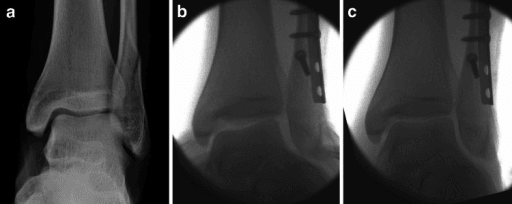
The corollary. This is why the global default is to fix the fibula and syndesmosis anatomically first and then reassess the medial side, rather than reflexively repairing every torn deltoid. Explore or repair the deltoid only if the mortise will not reduce, the medial clear space stays wide after lateral fixation, or the medial ankle remains grossly unstable.
Deltoid (and Soft-Tissue) Interposition Blocking Reduction
What gets trapped. In a pronation- or supination-external-rotation injury, most often the ruptured deltoid or the posterior tibial tendon, or else a small bony or periosteal fragment, can be dragged into the medial gutter, the space between the medial malleolus and the talus. There it physically blocks the talus from recentring.
How it presents. After anatomic fibular fixation the medial clear space stays widened and the talus stays laterally shifted: a mortise that will not close despite correct fibular length and rotation. The medial gutter looks widened on imaging, and in a frank medial dislocation a skin "dimple sign" over the medial malleolus, from a buttonholed, trapped structure, may be seen.
Why it matters. A persistently widened medial gutter is not acceptable. Leaving interposed tissue causes malreduction, chronic medial gutter pain and early arthritis, and it is one of the few absolute indications to open the medial side.
What to do. Open the medial gutter, identify and extract the interposed deltoid, posterior tibial tendon or fragment, and confirm that the talus now recentres, with the medial clear space normalising on fluoroscopy. Then repair the deltoid with suture anchors if a residual gap remains.
Surgical Technique
Approach. A curvilinear incision 6-8cm long, centred over the medial malleolus. Identify and protect the saphenous vein and nerve anteriorly, and raise full-thickness flaps to expose the medial malleolus and deltoid. Assess the level of ligament disruption: tibial avulsion, midsubstance tear, or avulsion from the tarsal insertion.
Suture anchor repair. The preferred modern technique.
- Anchors at the anatomic origin on the medial malleolus, chosen by the component injured - the anterior colliculus for superficial components, the intercollicular groove for deep components
- 3.0-3.5mm anchors with high-strength suture (2-0 FiberWire or equivalent)
- Mattress or modified Mason-Allen configuration for optimal strength
- Repair with the ankle in neutral dorsiflexion and 5 degrees of hindfoot inversion
- Repair the deep layer first, as it is critical for stability, then the superficial layer
Bone tunnel repair. The alternative. Drill 2.5-3.0mm tunnels through the medial malleolus at the anatomic origins, pass heavy non-absorbable suture through the ligament tissue, and tie the sutures over the bony bridge. It is more time-consuming than anchors, with comparable outcomes.
Augmentation. An internal brace of suture tape can augment the repair in high-demand athletes, placed from the medial malleolus to the sustentaculum or medial talus in the anatomic orientation of the deltoid. It acts as a temporary "internal splint" during healing; the evidence is limited, but the concept is biomechanically sound.
Concurrent procedures. Address associated injuries at the same sitting. Fix the syndesmosis first, then reassess the medial clear space, and repair the deltoid only if it remains widened. Repair the spring ligament if an injury is identified intraoperatively, and perform medial malleolus ORIF if the fracture pattern is present.
Chronic reconstruction. Chronic deltoid insufficiency with poor tissue quality or failed previous treatment requires reconstruction rather than repair. The medial-ankle-instability topic covers the reconstruction techniques, which use autograft or allograft to recreate the anatomic ligament complex.
Complications and Sequelae
Compartment syndrome. Rare but catastrophic, with a 2-5% incidence in high-energy deltoid injuries with associated fractures. It presents with pain out of proportion, pain on passive toe extension, tense swelling and paraesthesiae, and requires immediate fasciotomy of all four compartments. Prevent it with careful monitoring and a low threshold for compartment pressure measurement.
Vascular injury. Posterior tibial artery injury is extremely rare with isolated deltoid injury but possible with high-energy trauma. Assess distal pulses, capillary refill and Doppler signals, and obtain urgent vascular surgical consultation if perfusion is compromised.
Nerve injury. Tibial nerve or plantar branch injury presents with plantar foot numbness and toe flexion weakness, usually from direct trauma rather than iatrogenic injury. Saphenous nerve injury and wound complications are the other acute problems. Document neurovascular status pre-operatively for medicolegal protection.
Chronic medial ankle pain. A recognised sequel of inadequately treated injury, though its frequency has never been measured in a defined cohort. The differential includes deltoid scarring or fibrosis, spring ligament insufficiency, medial ankle impingement, PTT tendinopathy and subtalar arthritis.
Medial ankle instability. A recognised outcome of grade 3 injuries treated conservatively, with no published incidence. It presents with recurrent giving way, difficulty on uneven ground and activity limitation, and objective instability is demonstrated on stress radiographs.
Post-traumatic arthritis. The long-term consequence that matters most, and the reason anatomic reduction is the priority. Its incidence after deltoid injury specifically has not been established, and figures quoted in reviews are extrapolated from ankle-fracture cohorts generally. It results from initial cartilage damage, chronic instability causing abnormal joint mechanics, or malreduction of associated fractures, and prevention requires anatomic fracture reduction, addressing instability and optimising initial treatment.
Valgus hindfoot deformity. Progressive deformity from untreated chronic deltoid insufficiency combined with spring ligament attenuation, leading to a stage 4 PTTD-type presentation. Prevention requires early identification and reconstruction of chronic instability before a fixed deformity develops.
Missed syndesmotic injury is the most common and consequential complication of deltoid injury management. Approximately 40-60% of deltoid injuries have an associated syndesmotic disruption. Failure to identify and treat the syndesmotic component leads to chronic pain, instability and accelerated arthritis, so every deltoid injury needs syndesmotic assessment by clinical examination, radiographic measurements and a low threshold for MRI.
Postoperative Care
Rehabilitation after repair. Protection gives way to loading in four phases:
- 0-2 weeks - non-weight-bearing in a splint or cast, elevation and ice for swelling, wound care
- 2-6 weeks - progressive weight-bearing in a CAM boot, active range of motion exercises, avoiding forced eversion
- 6-12 weeks - full weight-bearing, progressive resistance exercises and proprioceptive training
- 3-6 months - sport-specific training, external support for activities, and a gradual return to cutting and pivoting
Return to sport requires full strength (less than 10% deficit), normal range of motion, negative stress tests and passed functional hop testing.
Outcomes and Prognosis
What is known. There is no outcome study stratified by deltoid injury grade, so grade-specific success rates are not available and are not quoted here. The published figures concern fracture-associated injury. Of the stress-positive Weber B fractures managed non-operatively after MRI in Koval's series, all united and 14 of 15 patients scored AOFAS 100 at one year (n=21, 15 followed).
Favourable factors. Isolated grade 1-2 injury, no associated syndesmotic injury, age less than 40 years, normal BMI, sedentary occupation, good rehabilitation compliance, and injury to the superficial component only.
Unfavourable factors. Grade 3 complete rupture, deep component injury, associated syndesmotic disruption, age greater than 50, BMI greater than 30, manual labour or competitive athletics, poor rehabilitation access or compliance, and delayed diagnosis (greater than 4 weeks). Deep component injury predicts worse outcomes (72% success against 89% for a superficial-only injury) and higher rates of chronic symptoms, and combined superficial and deep injury has the worst prognosis.
Return to activity.
- Grade 1 - full activity at 4-6 weeks
- Grade 2 - 10-14 weeks
- Grade 3 treated conservatively - 16-20 weeks
- Grade 3 requiring surgery - 4-6 months
- High-level athletics after a complete rupture - may require 6-9 months regardless of treatment
Guidelines, Registries & Global Practice
Global Epidemiology and Guideline Positions
Isolated deltoid injury is rare (roughly 3-5% of ankle sprains). The far more common scenario is deltoid rupture as the medial component of a rotational ankle fracture: a deltoid tear (rather than a medial malleolus fracture) is found in a substantial proportion of supination-external-rotation (SER) and pronation-external-rotation (PER) fractures, creating the bimalleolar-equivalent pattern.
No single society publishes a dedicated standalone deltoid-injury guideline; recommendations are embedded in ankle-fracture and ankle-instability guidance. The cross-society consensus is consistent worldwide.
- Region
- Global
- Position on deltoid injury
- Anatomic fibular (and syndesmotic) reduction is primary; explore/repair deltoid mainly if reduction is blocked or MCS stays wide
- Region
- US
- Position on deltoid injury
- Bimalleolar-equivalent concept; limited evidence repair improves outcomes over ORIF alone
- Region
- UK
- Position on deltoid injury
- Stability-based management; weight-bearing/stress assessment to define unstable Weber B before surgery
- Region
- Europe
- Position on deltoid injury
- Chronic medial instability assessed and reconstructed within a global ankle-instability framework
Controversies and Areas of Uncertainty
The deltoid is one of the most genuinely contested soft-tissue topics in foot and ankle surgery. A consultant-level answer acknowledges the equipoise rather than overstating certainty.
To repair or not to repair? Both syntheses of the same eight studies agree that the medial clear space is better after repair, with reduced malreduction, in five of six studies reporting it. On function they disagree: Guo's meta-analysis (388 patients) found the AOFAS score statistically superior after repair, James and Dodd's systematic review found functional outcomes equivalent, and the JAAOS review concludes there is limited evidence of superior clinical outcomes with added repair. High-quality RCT evidence is lacking. Repair reliably improves the radiograph, and whether that converts into a better ankle is unproven; say that to a patient or an examiner, rather than a percentage.
Does a torn deltoid mean an unstable syndesmosis? No. In Massri-Pugin's cadaveric work, isolated deltoid disruption, even with the AITFL, did not destabilise the syndesmosis in the coronal plane; instability required additional interosseous-ligament injury. A deltoid tear is a marker of energy, and the syndesmosis still has to be tested on its own merits.
Deltoid Repair vs No Repair in Acute Ankle Fracture (Meta-analysis)
Management of Deltoid Injury in Acute Ankle Fracture (Systematic Review, 3 RCTs)
Role of the Deltoid Ligament in Syndesmotic Instability (Cadaveric)
MRI-Guided Selective Surgery After Positive Stress Test (Weber B)
Gravity Stress Radiographs for Deep Deltoid Integrity in SER Fractures
Deltoid Rupture in Ankle Fracture: Diagnosis and Management (Review)
MCQ Practice Points
Q: What are the components of the deltoid ligament and their primary functions?
A: The deltoid ligament has superficial and deep components. Superficial (4 bands): tibionavicular, tibiospring, tibiocalcaneal, posterior tibiotalar - resists eversion/abduction. Deep (2 bands): anterior tibiotalar, posterior tibiotalar (strongest) - primary restraint to external rotation and lateral talar shift. The deep deltoid is the key stabilizer; isolated superficial injury rarely causes instability.
Q: When is deltoid ligament repair indicated in ankle fracture surgery?
A: Deltoid repair is rarely indicated in most ankle fractures. Indications: Medial clear space widening persists (greater than 4mm or greater than superior clear space) after lateral malleolus fixation; Deltoid interposition blocking reduction (interposed tissue visible on stress views); Grossly unstable medial ankle despite lateral fixation. Most cases: Anatomic fibular reduction restores medial stability without direct deltoid repair.
Q: How do you assess deltoid ligament integrity intraoperatively?
A: Intraoperative assessment: (1) Medial clear space measurement on mortise fluoroscopy after fibular fixation - should be less than 4mm and equal to superior clear space; (2) External rotation stress test - widening indicates deep deltoid incompetence; (3) Direct visualization through medial incision if concern for interposed tissue. If medial clear space normalizes with fibular fixation, deltoid is functionally competent even if torn.
Q: What is the clinical significance of isolated deltoid ligament injury without fracture?
A: Isolated deltoid injury is rare and usually occurs with: High ankle sprain (syndesmotic injury pattern); Ankle dislocation without fracture. Treatment: Most heal with non-operative management (immobilization 4-6 weeks). Surgery considered for: Chronic medial instability with valgus tilting; Associated syndesmotic instability requiring fixation; Deltoid avulsion with bone fragment amenable to fixation. Chronic deltoid insufficiency can lead to progressive valgus deformity.
Q: How does deltoid ligament status affect the treatment of Weber B ankle fractures?
A: Weber B fractures are classified as stable (deltoid intact) or unstable (deltoid incompetent). Assessment: Gravity stress radiographs or external rotation stress views - medial clear space widening greater than 4mm indicates deltoid rupture. Stable Weber B (intact deltoid): Non-operative treatment in walking boot. Unstable Weber B (torn deltoid): Requires ORIF of fibula to restore ankle stability. The deltoid determines fracture stability, not just fracture pattern.
Clinical Decision Scenarios
Practise clinical reasoning and management decisions out loud
“A 26-year-old recreational footballer presents to emergency department 2 hours after twisting injury. Mechanism was landing from jump with foot pronated and externally rotated. Examination shows medial ankle swelling, tenderness over deltoid and anterolateral ankle joint line. External rotation stress test is painful medially and anteriorly. Weight-bearing radiographs show medial clear space of 4.5mm. Tibiofibular clear space is 5.5mm (borderline). MRI not immediately available. How do you proceed?”
“A 42-year-old presents 18 months following ankle sprain initially treated conservatively with 4 weeks CAM boot. She has persistent medial ankle pain with walking and inability to return to running. Examination shows medial ankle tenderness, mild valgus hindfoot alignment, positive but not severe external rotation stress test. Weight-bearing radiographs show medial clear space of 4.8mm. MRI demonstrates chronic deltoid thickening with partial thickness tears of superficial components, intact deep components, and grade 2 spring ligament injury. Failed 6 months physical therapy. What is your management approach?”
“A 38-year-old labourer sustains a Weber B fibular fracture with no medial malleolus fracture. Mortise radiograph shows a medial clear space of 6mm. The examiner asks: this is a bimalleolar-equivalent injury. After you fix the fibula, the medial clear space is still 4.5mm. Would you repair the deltoid, and what does the evidence say?”
Immediate Answer Opener
- Deltoid ligament injuries range from grade 1 sprains to complete ruptures, representing 3-5% of ankle sprains
- Deltoid is 2-3 times stronger than lateral ligaments, making isolated injury rare
- Most occur with pronation-external rotation mechanisms causing combined deltoid-syndesmotic injuries
- Grading: Grade 1 (strain), Grade 2 (partial tear), Grade 3 (complete rupture)
- Medial clear space greater than 4mm on weight-bearing radiographs indicates pathology
- Treatment predominantly conservative; surgery for associated fractures/syndesmosis or chronic instability
Anatomy Quick Reference
- Superficial deltoid: Tibionavicular, tibiocalcaneal, tibiospring ligaments from anterior colliculus
- Superficial component restrains valgus and eversion
- Deep deltoid: Anterior and posterior tibiotalar, tibiocalcaneal from intercollicular groove
- Deep component critical for rotational stability and prevents lateral talar translation
- Posterior deep tibiotalar is strongest component
- Deltoid 2-3x stronger than ATFL (800-1200N vs 300-400N tensile strength)
- Provides 40-50% of ankle stability with intact osseous anatomy
Grading and Injury Mechanisms
- Grade 1: Intact fibers with edema, mild tenderness, minimal swelling, conservative 2-3 weeks
- Grade 2: Partial tear, moderate-severe tenderness and swelling, mild instability, CAM boot 4-6 weeks
- Grade 3: Complete rupture, extensive ecchymosis, gross instability, often associated injuries
- Pronation-external rotation: combined with syndesmosis injury (most common mechanism)
- Pronation-abduction: Associated medial malleolus fracture
- Direct valgus: Rare isolated deltoid injury
Clinical Examination Essentials
- External rotation stress test: Knee flexed 90°, externally rotate foot, positive if increased rotation and medial pain
- Squeeze test: Compress mid-calf, positive if ankle syndesmotic pain
- Cotton test: Lateral translation of talus, positive if excessive (combined deltoid-syndesmosis)
- Palpation: Medial malleolus, deltoid course, spring ligament, PTT
- Always compare stress testing to contralateral ankle for asymmetry
Imaging Interpretation
- Medial clear space: Normal less than 4mm, pathologic if greater than 4mm or asymmetric to superior clear space
- Tibiofibular clear space: Normal less than 6mm
- Tibiofibular overlap: Normal greater than 6mm on AP view
- MRI grading: Grade 1 intact with edema, Grade 2 partial tear, Grade 3 complete discontinuity
- MRI must assess syndesmosis (AITFL, PITFL, IOL), spring ligament, PTT, bone bruising
- Weight-bearing views essential for detecting dynamic instability
Conservative Management Protocol
- Grade 1: PRICE, CAM boot or brace WBAT, ROM exercises week 1-3, return to activity 4-6 weeks
- Grade 2: CAM boot NWB progressing to partial weeks 2-3, immobilization 4-6 weeks, structured PT, return to sport 10-14 weeks
- Grade 3 isolated: Controversial management, NWB cast 2-3 weeks, progressive WB 4-6 weeks, extended rehab 3-6 months
- Serial radiographs required to monitor for instability development
Surgical Indications and Techniques
- Indications: Associated fracture requiring ORIF, syndesmotic injury with persistent MCS greater than 4mm, chronic instability failed 6 months conservative, irreducible ankle
- Approach: Curvilinear medial incision protecting saphenous nerve
- Suture anchors at anatomic origins: Anterior colliculus for superficial, intercollicular groove for deep
- Position: Ankle neutral dorsiflexion, 5° hindfoot inversion during repair
- Consider internal brace augmentation in athletes
- Address spring ligament if injured
Complications and Outcomes
- No grade-stratified outcome series exists - do not quote conservative success rates by grade
- Deltoid repair in fracture: the medial clear space improves consistently; whether function improves is disputed between the two syntheses of the same 8 studies
- Missed syndesmotic injury: Most common and consequential complication
- Chronic medial pain and persistent instability are recognised sequelae with no published incidence
- Post-traumatic arthritis: the outcome anatomic reduction exists to prevent; no deltoid-specific incidence published
- Prognostic factors: Grade, associated injuries, age, BMI, occupation, rehab compliance, deep vs superficial injury
Evidence Base
Key Studies
- 8 comparative studies, 388 patients (Weber B/C)
- Repair improved post-op and final medial clear space, AOFAS score and complication rate
- No difference in VAS pain
- Functional outcomes equivalent between repair and no repair
- Radiographic MCS and malreduction favoured repair in 5 of 6 studies
- Finding
- Repair improves MCS/AOFAS, not pain
- Implication
- Selective repair, not routine
- Finding
- Function equal, radiographs favour repair
- Implication
- Equipoise; RCT needed
- Finding
- Deltoid alone does not destabilise syndesmosis
- Implication
- Assess syndesmosis separately
- Finding
- 90% stress-positive Weber B heal nonop
- Implication
- MCS overestimates instability