Aggressive Fibromatosis | Locally Aggressive Benign | Surveillance-First Approach
- Desmoid tumors NEVER metastasize - benign but locally aggressive
- Active surveillance is first-line: in the prospective GRAFITI trial 28% regressed and 32% stabilised, so roughly 60% need no treatment
- CTNNB1 mutation (beta-catenin) drives 85-90% of sporadic desmoids
- Positive surgical margins acceptable - function preservation priority
- Recurrence 20-40% even with complete excision
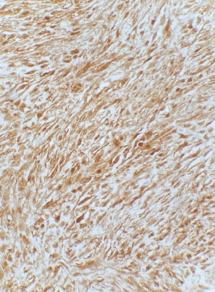
- “Nuclear beta-catenin staining SUPPORTS the diagnosis but is neither fully sensitive (~80%) nor specific - it does not replace morphology and clinical context
- “FAP-associated desmoids carry germline APC mutations, not somatic CTNNB1 - and a mesenteric desmoid in a young patient mandates colonoscopy
- “Low-signal bands on BOTH T1 and T2 reflect dense collagen and are characteristic, but not pathognomonic - biopsy is still required
- “S45F is the adverse CTNNB1 genotype; T41A is the commonest and more indolent
- “Do NOT re-resect positive margins - margin status does not predict recurrence, unlike sarcoma
Overview and Epidemiology
Desmoid tumour is a rare benign fibroblastic neoplasm: a monoclonal proliferation of myofibroblasts that grows by local infiltration and has no metastatic potential. Its difficulty lies elsewhere, in an unpredictable natural history, real local aggressiveness, and a high recurrence rate after resection.
Two names, one tumour. "Desmoid" comes from the Greek desmos, a band or tendon, for the collagenous, band-like consistency of the lesion. "Aggressive fibromatosis" emphasises the locally aggressive behaviour despite benign histology. The terms are synonymous, and both appear in examination questions.
The paradigm shift. Over the past decade the historical aggressive surgical approach has given way to surveillance first, because most desmoids need no treatment. In the prospective GRAFITI trial 28% regressed and 32% stabilised on active surveillance, so roughly 60% required no intervention; the earlier retrospective Fiore series reported a 5-year progression-free survival of about 50%. The two figures are not in tension: they measure different endpoints, and the prospective one is the more favourable. Treatment-related morbidity may exceed disease-related morbidity in many patients, and that recognition is what changed practice.
Who. The peak is at 20-40 years, median 30. Women predominate 2-3:1 overall, with a strong female bias in abdominal wall tumours, attributed to hormonal influence, and a more equal distribution in the extremities.
Where. Location shapes both presentation and management:
- Extra-abdominal, 60% - shoulder and thigh most common; head and neck account for 5-10% of extra-abdominal cases
- Abdominal wall, 25% - the rectus, with a postpartum association
- Intra-abdominal, 15% - the mesentery, where FAP-associated tumours arise
Molecular Pathophysiology and Genetics
The pathway. Desmoid tumorigenesis is driven by aberrant activation of Wnt-beta-catenin signalling, reached by two distinct genetic routes. In the normal cell beta-catenin is captured by the destruction complex (APC-Axin-GSK3beta), phosphorylated, and so marked for ubiquitin-mediated degradation, which keeps cytoplasmic levels low. When beta-catenin cannot be phosphorylated, or the complex that would phosphorylate it has lost APC, the protein accumulates, translocates to the nucleus, binds the TCF/LEF transcription factors and activates target genes that promote fibroblast proliferation. That nuclear accumulation is what beta-catenin immunostaining detects.
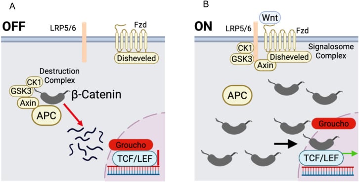
Sporadic tumours: CTNNB1, 85-90%. Mutation of the CTNNB1 gene on chromosome 3p21, which encodes beta-catenin, is the hallmark of the sporadic desmoid. The mutations sit in exon 3, predominantly at codons 41 (T41A) or 45 (S45F), and create a beta-catenin that cannot be phosphorylated and therefore cannot be degraded.
Genotype and behaviour. Get the direction right; it is regularly examined.
- Three hotspots account for almost all sporadic mutations: T41A (the commonest), S45F and S45P
- S45F is the adverse genotype. It predicts progression on surveillance and higher recurrence after surgery; in the prospective GRAFITI cohort it carried a hazard ratio of 6.24 (95% CI 1.92-20.30) for needing to start active treatment, alongside tumour size of 5 cm or more (HR 2.38)
- T41A behaves comparatively well: the most frequent mutation, and associated with a more indolent course
- Wild-type CTNNB1 in a convincing desmoid should prompt a search for an APC germline mutation
Practically, an S45F tumour warrants closer surveillance intervals and a lower threshold to treat; it does not by itself mandate surgery.
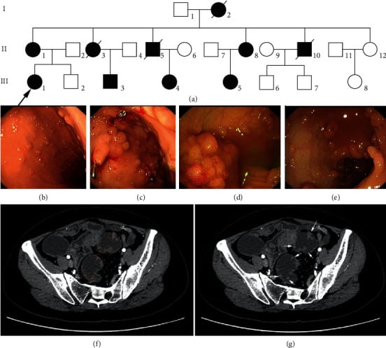
FAP-associated tumours: APC, 5-10%. A germline mutation of APC on chromosome 5q21-22, inherited as an autosomal dominant trait, causes familial adenomatous polyposis and its desmoids. APC is the tumour-suppressor component of the destruction complex, so its loss prevents beta-catenin degradation and produces the same nuclear accumulation as a CTNNB1 mutation. The tumours that result behave differently from sporadic disease:
- Occur in 10-20% of FAP patients
- Typically intra-abdominal (mesenteric), and often multifocal
- Associated with prior abdominal surgery, classically colectomy
- Generally more aggressive than sporadic desmoids
- The leading cause of death in FAP after prophylactic colectomy
APC genotype. Mutations beyond codon 1400 carry a higher desmoid risk; codons 1310-1580 carry the greatest predisposition; 3' APC mutations reach a desmoid incidence of up to 25%. FAP patients need counselling about desmoid risk before any abdominal surgery.
Risk factors. Beyond FAP, the recurring themes are injury and hormones.
- Mechanism
- Germline APC mutation
- Relative Risk
- 10-20% lifetime risk
- Clinical Relevance
- Screen FAP patients, genetic counseling
- Mechanism
- Wound healing response trigger
- Relative Risk
- 30% of patients report
- Clinical Relevance
- Avoid unnecessary surgery in FAP
- Mechanism
- Hormonal influence (estrogen)
- Relative Risk
- 40% of abdominal wall cases
- Clinical Relevance
- Counsel postpartum women on surveillance
- Mechanism
- Hormonal factors suspected
- Relative Risk
- Female predominance (see demographics)
- Clinical Relevance
- Consider hormonal therapy (tamoxifen)
Pathology and Histology
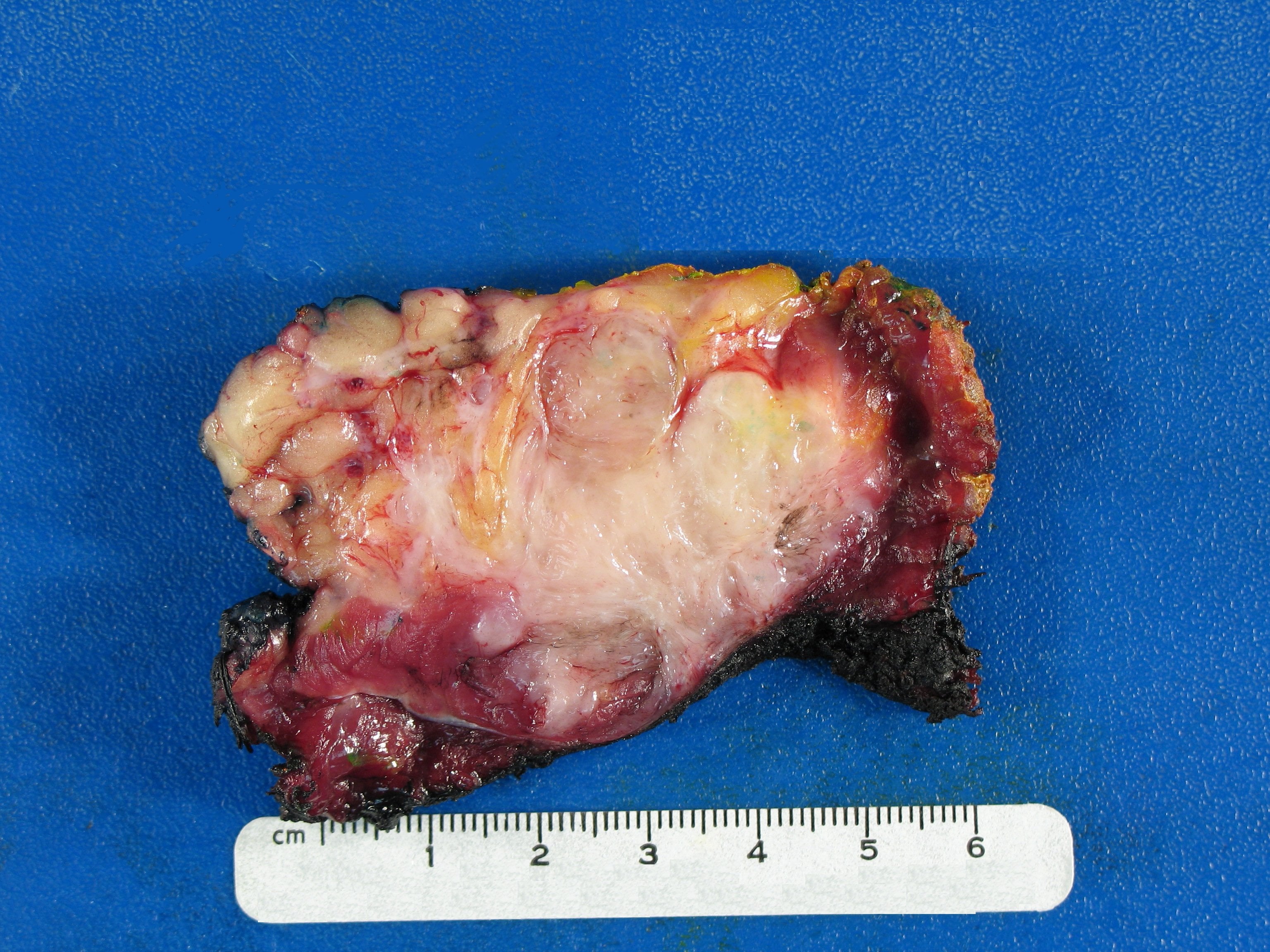
Macroscopic. A firm to rubbery, poorly circumscribed mass that resembles scar tissue or keloid, with a whorled, glistening white-grey cut surface reflecting its collagen. There is no true capsule: the borders are irregular and infiltrative, the tumour follows fascial planes and invades between muscle fibres. Size at diagnosis is typically 5-15 cm, and can be larger.
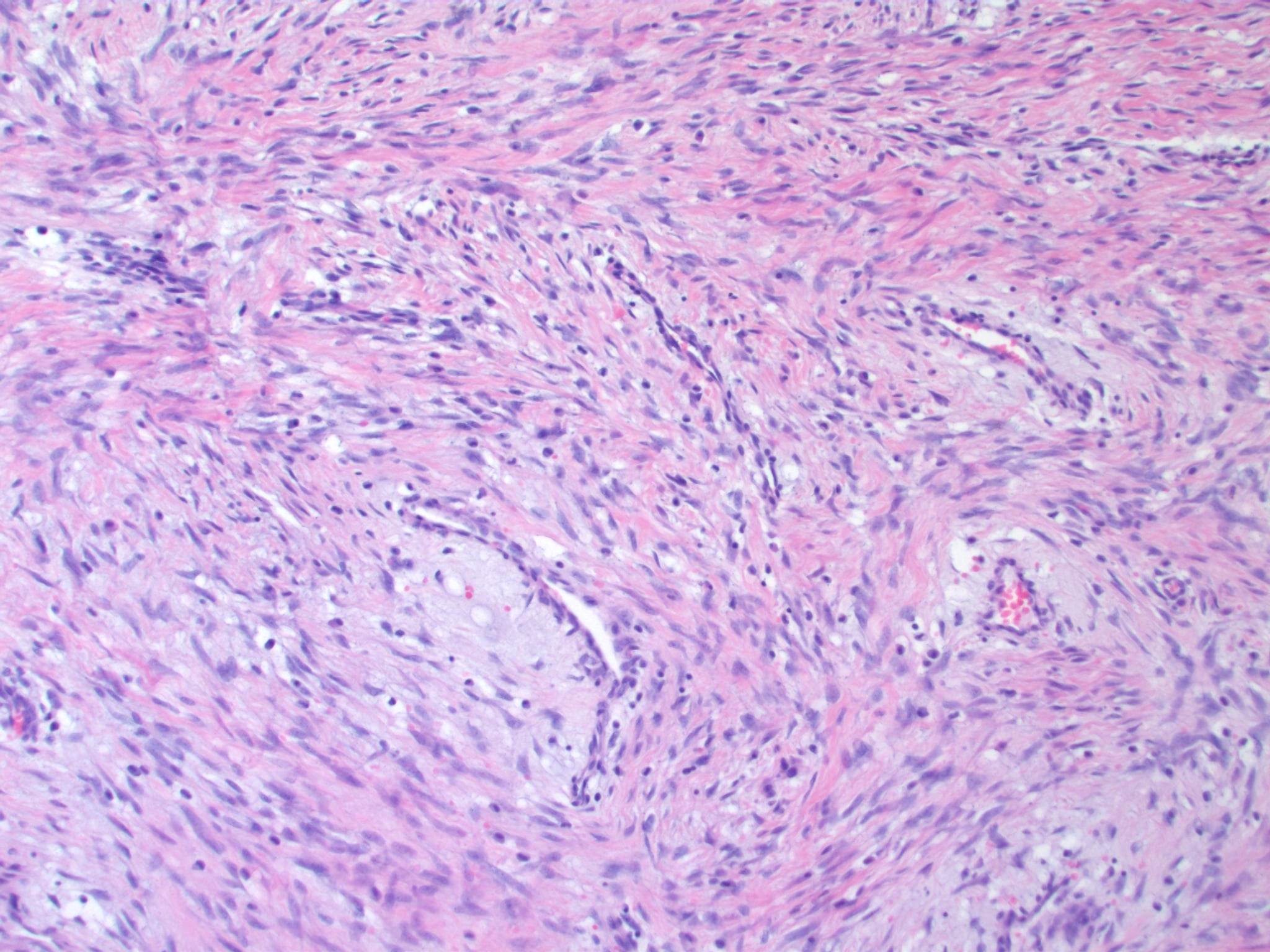
Low power. Long, sweeping, infiltrative fascicles of fibroblasts in collagen, with irregular margins that invade adjacent tissue and dissect between muscle fibres. Vascularity is variable and often low.
High power. Uniform spindle cells, myofibroblasts, with bland cytology and minimal nuclear atypia; the nuclei are vesicular with small nucleoli. Mitoses are rare to absent and necrosis is absent, which is the key to excluding sarcoma. Collagen is abundant and becomes keloid-like in older lesions.
Immunohistochemistry. The panel confirms the myofibroblastic line and excludes the mimics.
- Expression
- Positive in about 80%
- Significance
- Supports the diagnosis - must be nuclear not cytoplasmic; not specific
- Expression
- Positive (variable)
- Significance
- Confirms myofibroblastic differentiation
- Expression
- Low (under 5%)
- Significance
- Confirms benign nature
- Expression
- Negative
- Significance
- Excludes smooth muscle tumor
- Expression
- Negative
- Significance
- Excludes neural tumor
- Expression
- Negative
- Significance
- Excludes solitary fibrous tumor
Reading the beta-catenin stain. Cytoplasmic beta-catenin is a normal, non-specific finding; it is nuclear accumulation that reflects Wnt pathway activation, and nuclear staining is the pattern to look for. It is only about 80% sensitive and it is not specific, so on its own it confirms nothing: it must sit beside the morphology and the clinical context. Negative staining should prompt reconsideration of the diagnosis, and FAP-associated tumours show the same nuclear pattern through APC loss.
BLANDHistology Differential Diagnosis
Hook:BLAND histology distinguishes desmoid from aggressive sarcoma!
Clinical Presentation
Extra-abdominal, 60%. A painless, slowly enlarging mass over months to years, prior trauma to the area (sports injury, surgery) in 30%, found incidentally or noticed by the patient: firm, non-mobile, fixed to the underlying structures. Growth is slow but may run in periods of rapid enlargement followed by stabilisation, and prior trauma to the area (sports injury, surgery) is reported in 30%. Pain appears in 30%, when nerve compression develops, and a large tumour or one involving the shoulder or thigh limits function. The usual sites:
- Shoulder girdle - deltoid, pectoralis, scapular muscles
- Thigh - quadriceps, adductors
- Chest wall, back, gluteal region
- Head and neck, rarely
Abdominal wall, 25%. A firm mass in the anterior abdominal wall, usually in the rectus, and usually painless, so that cosmetic concern predominates. The patient is often a postpartum woman, and a history of caesarean section or prior abdominal surgery is present in 30%. The risk factors are the postpartum period (hormonal change plus rectus trauma), prior caesarean or abdominal surgery, multiple pregnancies and female sex.
Intra-abdominal, 15%. Abdominal pain, bloating and early satiety, with obstructive symptoms when the tumour is mesenteric and a palpable mass when it is large. The tumour may present through its complications: small bowel obstruction, ureteric obstruction with hydronephrosis, and rarely portal vein compression or gastrointestinal bleeding. A history of FAP is present in 50%.
Every patient with an intra-abdominal desmoid is screened for FAP:
- Family history of colorectal cancer or polyposis
- Personal history of colonic polyps
- Examination for extracolonic features: osteomas (skull, mandible), supernumerary teeth, CHRPE (congenital hypertrophy of the retinal pigment epithelium), epidermoid cysts
- Colonoscopy if suspicious
- Genetic testing for an APC germline mutation if clinical suspicion
Examination. The mass is firm to hard and non-tender unless a nerve is compressed, with poorly defined margins and an infiltrative feel, fixed to the underlying fascia or muscle rather than freely mobile. There are no overlying skin changes, unlike malignancy, and no lymphadenopathy, because the tumour is benign. In the abdominal wall, Carnett's sign (pain increasing with contraction of the abdominal muscles) separates an abdominal-wall mass from intra-abdominal pathology. Complete the assessment with the range of motion for an extremity lesion, a neurological examination where nerve compression is suspected, and a vascular examination where the tumour lies near major vessels.
Investigations and Imaging
MRI is the investigation of choice for diagnosis, staging and surveillance.
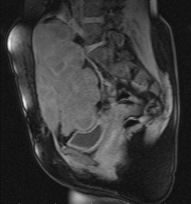
T1. The tumour is isointense to muscle, with poorly defined, irregular margins and extension into adjacent muscle. The fascia sign, or tail sign, is a linear tail of tumour running along the fascial planes; it is highly suggestive of desmoid and reflects the tumour's predilection for aponeurotic structures. T1 is the sequence for anatomical detail and surgical planning: the relationship to neurovascular structures, the extent of muscle involvement, and any bone contact, which is extraperiosteal, without invasion.
T2. Signal varies with cellularity and collagen content, so the overall appearance is heterogeneous: hypercellular areas are intermediate to high, fibrous areas low. Low-signal bands of dense collagen create a characteristic striped appearance that is highly specific for desmoid but, as the summary warns, not pathognomonic; biopsy is still required. The more low-signal tissue, the more fibrous and less cellular the tumour; high T2 signal marks the more cellular, potentially more active tumour. T2 is the sequence for tissue characterisation and diagnosis.
Post-contrast. Enhancement is heterogeneous, because the cellular (high T2) areas enhance and the fibrous bands do not; peripheral enhancement may be seen. There is no necrosis, and its absence helps to exclude sarcoma. Contrast is helpful but not always required for diagnosis.
The triad. Fascia sign on T1, low-signal bands on T2 and heterogeneous enhancement: the three together are highly specific for desmoid.
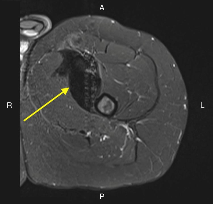
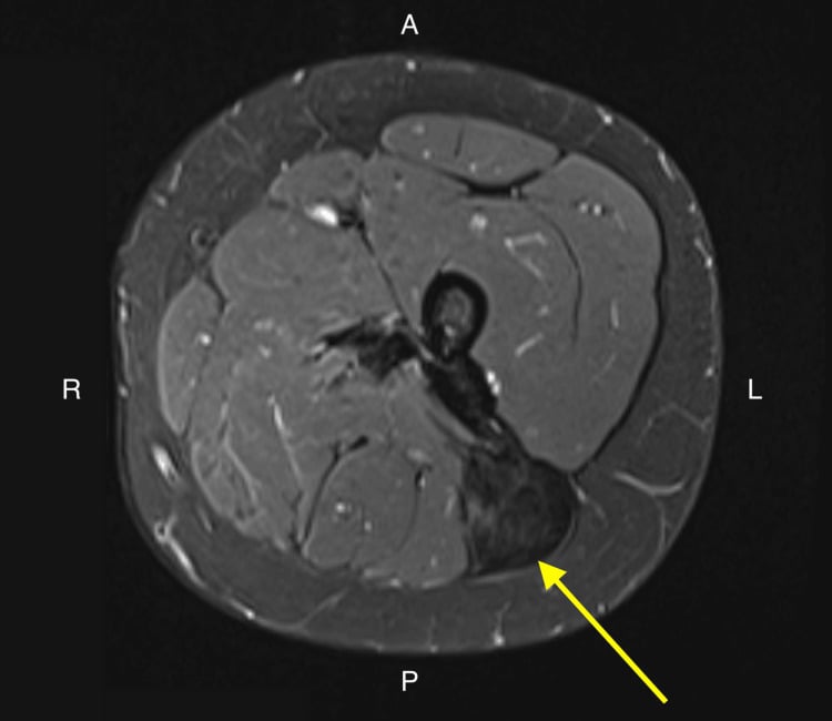
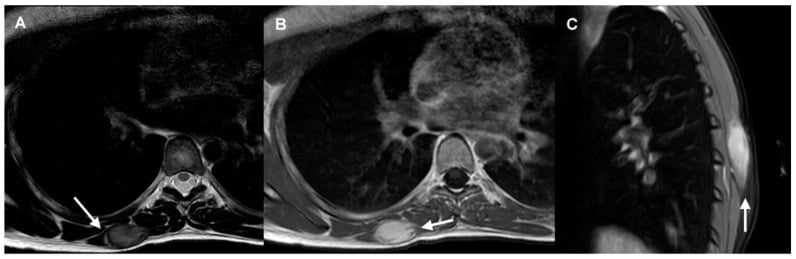
Judging activity and response on MRI. Judging treatment success by size alone, conventional RECIST, underestimates benefit, because a desmoid can become biologically inactive without shrinking. As the tumour matures into a quiescent fibrotic lesion the MRI shows falling T2 signal (low-signal collagen replacing cellular high-signal tissue), reduced contrast enhancement and, usually, loss of symptoms, while the measured diameter stays the same. Stable size with falling T2 signal is a good response; rising T2 signal or enhancement signals reactivation. So assess response by symptom and functional change together with T2-signal and enhancement change, not by size alone, which is why size-based RECIST is a poor endpoint for desmoid trials. Be patient: responses to systemic therapy and radiotherapy are slow (often months, to a median of 6-12 months) and continue for years; do not declare failure early, and tumours may keep regressing after treatment stops.
Ultrasound has a limited role. It shows a hypoechoic mass with infiltrative margins in a superficial lesion and can guide a core biopsy of a superficial tumour, but it cannot adequately assess extent.
CT characterises soft tissue less well than MRI. Its role is in intra-abdominal disease, assessing bowel obstruction and organ involvement, and in guiding percutaneous biopsy.
PET-CT shows variable FDG uptake and is not reliable for diagnosis. It may help in intra-abdominal cases and has a limited role in routine work-up.
Biopsy. Histological confirmation before treatment is essential: it excludes sarcoma and provides tissue for immunohistochemistry. The image-guided (ultrasound or CT) core needle biopsy is the technique of choice, taking 3-4 cores with a 14-16 gauge needle, and the specimen goes for:
- H&E histology
- Nuclear beta-catenin immunostain
- Ki67 proliferation index
- Desmin and S100, to exclude other diagnoses
Core biopsy is diagnostic in over 90%, and false negatives are rare if adequate tissue is obtained; repeat the biopsy if it is non-diagnostic and suspicion remains high.
Do not perform an excisional biopsy. It violates oncological principles if the lesion proves to be a sarcoma, contaminates the tissue planes and makes subsequent surgery more difficult, and a core needle biopsy is both safe and diagnostic.
Do not operate without a histological diagnosis. A lipoma or a benign lesion cannot be assumed from imaging alone, a benign diagnosis must be confirmed before surveillance is offered, and a sarcoma requires a different surgical approach.
Differential Diagnosis
- Key Distinguishing Features
- Nuclear atypia, mitoses, necrosis present; beta-catenin negative
- Definitive Test
- Biopsy with IHC
- Key Distinguishing Features
- Rapid growth (weeks), self-limited; USP6 rearrangement
- Definitive Test
- Clinical course, biopsy
- Key Distinguishing Features
- Sternocleidomastoid mass in infants, birth trauma
- Definitive Test
- Age, location, spontaneous resolution
- Key Distinguishing Features
- Subscapular mass in elderly, specific location, fat on MRI
- Definitive Test
- MRI shows fat intermixed with fibrosis
- Key Distinguishing Features
- Heterotopic ossification, trauma history, zonal phenomenon
- Definitive Test
- MRI/CT shows peripheral ossification
Management Algorithm
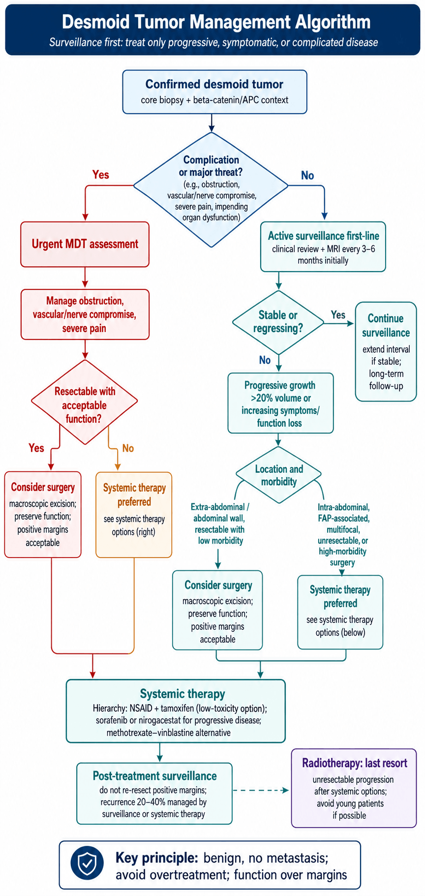
Why surveillance comes first. The natural history set out in the overview is the argument: most tumours stay still or shrink, surgery carries functional loss and a high recurrence rate, and treatment complications may exceed the morbidity of the disease. A benign tumour that never metastasises gives you time to watch, and progression after a period of stability remains manageable.
Who is suitable. Surveillance is the default for the patient who is asymptomatic or minimally symptomatic, without functional impairment or an acute complication such as obstruction or compression, who prefers it after an informed discussion and understands what monitoring involves. On the tumour side it suits:
- Any size
- Stable disease on serial imaging
- A location where surgery would cause major morbidity
- An extra-abdominal site (better prognosis)
- A first presentation rather than a recurrence
The schedule. Intervals lengthen as the tumour declares itself, but surveillance never ends, because late behaviour is unpredictable.
Active Surveillance Schedule
Intensive monitoring while the tumour declares its behaviour:
- Clinical examination at each visit
- MRI every 3-6 months, assessing size, signal characteristics and enhancement
- Document symptoms (pain, functional limitation)
- Photograph for the clinical record
If the tumour is stable:
- Clinical examination every 6 months
- MRI every 6-12 months, less often once a clear pattern of stability is established
- Patient education on the warning signs
Long-term follow-up continues indefinitely:
- Annual clinical examination
- MRI every 1-2 years or as clinically indicated
- Late growth remains possible; the patient self-monitors between visits
Triggers for intervention. Treatment is considered for:
- Progressive growth on serial MRI, greater than 20% volume increase
- Increasing symptoms - pain, functional impairment
- A developing complication - nerve compression, vascular compromise
- Bowel or ureteric obstruction in intra-abdominal disease
- Patient anxiety despite stable disease, through shared decision-making
Counselling. Tell the patient what the evidence says: the tumour is benign and never spreads to other organs, the odds favour it staying the same or shrinking without treatment, monitoring is a safe and active approach, treatment can begin at any point if the tumour progresses, and surveillance is a long-term commitment. Surgery, when it is chosen, carries recurrence and functional loss, and that is part of the same conversation.
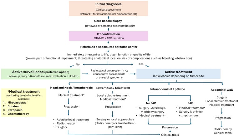
Systemic Therapy
Who needs it. Systemic therapy is reserved for:
- Unresectable tumours with documented progression
- Recurrent tumours not amenable to further surgery
- Symptomatic intra-abdominal desmoids
- Multifocal FAP-associated desmoids
- Patient preference to avoid surgery
Rank the options by their evidence. Two agents have a positive, placebo-controlled phase 3 randomised trial: nirogacestat (DeFi), which is also the only agent approved specifically for desmoid tumours, and sorafenib. Pazopanib has a randomised phase 2 (DESMOPAZ). Everything else - NSAIDs, anti-oestrogens, methotrexate-vinblastine, imatinib, interferon - rests on uncontrolled series.
This is the single most important idea in the systemic section, and it is a favourite discriminator. Desmoid tumours regress spontaneously in about a quarter to a third of patients - 28% regressed on pure observation in the prospective GRAFITI cohort - and in the sorafenib trial the placebo arm had an objective response rate of 20% and a 2-year progression-free survival of 36%.
So any single-arm series reporting "70-80% disease control" with a well-tolerated oral drug is reporting mostly the natural history of the disease, not the effect of the drug. Historical response rates quoted for tamoxifen, sulindac and interferon come from exactly that kind of uncontrolled data and cannot be compared with the randomised figures. When an examiner asks you to justify a systemic agent, cite the controlled comparison or say plainly that there isn't one.
Nirogacestat (gamma-secretase inhibitor) - the best-evidenced option
Mechanism. An oral gamma-secretase inhibitor. Gamma-secretase cleaves the Notch receptor to release its intracellular domain, so inhibiting it blocks Notch signalling, and Notch and Wnt/beta-catenin cross-talk in desmoid fibroblasts. It is not a direct beta-catenin inhibitor, which is a common misconception.
Efficacy (DeFi, phase 3, 70 vs 72 patients, nirogacestat 150 mg twice daily):
- Hazard ratio for progression or death 0.29 (95% CI 0.15-0.55, P less than 0.001)
- 2-year event-free 76% vs 44% with placebo
- Objective response 41% vs 8% (P less than 0.001), with complete response in 7% vs 0%
- Median time to response 5.6 months, faster than sorafenib's 9.6 months
- Significant improvements in pain, symptom burden, physical and role functioning and quality of life (all P at or below 0.01), the endpoints that matter most in a non-metastasising disease
Toxicity. Diarrhoea 84%, nausea 54%, fatigue 51%, hypophosphataemia 42%, maculopapular rash 32%, but 95% of adverse events were grade 1 or 2.
The counselling point examiners look for. Among women of childbearing potential, 27 of 36 (75%) developed adverse events consistent with ovarian dysfunction, which resolved in 20 of 27 (74%). This must be discussed before starting in a young woman, and desmoid tumours peak in exactly that group.
Status. The first agent ever approved specifically for desmoid tumours (FDA, 2023).
Radiotherapy
Radiotherapy is rarely used in modern desmoid management, and when it is used it is the last resort:
- An unresectable tumour failing systemic therapy
- A recurrent tumour not amenable to surgery or systemic therapy
- Symptomatic progression despite all other modalities
- Older patients, in whom late effects matter less
Technique. 50-56 Gy in 25-28 fractions, delivered daily over 5-6 weeks with conformal planning (IMRT or proton therapy) to minimise normal-tissue exposure.
Outcomes. Local control is 70-80% at 5 years, with a response (shrinkage or stabilisation) in 50-60%, and a median time to response of 6-12 months.
Toxicity is significant:
- Acute: skin reaction, fatigue
- Subacute: fibrosis at 3-12 months
- Chronic: fibrosis and joint stiffness in 40-50%, lymphoedema in 10-20%, muscle atrophy
- Radiation-induced sarcoma: rare, 0.5-1% at 10 or more years
Avoid radiotherapy in young patients if at all possible. The lifetime risk of a secondary malignancy is real and a radiation-induced sarcoma can appear decades later, and the fibrosis costs function through joint stiffness and muscle atrophy. Reserve it for older patients (over 50-60) with no other options, and always discuss the risks against the benefits thoroughly.
Image-Guided Cryoablation
Where it sits. Image-guided percutaneous ablation, chiefly cryoablation, now sits between active surveillance and surgery for extra-abdominal desmoids, and is increasingly used where an operation would cost function. CT- or MRI-guided percutaneous probes generate lethal ice-balls that destroy the tumour, and the visible ice-ball allows real-time monitoring of the ablation zone; high-intensity focused ultrasound (HIFU) is an alternative non-invasive thermal option.
Indications. A progressive or symptomatic extra-abdominal desmoid, especially where surgery would be morbid; recurrent disease; and an alternative to systemic therapy. It is repeatable and preserves the surgical and systemic options.
Advantages. Function-preserving and day-case, with good symptomatic relief and local-control rates in published series.
Cautions. Large tumours need staged or multiple sessions. Thermoprotection (hydro- or CO2-dissection, skin warming) is essential when the tumour abuts skin, bowel or a major nerve, to avoid cryo-injury. It is not suited to intra-abdominal or mesenteric disease near bowel, and it requires interventional-radiology expertise.
Surgical Management
Surgery is now reserved for selected cases after surveillance has failed.
- Clinical Scenario
- Growth on serial MRI with pain or functional limitation
- Surgical Goal
- Complete macroscopic excision, accept positive margins
- Clinical Scenario
- Bowel obstruction, vascular compromise, severe pain
- Surgical Goal
- Debulking or complete resection, emergency surgery
- Clinical Scenario
- Cannot exclude sarcoma despite biopsy
- Surgical Goal
- Complete excision for definitive diagnosis
- Clinical Scenario
- Informed patient chooses surgery despite stable disease
- Surgical Goal
- Shared decision-making, counsel on recurrence risk
Function over margins. This is the critical departure from sarcoma surgery, where wide margins are mandatory. Because the tumour is benign, margin status has no survival impact, a recurrence is manageable with surveillance or systemic therapy, and a positive margin is therefore acceptable wherever clearance would cost nerve, vessel or critical muscle. Function is the priority, and the surgical decision-making that follows serves long-term function rather than the pathology report.
A major nerve (median, ulnar, sciatic, femoral). Attempt preservation if a clearance of greater than 1 mm is possible; if the tumour encases the nerve, preserve the nerve and accept the positive margin. A major nerve is never sacrificed for a negative margin, because the functional loss is permanent and devastating.
A major vessel (femoral, popliteal, brachial). Dissect the tumour off the vessel if a plane exists; if it is adherent, preserve the vessel and accept the positive margin. Vascular reconstruction is possible but adds morbidity, so a positive margin is the better trade.
Muscle and bone. Non-critical muscle can be resected; functionally critical muscle is preserved where possible, so the deltoid, which the shoulder depends on, is kept while the sartorius can be sacrificed. A tumour abutting bone is resected extraperiosteally, and bone is not resected for margin in a benign tumour.
Frozen section rarely changes intraoperative management, because a positive margin is often anticipated and accepted; its value is in documenting margin status for adjuvant planning.
Preoperative planning. Review the MRI with radiology, identify the neurovascular structures at risk, plan an extensile incision for adequate exposure, and consent for the acceptance of a positive margin and the risk of recurrence.
The operation.
- Exposure - wide exposure of the entire tumour through an extensile incision with adequate working room, identifying and protecting the critical structures early
- Dissection - aim for complete macroscopic excision along tissue planes where they exist; the tumour is often densely adherent to fascia and muscle, and piecemeal resection is accepted if that is what preserves function
- Critical structures - dissect the tumour carefully from nerve and vessel; if a structure is encased, preserve it and leave tumour on its surface; neurolysis where a nerve is compressed but separable; never sacrifice a structure for margin
- Specimen - orient with sutures, document any margin concerns, and send for permanent sections
- Reconstruction - primary closure if tension-free, local flaps for soft-tissue cover, mesh reconstruction of the abdominal wall if the rectus has been excised, and drains for dead space
After surgery. Early mobilisation, drain removal once output is under 30 ml per 24 hours, wound monitoring, and then the final pathology for margin status, on which surveillance or adjuvant therapy is planned.
Outcomes. Recurrence after resection runs at 20-40% even after complete excision, and it is the commonest complication of surgery.
- Negative Margins
- 20-30%
- Positive Margins
- 30-50%
- Management
- Higher with positive margins but many do not recur
- Negative Margins
- 30-40%
- Positive Margins
- 40-60%
- Management
- Late recurrence possible, lifelong surveillance needed
- Negative Margins
- Median 24 months
- Positive Margins
- Median 18 months
- Management
- Can occur decades later, unpredictable
- Negative Margins
- Variable by site
- Positive Margins
- Better if nerve/vessel preserved
- Management
- Function preservation improves quality of life
Recurrence also varies by site:
- Extra-abdominal: 20-40%
- Abdominal wall: 20-30%
- Intra-abdominal: 40-60%, the worst prognosis
Why a positive margin is not re-resected. The crude figures above are higher with a positive margin, yet many patients with positive margins never recur, and in Crago's multivariate analysis the microscopic margin did not independently predict recurrence at all. Re-resection adds morbidity without clear benefit, recurrence is manageable with surveillance or systemic therapy, and function matters more than clearance. Examiners expect this principle stated plainly.
Managing a recurrence. The first line is active surveillance, exactly as for the primary tumour. Re-resection is for the recurrence that is progressive and symptomatic, systemic therapy is considered especially after multiple recurrences, and radiotherapy remains the last resort.
Complications
Of the disease. The complications follow the site.
- Incidence
- 20-30%
- Location
- Intra-abdominal desmoids
- Management
- Surgery, systemic therapy, nutritional support
- Incidence
- 10%
- Location
- Intra-abdominal/pelvic
- Management
- Stent, nephrostomy, systemic therapy
- Incidence
- 10-15%
- Location
- Extremity desmoids
- Management
- Surgery if progressive, pain management
- Incidence
- 30%
- Location
- Large extremity tumors
- Management
- Analgesia, physiotherapy, systemic therapy
Of surgery. Recurrence, covered above, is the commonest. The others:
- Wound infection: 10-15%
- Nerve injury: 5-10%, higher if the tumour is adherent
- Functional impairment: 20-40% (shoulder, thigh)
- Abdominal wall hernia: 10% where mesh reconstruction was needed
Of systemic therapy and radiotherapy. Each agent's toxicity is set out with the agent above: the GI upset and hot flushes of NSAIDs and tamoxifen, the rash, fatigue, hypertension and diarrhoea of sorafenib together with hand-foot syndrome in 50%, the myelosuppression and nausea of methotrexate-vinblastine, and the fibrosis, lymphoedema and secondary malignancy of radiotherapy.
Prognosis and Natural History
- Favourable
- Abdominal wall (best of all - 91% 5-year local recurrence-free survival after resection)
- Unfavourable
- Extremity (independent predictor of recurrence); intra-abdominal/mesenteric carries the greatest morbidity
- Favourable
- Older
- Unfavourable
- YOUNGER - young age was an independent predictor of recurrence in Crago's 495-patient series
- Favourable
- Smaller
- Unfavourable
- Larger - one of the three nomogram variables, and 5cm or more carried HR 2.38 for needing treatment on surveillance
- Favourable
- T41A (the commonest, more indolent)
- Unfavourable
- S45F - hazard ratio 6.24 for starting active treatment (GRAFITI)
- Favourable
- Unifocal, sporadic
- Unfavourable
- Multifocal, FAP-associated
- Favourable
- Not a reliable prognostic factor either way
- Unfavourable
- R1 resection did NOT predict recurrence on multivariate analysis - which is precisely why positive margins are not re-excised
Each of these is the opposite of the intuition carried over from sarcoma practice:
- Young age is BAD, not good. In Crago's series young age, extremity site and large size were the three independent predictors of recurrence, and they are the only three variables in the validated nomogram. Age, site, size.
- A positive margin does NOT predict recurrence. It was not significant on multivariate analysis, and adjuvant radiotherapy did not improve local recurrence-free survival either. This is the single biggest departure from sarcoma thinking, and it is why you do not go back for the margin.
- S45F is the bad genotype, not T41A. T41A is the commonest and the more indolent; S45F carries a more than six-fold hazard for requiring treatment.
Guidelines, Registries & Global Practice
Global Epidemiology
Desmoid-type fibromatosis is a rare tumour. Population-based and registry data give an estimated incidence of roughly 3 to 5 cases per million per year, with a female predominance and a peak in the third and fourth decades. The prospective Dutch GRAFITI cohort and large referral-centre series confirm that the majority of patients managed with active surveillance never require active treatment, and that genotype (S45F CTNNB1) and tumour size predict the need for intervention (Schut et al., Ann Surg 2023; DOI). The defining molecular feature, CTNNB1 mutation in approximately 85% of sporadic cases, is consistent across international cohorts (Gronchi et al., Ann Oncol 2014; DOI).
Side-by-Side Guideline Guidance
- Front-Line Strategy
- Active surveillance for most newly diagnosed tumours
- Surgery
- Reserved for progression; function over margins
- Systemic Therapy
- Sorafenib, nirogacestat, low-dose chemo, hormonal/NSAID for progression
- Evidence Basis
- Expert consensus + RCTs
- Front-Line Strategy
- Watchful waiting first-line; treat on documented progression
- Surgery
- Selective; avoid mutilating resection
- Systemic Therapy
- Stepwise escalation; refer to sarcoma unit
- Evidence Basis
- Level I-III
- Front-Line Strategy
- Observation acceptable for asymptomatic disease
- Surgery
- Resection if progressive/symptomatic and function preserved
- Systemic Therapy
- Sorafenib or nirogacestat category 1 for progression
- Evidence Basis
- Category 1-2A
- Front-Line Strategy
- Front-line wait-and-see algorithm
- Surgery
- Stepwise, anatomy-driven, referral-centre
- Systemic Therapy
- Treatment only on progression
- Evidence Basis
- Consensus (PMID 24325833)
Registry & Prospective Evidence
- Design
- 105 patients, active surveillance
- Key Finding
- Only 30% needed treatment at 3 years; 28% regressed
- Practice Impact
- Validates surveillance-first internationally
- Design
- 495 resected patients
- Key Finding
- Margin NOT predictive; site/age/size are
- Practice Impact
- Function-preserving surgery; selective systemic therapy
- Design
- 142 patients, nirogacestat vs placebo
- Key Finding
- ORR 41% vs 8%; 2-yr event-free 76% vs 44%
- Practice Impact
- First desmoid-specific approved agent
Practice Variation & Global Access
International practice has converged on a surveillance-first, multidisciplinary model coordinated through specialist sarcoma centres, though access to newer agents varies by jurisdiction. Suspected desmoid tumours should be discussed with a specialist sarcoma multidisciplinary team, with image-guided core needle biopsy arranged locally or at the referral centre. Tamoxifen is widely available and inexpensive, and nuclear beta-catenin immunohistochemistry is available through standard anatomical pathology services. Sorafenib and nirogacestat regulatory and reimbursement status differs between countries, so confirm local availability before counselling patients on systemic options.
Documentation. The record should show:
- Histological diagnosis before treatment (biopsy mandatory)
- MDT discussion for complex cases
- Informed consent: natural history (roughly 60% stable/regress - GRAFITI), recurrence risk, margin philosophy
- Surveillance protocol clearly documented
- Positive margin accepted: document the reason (function preservation)
Common litigation issues. The recurring failures are:
- Excising a mass without biopsy (missed sarcoma)
- Aggressive surgery without a trial of surveillance
- Nerve sacrifice for negative margins (unacceptable)
- Inadequate surveillance leading to late detection of recurrence
MCQ Practice Points
Q: What is the diagnostic immunohistochemical marker for desmoid tumor? A: Nuclear beta-catenin - Nuclear (not cytoplasmic) accumulation of beta-catenin is seen in about 80% of desmoid tumors and supports, but does not by itself confirm, the diagnosis. This reflects CTNNB1 mutation preventing beta-catenin degradation, leading to nuclear translocation and Wnt pathway activation. Cytoplasmic beta-catenin is non-specific.
Q: What is the first-line management for asymptomatic extra-abdominal desmoid tumor? A: Active surveillance - Modern evidence supports a surveillance-first approach as roughly 60% of desmoids remain stable or regress spontaneously without intervention (GRAFITI: 28% regressed, 32% stable). Surgery has high morbidity (recurrence 20-40%, functional loss) and should be reserved for progressive symptomatic tumors. MRI every 3-6 months for monitoring.
Q: A desmoid tumor is resected with positive margins. What is the appropriate next step? A: Active surveillance, do NOT re-resect - Unlike sarcoma, positive margins in desmoid do not mandate re-resection. Positive margins increase recurrence from 20-30% to 30-50% but many patients never recur. Function preservation is priority over margin clearance. Recurrence is manageable with surveillance or systemic therapy.
Q: What percentage of FAP patients develop desmoid tumors? A: 10-20% lifetime risk - Desmoid tumors occur in 10-20% of patients with familial adenomatous polyposis. These are typically intra-abdominal (mesenteric), often triggered by prior colectomy, and represent leading cause of mortality in FAP after prophylactic colectomy. APC germline mutation rather than CTNNB1 somatic mutation.
Q: What MRI finding is pathognomonic for desmoid tumor? A: Low-signal T2 bands - Low-signal bands on T2-weighted MRI represent dense collagen bundles and are highly specific for desmoid tumor. Combined with fascia sign (tail along fascia on T1) and infiltrative margins, these features are diagnostic. Heterogeneous enhancement due to mix of cellular and fibrous areas.
Exam Viva Scenarios
Practise clinical reasoning and management decisions out loud
“A 32-year-old woman presents 6 months postpartum with a 6cm mass in the right rectus abdominis. Core biopsy shows bland spindle cells with nuclear beta-catenin positivity, consistent with desmoid tumor. MRI shows infiltration of rectus muscle. She is asymptomatic. How would you manage this patient?”
“During resection of a shoulder girdle desmoid tumor, you find that the tumor densely encases the axillary nerve. You can either preserve the nerve with positive margins or resect the nerve en bloc for negative margins. What do you do and why?”
“A 28-year-old woman with known FAP status post total colectomy 2 years ago presents with abdominal pain and intermittent small bowel obstruction. CT shows a 12cm mesenteric mass consistent with desmoid tumor. What is your management approach?”
Key Definition
- Benign fibroblastic tumor - NEVER metastasizes (0% metastatic potential)
- Locally aggressive with infiltrative growth pattern
- Also called aggressive fibromatosis
- Unpredictable: ~60% stable/regress, 20-30% progress
Molecular Genetics (High Yield)
- CTNNB1 mutation 85-90% (sporadic) - exon 3, codons 41/45
- APC mutation 5-10% (FAP-associated) - germline
- Both cause nuclear beta-catenin accumulation via Wnt pathway
- Nuclear beta-catenin IHC supports the diagnosis (~80% sensitive, not pathognomonic)
Location Distribution
- Extra-abdominal 60% (shoulder, thigh most common)
- Abdominal wall 25% (rectus, postpartum women)
- Intra-abdominal 15% (mesentery, FAP-associated)
- FAP-desmoids: intra-abdominal, multifocal, worse prognosis
MRI Triad (Pathognomonic)
- T1: Fascia sign (tail along fascial planes)
- T2: Low-signal bands (collagen bundles) - diagnostic
- Post-contrast: Heterogeneous enhancement (cellular areas enhance)
Management Algorithm
- First-line: Active surveillance (~60% stable/regress)
- MRI every 3-6 months, intervene if progressive/symptomatic
- Surgery: Function over margins, positive margins acceptable
- Systemic: nirogacestat and sorafenib have placebo-controlled trials; NSAIDs/tamoxifen rest on uncontrolled data
- Radiotherapy: Last resort (secondary malignancy risk)
Surgical Philosophy
- Function preservation PRIORITY over negative margins
- Positive margins acceptable (unlike sarcoma management)
- Do NOT sacrifice major nerve/vessel for margins
- Do NOT re-resect positive margins (recurrence manageable)
- Recurrence 20-40% regardless of margin status
Natural History
- 10-20% spontaneous complete regression
- ~60% stable or regressive disease without treatment (GRAFITI)
- 20-30% progressive growth requiring treatment
- Recurrence after surgery: 20-40% at 5-10 years
FAP-Associated Features
- 10-20% of FAP patients develop desmoids
- Intra-abdominal location (mesentery), multifocal
- Triggered by abdominal surgery (colectomy)
- Leading cause of death in FAP after prophylactic colectomy
- Systemic therapy first-line (surgery triggers more tumors)
Exam Pearls
- NEVER metastasizes - benign but locally aggressive
- Nuclear (not cytoplasmic) beta-catenin diagnostic
- Surveillance first-line - avoid overtreatment
- Positive margins acceptable - function priority
- Unpredictable behavior - can regress spontaneously
Evidence Base and Key Studies
Front-Line Conservative Approach (Fiore, IGR/INT)
- 142 desmoid patients (Institut Gustave Roussy and Istituto Nazionale Tumori) managed front-line without surgery or radiotherapy
- 83 patients had a deliberate wait-and-see policy, 59 had front-line medical therapy
- 5-year progression-free survival 49.9% (wait-and-see) vs 58.6% (medical therapy), not significantly different (P=0.32)
- Similar results for primary and recurrent disease; no clinical variable independently predicted PFS
- All progressing patients were subsequently salvaged safely
Prognostic Nomogram - Margin Not Predictive (Crago, MSKCC)
- 495 patients resected for primary or recurrent desmoid; 100 of 439 with complete gross resection recurred (23%)
- 5-year local recurrence-free survival 69% overall; abdominal wall tumours best at 91%
- On multivariate analysis recurrence was driven by extremity site, young age and large size - NOT by microscopic margin (R1)
- Adjuvant radiotherapy did not improve local recurrence-free survival
- Nomogram of size, site and age (concordance 0.70) validated in an international multi-institutional dataset
Sorafenib for Progressive Desmoid Tumors (Phase 3, Alliance A091105)
- Double-blind randomised placebo-controlled trial, 87 patients with progressive, symptomatic or recurrent desmoids
- 2-year progression-free survival 81% sorafenib vs 36% placebo (HR 0.13, 95% CI 0.05-0.31, P less than 0.001)
- Objective response rate 33% sorafenib vs 20% placebo before crossover
- Most common grade 1-2 events: rash 73%, fatigue 67%, hypertension 55%, diarrhoea 51%
- Median time to response approximately 9.6 months on sorafenib
Nirogacestat (Gamma-Secretase Inhibitor) - DeFi Phase 3
- International double-blind randomised placebo-controlled trial, 142 adults with progressing desmoid tumours
- 2-year event-free likelihood 76% nirogacestat vs 44% placebo (HR for progression/death 0.29, 95% CI 0.15-0.55, P less than 0.001)
- Objective response rate 41% vs 8% (complete response 7% vs 0%)
- Significant improvements in pain, symptom burden and quality of life
- Common events: diarrhoea 84%, nausea 54%, fatigue 51%; 75% of women of childbearing potential had ovarian dysfunction (usually reversible)
Pazopanib versus Methotrexate-Vinblastine (DESMOPAZ)
- Non-comparative randomised open-label phase 2, 72 adults with centrally confirmed PROGRESSIVE desmoid tumours, allocated 2:1 to pazopanib 800mg daily for up to 1 year or weekly intravenous methotrexate-vinblastine
- 83.7% (95% CI 69.3-93.2) of the pazopanib group were progression-free at 6 months, versus 45.0% (95% CI 23.1-68.5) with methotrexate-vinblastine
- Grade 3-4 events with pazopanib were hypertension (21%) and diarrhoea (15%); with methotrexate-vinblastine, neutropenia (45%) and transaminitis (18%)
- Methotrexate-vinblastine was, until this trial, the only chemotherapy regimen ever assessed for desmoid tumours in a trial setting
Prospective Active Surveillance - GRAFITI Trial
- Nationwide Dutch multicentre prospective cohort, 105 patients with non-intra-abdominal desmoid on initial active surveillance
- At 3 years cumulative incidence of starting active treatment was only 30%; progression-free survival 58%
- During surveillance 28% regressed, 32% were stable; 40% had initial progression that frequently later stabilised or regressed
- Tumour 5 cm or larger (HR 2.38) and S45F CTNNB1 mutation (HR 6.24) predicted need for active treatment
- Most patients never required active treatment
Italian-French Sarcoma Group Position Paper (Consensus Guidance)
- Joint position paper of the Italian and French Sarcoma Groups proposing a stepwise algorithm for sporadic desmoid-type fibromatosis
- Endorses a front-line wait-and-see approach with treatment reserved for documented progression
- Confirms approximately 85% CTNNB1 prevalence and the central role of Wnt/beta-catenin signalling
- Emphasises individualised, anatomy-driven decisions and mandatory referral-centre counselling
- Recognises the absence of an evidence-based single standard of care