Eaton-Littler Staging of Thumb CMC Arthritis
Stage I = ligament reconstruction (preserve the joint). Stage IV = trapeziectomy (the joint is destroyed and the adjacent scaphotrapezial joint is also arthritic). The middle stages (II–III) are where debate lives — examiners want you to demonstrate that you understand the transition from joint-preserving to joint-sacrificing surgery. Always mention the beak ligament (anterior oblique ligament) as the primary stabiliser that fails early, allowing dorsal subluxation and the cascade of osteoarthritis.
Anatomy and Pathomechanics
Understanding why the thumb CMC joint degenerates is essential for viva questions:
- The trapeziometacarpal (TM) joint is a saddle (sellar) joint — it permits flexion-extension and abduction-adduction with a degree of rotation. This freedom of motion comes at the cost of intrinsic instability.
- The beak ligament (anterior oblique ligament — AOL) runs from the palmar ridge of the first metacarpal base to the tubercle of the trapezium. It is the primary restraint to dorsal subluxation during pinch and grip.
- With age and repetitive loading, the AOL attenuates or tears, allowing the metacarpal base to sublux dorsally. This concentrates contact forces on the dorsal articular surface, leading to cartilage loss and osteophyte formation in a predictable pattern.
- The dorsoradial ligament (DRL) and posterior oblique ligament (POL) are secondary stabilisers but are less often discussed in the Eaton-Littler context.
- Pan-trapezial arthritis (Stage IV) means the scaphotrapezial (ST) joint is also degenerate. This matters because trapeziectomy alone addresses both joints; arthrodesis of only the TM joint would leave painful ST arthritis un-treated.
The beak ligament fails firstThe ligaments around the thumb CMC
Hook:For exams: beak (AOL) ligament failure → dorsal subluxation → the start of the Eaton-Littler cascade.
The staging is radiographic, but the examiner wants the clinical syndrome the radiograph explains:
- Epidemiology — very common, especially post-menopausal women (40s onward), often bilateral, associated with generalised osteoarthritis and ligamentous laxity.
- Presentation — insidious thumb-base pain worse with pinch and grip (opening jars, turning keys/door handles, writing), with aching, weakness and loss of dexterity.
- Examination — tenderness over the TM joint; a positive grind/torque test (axial load plus rotation reproduces pain and crepitus) and lever/shear test; visible squaring ("shoulder sign") of the thumb base from dorsoradial metacarpal subluxation; and thenar wasting.
- The compensatory deformity (high-yield) — advanced disease produces a first-metacarpal adduction contracture (a narrowed first web) with compensatory metacarpophalangeal (MCP) hyperextension — the "zigzag"/collapse deformity. The surgical corollary: at trapeziectomy you must also release the adducted first web and correct a significant MCP hyperextension (e.g. more than ~30°) — by volar capsulodesis, sesamoid arthrodesis, EPB transfer or temporary MCP pinning/fusion — or the deformity and pain recur.
Radiographic Technique and Pitfalls
Accurate staging depends on good imaging. A standard hand radiograph is often inadequate for the thumb CMC joint.
- Robert's view (hyperpronated PA): profiles the thumb CMC joint free of overlap. This is the single most useful view for staging.
- True lateral of the thumb CMC: obtained with the hand resting flat on the cassette and the thumb abducted; the X-ray beam is directed perpendicular to the trapeziometacarpal joint. Essential for assessing joint-space narrowing and dorsal subluxation.
- Stress views (forceful radial abduction of the thumb against resistance): may demonstrate dynamic instability in Stage I where static films look near-normal.
- Pitfalls: overlap of the trapezium and scaphoid on a poorly positioned lateral can falsely suggest ST joint involvement or miss it. Always confirm Stage IV on a well-positioned view before committing to pan-trapezial surgery.
- CT and MRI are not part of the standard Eaton-Littler staging (which is a plain-radiograph system) but may be used to assess the beak ligament pre-operatively or to stage the ST joint when plain films are equivocal.
Surgical Decision-Making by Stage
- Joint-Preserving Options
- Beak ligament reconstruction (FCR or APL transfer)
- Joint-Sacrificing Options
- Not indicated
- Key Decision Point
- Confirm instability on stress views; conservative measures first
- Joint-Preserving Options
- Ligament reconstruction ± temporary K-wire
- Joint-Sacrificing Options
- Arthroscopic debridement or interposition
- Key Decision Point
- Young high-demand patient favours ligament reconstruction
- Joint-Preserving Options
- Ligament reconstruction controversial (joint already damaged)
- Joint-Sacrificing Options
- Trapeziectomy with ligament interposition (FCR/APL) or suspensionplasty
- Key Decision Point
- Joint is too arthritic to preserve reliably — most surgeons now offer trapeziectomy
- Joint-Preserving Options
- Not appropriate — both joints diseased
- Joint-Sacrificing Options
- Complete trapeziectomy (addresses TM and ST); TM arthrodesis only if ST joint is truly spared (rare in Stage IV)
- Key Decision Point
- Arthrodesis for high-demand young manual workers; trapeziectomy for older patients
Preserve · Bridge · RemoveSurgical algorithm simplified
Hook:Exception: arthrodesis for young, high-demand patients with Stage III disease and a normal ST joint.
Never offer ligament reconstruction for Stage IV disease — the scaphotrapezial joint is also arthritic and will remain painful. Similarly, do not fuse the TM joint in a patient with Stage IV disease unless you simultaneously address the ST joint; a TM arthrodesis leaves a painful ST joint un-treated.
The examiner expects the full menu for Stage III/IV and the specific complications (a viva favourite):
- The options spectrum — trapeziectomy (excisional arthroplasty) alone (the Davis RCT showed no added benefit from interposition or LRTI); trapeziectomy + ligament reconstruction and tendon interposition (LRTI, an FCR slip); trapeziectomy + suspensionplasty (suture suspension, a suture-button/"tightrope", or APL suspension); trapeziometacarpal joint replacement / implant arthroplasty (modern dual-mobility total TM implants — faster recovery but loosening/survivorship concerns — and pyrocarbon implants); and arthrodesis (TM fusion) for the young, high-demand manual worker with an isolated, healthy ST joint.
- Complications of trapeziectomy — metacarpal subsidence / proximal migration with impingement on the scaphoid (scaphometacarpal pain), persistent weakness of pinch and grip, persistent or recurrent pain, injury to the superficial (dorsal) radial sensory nerve → painful neuroma/dysaesthesia, FCR/scar problems and stiffness. Implants add loosening/dislocation; arthrodesis adds nonunion (around 10%), hardware problems and later peritrapezial/ST pain.
The Eaton-Littler Classification
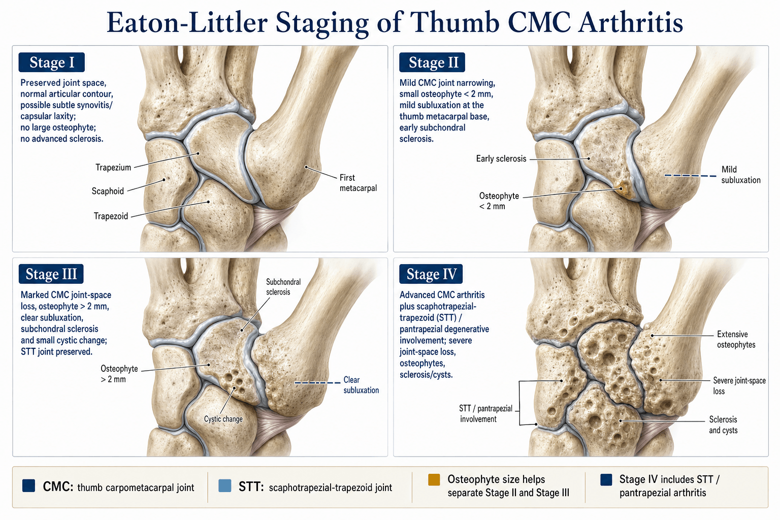
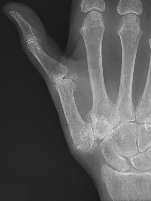
The classification is based on true lateral and AP radiographs of the thumb CMC joint (Robert's view is ideal). Four stages are defined by joint-space narrowing, osteophyte size, subchondral sclerosis, and involvement of the adjacent scaphotrapezial (ST) joint.
- Joint Space
- Normal or slightly widened; subchondral sclerosis may be present
- Osteophytes
- None
- Scaphotrapezial Joint
- Normal
- Typical Management
- Ligament reconstruction (beak ligament)
- Joint Space
- Narrowed; mild subluxation may be seen
- Osteophytes
- Less than 2 mm
- Scaphotrapezial Joint
- Normal
- Typical Management
- Ligament reconstruction ± flexor carpi radialis sling
- Joint Space
- Markedly narrowed or obliterated
- Osteophytes
- Greater than 2 mm
- Scaphotrapezial Joint
- Normal
- Typical Management
- Trapeziectomy with ligament interposition or suspensionplasty
- Joint Space
- Obliterated (pan-trapezial disease)
- Osteophytes
- Greater than 2 mm
- Scaphotrapezial Joint
- Involved (narrowed, sclerosis, osteophytes)
- Typical Management
- Trapeziectomy (complete); arthrodesis if high-demand young patient
SNOO — Space, Narrowing, Osteophytes, Other jointRemember the four stages
Hook:SNOO walks you up the stages: Space → Narrowing → Osteophytes → Other (ST) joint.
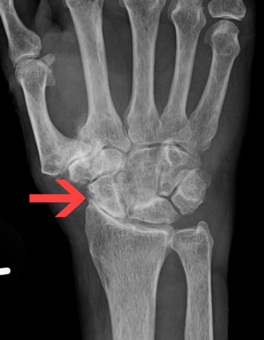
The osteophyte threshold that separates Stage II from Stage III is 2 mm — examiners may give you a radiograph and ask you to stage it. Measure the largest osteophyte: less than 2 mm is Stage II, greater than 2 mm is Stage III. If the scaphotrapezial joint is also narrowed, it becomes Stage IV regardless.
Differential Diagnosis of Thumb Base Pain
The grind test (axial compression and rotation of the thumb CMC) reproduces pain and crepitus in CMC arthritis, but consider:
- De Quervain's tenosynovitis — Finkelstein's test positive; pain is over the first dorsal compartment, not the CMC joint. May coexist with CMC arthritis.
- Scaphoid fracture or non-union — snuffbox tenderness; radiographic scaphoid pathology.
- ST joint arthritis without TM involvement — rare but possible; pain is more proximal. Eaton-Littler would classify this as outside the system (the TM joint is normal).
- Flexor carpi radialis tendinitis — pain volar and radial at the wrist; may mimic CMC pain but the grind test is negative.
- Carpal tunnel syndrome — paraesthesia in the median nerve distribution; night symptoms. Co-exists commonly with thumb CMC arthritis (both conditions increase with age).
- Trigger thumb — clicking or locking of the IP joint with a palpable nodule at the A1 pulley; distinct from CMC pathology.
Non-Operative Management
Not every patient with thumb CMC arthritis needs surgery. Non-operative treatment is first-line for Stage I and acceptable for Stage II–III in low-demand or medically unfit patients:
- Thumb spica splinting — rest the CMC joint, particularly at night. A custom thermoplastic splint immobilising the CMC while leaving the IP free is most effective.
- Activity modification — avoid sustained lateral pinch and power grip; use ergonomic tools with larger grips.
- NSAIDs — topical or oral for symptomatic relief.
- Hand therapy — thenar strengthening (particularly opponens and abductor pollicis brevis) to compensate for CMC instability.
- Intra-articular corticosteroid injection — provides temporary relief (typically 3–6 months). Useful as a diagnostic adjunct and for patients not ready for surgery. Repeat injections carry diminishing returns and may accelerate cartilage loss.
- Hyaluronic acid injection — some evidence for short-term pain relief in Stage I–II but not superior to corticosteroid in most trials.
Viva practice
Exam Viva
Practise clinical reasoning and management decisions out loud
“A 58-year-old right-hand-dominant woman presents with a two-year history of gradually worsening pain at the base of her right thumb. She has pain with lateral pinch (e.g. turning a key) and on examination has a positive grind test with mild thenar wasting. Radiographs show narrowing of the thumb CMC joint with a 2.5 mm osteophyte at the metacarpal base; the scaphotrapezial joint appears preserved. How would you classify and manage this?”
“A 42-year-old builder presents with pain at the base of his dominant thumb. Stress radiographs demonstrate dorsal subluxation of the first metacarpal on the trapezium but the joint space is preserved with no osteophytes. He has failed six months of splinting and activity modification. What is the diagnosis, how do you stage it, and what is your surgical plan?”
Exam cheat sheet
The four stages (radiographic)
- Stage I: joint space preserved or slightly widened; no osteophytes; stress views may show subluxation
- Stage II: joint narrowed; osteophytes less than 2 mm; scaphotrapezial joint normal
- Stage III: joint markedly narrowed or obliterated; osteophytes greater than 2 mm; scaphotrapezial joint normal
- Stage IV: pan-trapezial disease — TM joint obliterated AND scaphotrapezial joint involved
Surgical algorithm by stage
- Stage I: ligament reconstruction (beak ligament / AOL) — joint preserving
- Stage II: ligament reconstruction or arthroscopic debridement — still joint preserving
- Stage III: trapeziectomy (with or without interposition / suspensionplasty) — joint sacrificing
- Stage IV: complete trapeziectomy (addresses both TM and ST joints); arthrodesis if young and high-demand with isolated TM disease
Key anatomy and exam pearls
- Beak ligament (anterior oblique ligament) is the primary restraint to dorsal subluxation
- Robert's view is the best radiograph for staging the thumb CMC joint
- The grind test reproduces pain and crepitus in CMC arthritis
- Osteophyte threshold separating Stage II from III is 2 mm
- Stage IV always involves the scaphotrapezial joint — confirm on a well-positioned lateral
Evidence
Ligament reconstruction for the painful thumb carpometacarpal joint
- The original Eaton-Littler paper describing volar (beak / anterior oblique) ligament reconstruction using a slip of flexor carpi radialis for the unstable, early-arthritic thumb CMC joint.
- Set out the radiographic staging of trapeziometacarpal arthritis on which treatment is based.
- Provided the rationale for joint-preserving ligament reconstruction in early (pre-arthritic / Stage I) disease.
Excision of the trapezium for osteoarthritis of the trapeziometacarpal joint: a study of the benefit of ligament reconstruction or tendon interposition
- Randomised controlled trial of 183 thumbs across THREE arms: simple trapeziectomy, trapeziectomy + palmaris longus interposition, and trapeziectomy + flexor carpi radialis ligament reconstruction and tendon interposition (LRTI).
- At 1 year, 82% had good pain relief and 68% regained sufficient strength for normal activities; outcomes and range of movement were NOT influenced by the type of surgery.
- Key-pinch improved from 3.5 to 4.6 kg regardless of technique, and complications were distributed evenly among the three groups.
According to PubMed, the staging and volar ligament reconstruction are from Eaton & Littler 1973 (J Bone Joint Surg Am 1973;55(8):1655-66; PMID 4804988), with the pan-trapezial Stage IV from the later Eaton-Glickel modification. The three-arm RCT showing no benefit of interposition/LRTI over simple trapeziectomy is Davis et al. 2004 (DOI 10.1016/j.jhsa.2004.06.017); the long-term LRTI durability data are Tomaino et al. 1995 (DOI 10.2106/00004623-199503000-00003); and the arthrodesis-versus-LRTI comparison is Hartigan et al. 2001 (DOI 10.2106/00004623-200110000-00002).