Post-traumatic vs Inflammatory | Conservative First | TEA for End-Stage | Ulnar Nerve
- RA is most common inflammatory cause - bilateral involvement, systemic disease
- Post-traumatic follows fractures, dislocations, or chronic instability
- Conservative management first - NSAIDs, injections, activity modification
- TEA indicated when conservative fails, age over 60, low demand
- Ulnar nerve commonly affected - assess and document it before every elbow operation
- “RA elbow arthritis: bilateral, systemic, medical management first
- “Post-traumatic: history of fracture/dislocation, may have instability
- “TEA best outcomes in RA (90% 10-year survival) vs trauma (75%)
- “Ulnar nerve must be assessed and protected in all elbow procedures
Overview and Epidemiology
Elbow arthritis covers the inflammatory and degenerative conditions of the elbow that cause pain, stiffness and loss of function. It differs from the hip and knee, where primary osteoarthritis predominates: at the elbow the cause is most commonly post-traumatic or inflammatory, usually rheumatoid arthritis.
Who. Symptomatic disease affects less than 1% of the population, far fewer than hip or knee arthritis, and peaks at 50-70 years.
- Rheumatoid arthritis - the most common inflammatory cause, typically bilateral, and more common in women
- Post-traumatic arthritis - common after fractures, dislocations or chronic instability, with an equal sex distribution
- Primary osteoarthritis - rare, usually in manual labourers with repetitive stress
- Other causes - haemophilic, crystalline and neuropathic arthropathy
Why it matters. Elbow arthritis cuts into eating, personal hygiene and work. Unlike lower-limb arthritis it involves the dominant and non-dominant arms alike, and bilateral rheumatoid disease causes severe functional impairment.
The elbow tolerates less cartilage loss than the hip or knee before it becomes symptomatic, and the treatment options are more limited. Total elbow arthroplasty is less common than hip or knee replacement because of higher complication rates and stricter patient selection.
Anatomy and Pathophysiology
The joint. The elbow has three articulations:
- Ulnohumeral - the primary hinge, flexion-extension 0-150°
- Radiocapitellar - valgus stability and load transmission
- Proximal radioulnar - forearm rotation, pronation and supination
The stabilisers. The medial collateral ligament is the primary valgus stabiliser and the lateral collateral ligament the primary varus stabiliser. The anterior and posterior capsule add their constraint, and biceps, triceps and brachialis stabilise the joint dynamically.
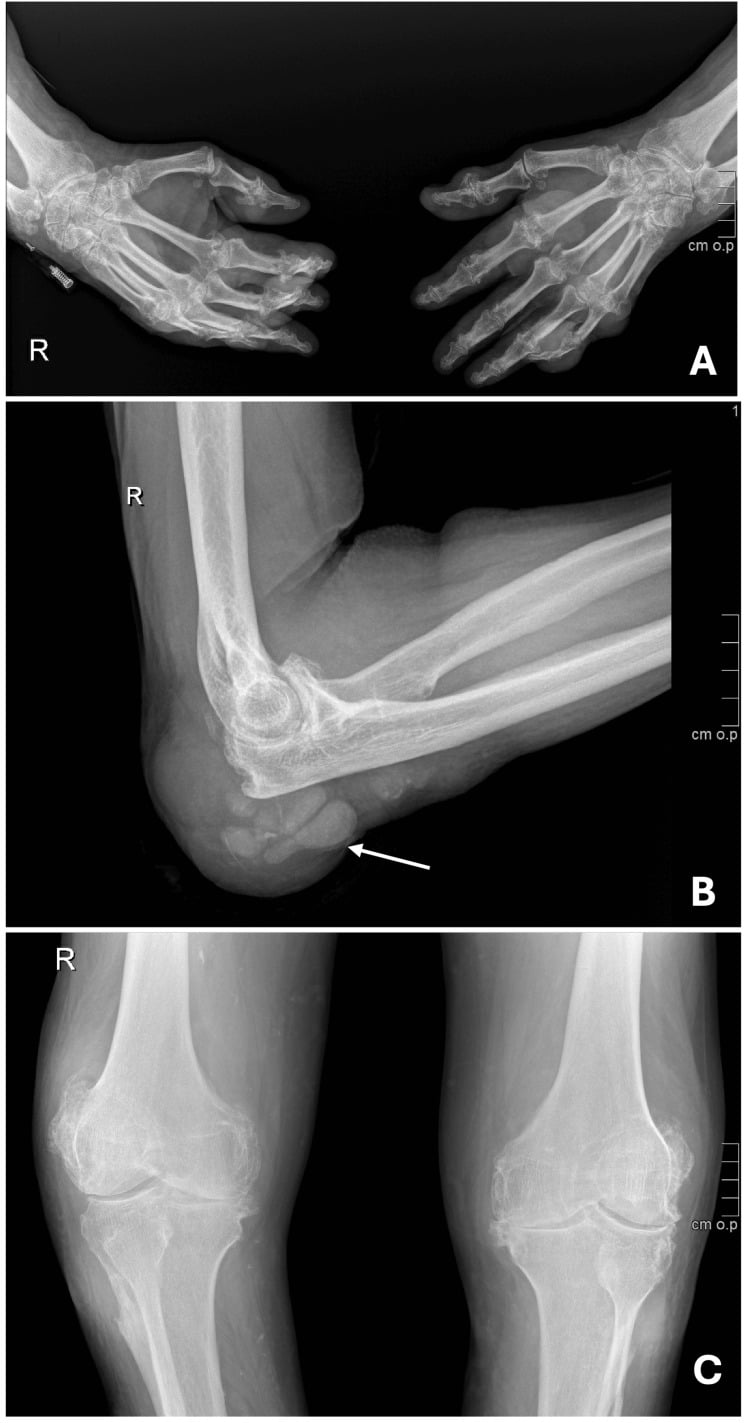
Rheumatoid arthritis. Synovial inflammation destroys the cartilage, and pannus erodes the articular surfaces. Ligamentous laxity follows, and with it instability.
Post-traumatic arthritis. The injury damages cartilage at the outset. Malalignment then distributes load abnormally, instability exposes the joint to recurrent trauma, and osteochondral defects progress to degeneration.
Primary osteoarthritis. Repetitive stress wears the cartilage. The disease is usually unilateral, and loose bodies are common.
Classification Systems
Classify by cause first: the cause guides the treatment and predicts the outcome.
Type I, inflammatory. Bilateral and systemic, driven by synovial inflammation.
- Rheumatoid arthritis (most common)
- Systemic lupus erythematosus
- Psoriatic arthritis
- Juvenile idiopathic arthritis
Type II, post-traumatic. Unilateral, with a history of trauma, and there may be malalignment.
- After fractures of the distal humerus, radial head or olecranon
- After simple or complex dislocations
- Chronic instability - recurrent dislocations, ligament deficiency
Type III, primary degenerative. Usually unilateral, each with its own specific risk factors.
- Primary osteoarthritis (rare)
- Haemophilic arthropathy
- Neuropathic (Charcot)
Clinical Assessment
History. The questions that sort the cause and the treatment:
- Pain - where it is, its character, and whether it comes with activity or at rest
- Stiffness - loss of flexion-extension or of pronation-supination
- Instability - a feeling of giving way, recurrent dislocations
- Previous fractures or dislocations
- Systemic features of rheumatoid arthritis - morning stiffness, bilateral involvement
Look and feel. Inspect for swelling, deformity, scars, muscle atrophy (especially of triceps) and the carrying angle, cubitus valgus or varus. Palpate for joint-line tenderness, loose bodies and the synovial thickening of rheumatoid disease, and examine the ulnar nerve in the cubital tunnel, posterior to the medial epicondyle.
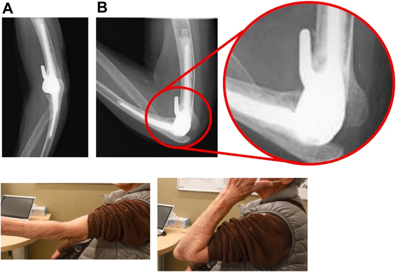
Movement. Normal flexion-extension is 0-150° and the functional arc 30-130°; pronation and supination are normally 80° each. Decide whether stiffness is capsular or a mechanical block.
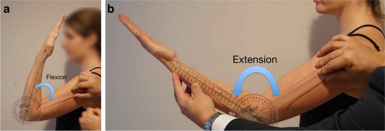
Special tests. Each stresses one structure:
- Valgus stress - MCL integrity
- Varus stress - LCL integrity
- Tinel's sign at the cubital tunnel - the ulnar nerve
- Apprehension and pivot shift - instability
Neurology. Assess every arthritic elbow for instability and for ulnar nerve involvement. The ulnar nerve is commonly affected, so examine and document it before every elbow operation: sensation in the ulnar 1.5 digits, and motor power in the interossei and FDP. Test the median nerve (sensation, thenar muscles) and the radial nerve (sensation, wrist and finger extension) as well.
Investigations
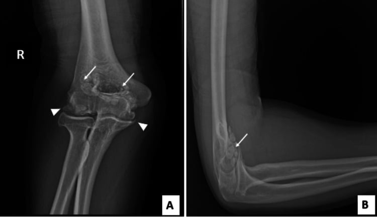
Radiographs. AP and lateral views show joint-space narrowing, osteophytes and loose bodies. Stress views assess instability, and views of the contralateral elbow give a reference.
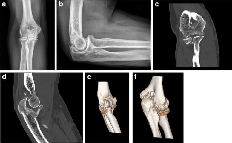
CT. More sensitive than radiographs for loose bodies, and with 3D reconstruction it assesses bone stock and version. It is used for preoperative planning before TEA or debridement.
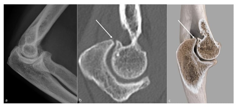
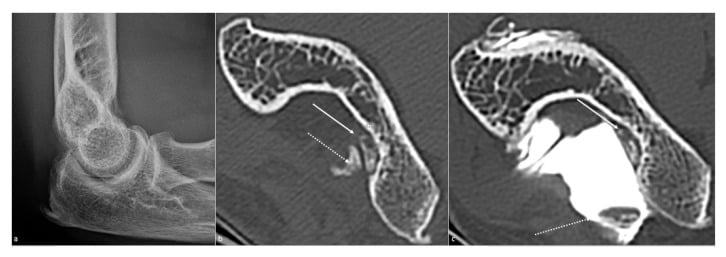
MRI. Shows early cartilage change and osteochondral defects, rheumatoid pannus and synovitis, the integrity of the MCL and LCL, and the ulnar nerve in the cubital tunnel.
Blood tests. Rheumatoid factor, anti-CCP, ESR and CRP for the rheumatoid work-up; infection markers if septic arthritis is a concern; and bone health if TEA is being considered.
Neurophysiology. EMG and nerve conduction studies assess ulnar nerve function if the patient is symptomatic. Investigate the brachial plexus if there are neurological symptoms.
Differential Diagnosis
- Key Distinguishing Features
- Bilateral, symmetrical, synovitis, morning stiffness, systemic features
- Confirmatory Workup
- RF, anti-CCP, ESR/CRP; periarticular erosions on X-ray
- Key Distinguishing Features
- Prior fracture/dislocation, often unilateral, may have malalignment or instability
- Confirmatory Workup
- History; X-ray asymmetric joint space loss, deformity, loose bodies
- Key Distinguishing Features
- Manual worker/athlete, dominant arm, locking from osteophytes, terminal-arc pain
- Confirmatory Workup
- X-ray: olecranon/coronoid osteophytes, loose bodies, preserved mid-arc space
- Key Distinguishing Features
- Acute hot swollen joint, fever, severe pain on micro-movement
- Confirmatory Workup
- Aspiration (WCC, Gram stain, culture, crystals), CRP — urgent
- Key Distinguishing Features
- Acute self-limiting attacks, tophi, chondrocalcinosis
- Confirmatory Workup
- Aspirate for crystals under polarised light; serum urate
- Key Distinguishing Features
- Ulnar paraesthesia/weakness without true joint destruction
- Confirmatory Workup
- Tinel/elbow flexion test; nerve conduction studies
Management
The decision. Conservative management is first-line for every patient and should be exhausted before surgery is considered. Surgery, debridement or TEA, is reserved for failed conservative management in the appropriate patient, and the options depend on age, demand and disease severity.
Medical control of rheumatoid disease. Medical management is first-line in rheumatoid arthritis, and continues until pain is uncontrolled or function severely limited.
- DMARDs - methotrexate, sulfasalazine
- Biologics - anti-TNF, anti-IL6
- Corticosteroids - oral or intra-articular
Non-pharmacological measures. Activity modification, physiotherapy to maintain range and strength, an elbow brace for support, and weight management. With injections, activity modification is the first line for post-traumatic arthritis.
Injections. A corticosteroid injection gives temporary relief, for 3-6 months; hyaluronic acid has limited evidence. Allow a maximum of 2-3 injections in a lifetime.
- Aetiology
- Inflammatory (RA)
- Treatment
- TEA both sides (staged)
- Key Consideration
- Best outcomes - 90% 10-year survival
- Aetiology
- Post-traumatic OA
- Treatment
- Conservative, debridement if needed
- Key Consideration
- TEA too young - wait until over 60 if possible
- Aetiology
- Acute fracture
- Treatment
- Primary TEA
- Key Consideration
- Faster rehab than ORIF
- Aetiology
- Post-traumatic OA
- Treatment
- Avoid TEA, consider interposition or fusion
- Key Consideration
- TEA contraindicated - high failure risk
Surgical Technique
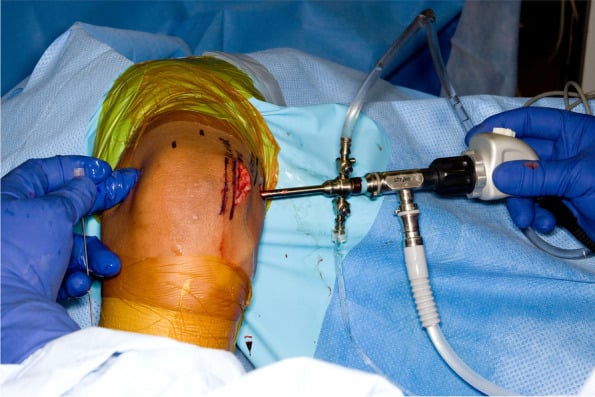
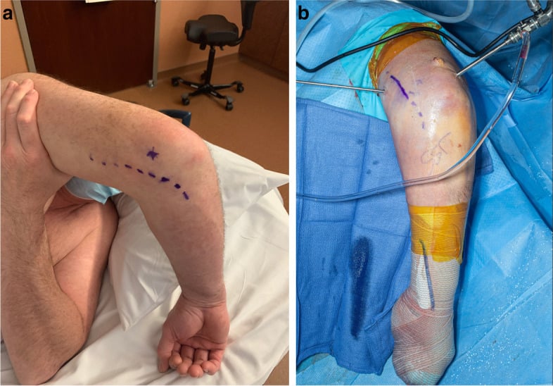
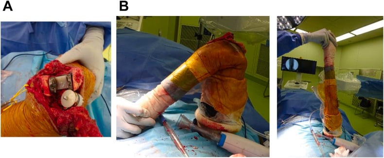
Set-up. Standard portals are anteromedial, anterolateral and posterolateral. Lateral positioning permits posterior arthroscopy.
The steps.
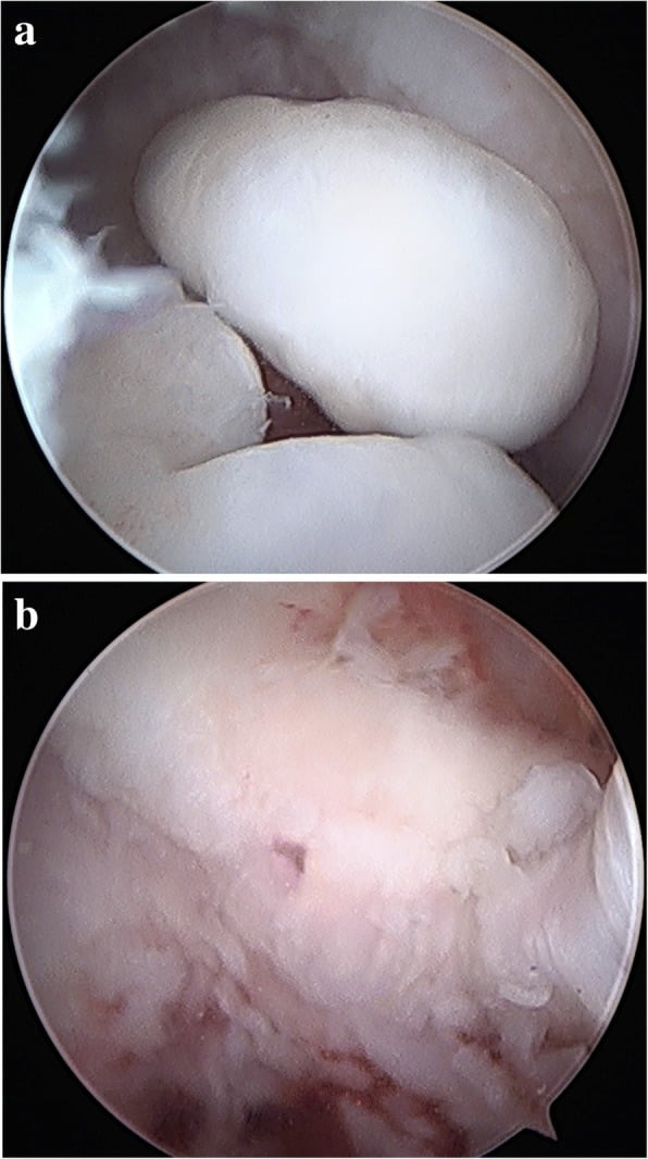
- Remove the loose bodies
- Debride the osteophytes on the coronoid and olecranon
- Release the capsule if the elbow is stiff
- Excise the radial head if it is arthritic
Complications
- Incidence
- 3% significant (TEA, in situ release)
- Risk Factors
- Cubital tunnel disease, previous surgery, transposition
- Management
- Observation if neuropraxia, exploration if transection, may need transposition
- Incidence
- 15% at 10 years
- Risk Factors
- High demand, trauma indication, young age
- Management
- Revision TEA with longer stems, bone graft
- Incidence
- 5-10%
- Risk Factors
- Triceps-reflecting approach, inadequate repair
- Management
- Extension lag, may need revision repair or tendon transfer
- Incidence
- 2-3%
- Risk Factors
- Previous surgery, RA, immunosuppression
- Management
- Debridement, antibiotics, may need explant
- Incidence
- 2-4%
- Risk Factors
- Unlinked designs, ligament deficiency
- Management
- Revision to linked design or ligament reconstruction
- Incidence
- 5-10%
- Risk Factors
- Trauma history, extensive dissection
- Management
- Prophylaxis with indomethacin, may need excision
The ulnar nerve. Routine anterior transposition versus simple in situ release, or leaving the nerve undisturbed, remains debated. The evidence favours selective transposition for stiff elbows or preoperative symptoms rather than transposing every case.
Significant ulnar nerve complications occurred in 3% of arthroplasties in the Dachs series, which used routine in situ release; higher rates in older reviews count any transient symptom. The nerve must be identified and protected at the start of surgery. Transposition is not always required and may increase nerve handling and operative time without clear benefit - reserve it for the elbow with marked preoperative flexion loss, for symptomatic preoperative neuropathy, or when the nerve tracks abnormally against the implanted prosthesis.
Postoperative Care and Rehabilitation
- Splint at 90° of flexion
- Elevation to reduce swelling
- Pain control
- Ulnar nerve monitoring
- Splint removed at 2 weeks
- Gentle passive motion, avoiding forced extension
- Active-assisted motion
- No active extension until 6 weeks, to protect the triceps
- Active extension strengthening
- Progressive range-of-motion exercises
- Light activities, under 2 kg
- Return to daily living
- Weight restrictions: under 5 kg single lift, under 2 kg repetitive
- Avoid impact activities
- Regular follow-up for loosening
Outcomes and Prognosis
Conservative treatment. In rheumatoid arthritis most patients achieve adequate control with modern treat-to-target medical management, which is why the end-stage rheumatoid elbow is now uncommon. In post-traumatic arthritis 40-50% improve with conservative measures, and in primary OA 50-60% respond. The benefit lasts a variable time and may delay surgery for years.
Arthroscopic debridement. Most patients improve symptomatically, and debridement may delay the need for TEA. The relief is temporary, put at 2-5 years, although none of the series cited here quantifies the response rate or its duration.
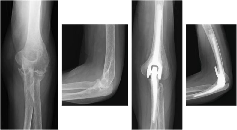
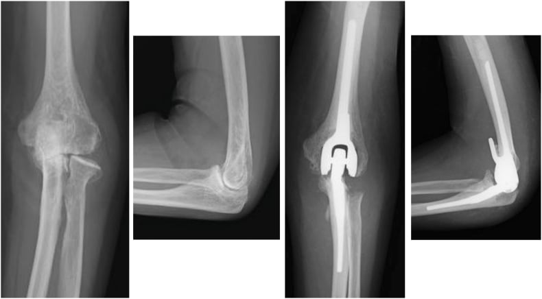
Total elbow arthroplasty. Rheumatoid patients do best, with 90% 10-year survival, 90% pain relief and a 100° functional arc, the 30-130° flexion arc for daily activities. Trauma patients have higher failure rates: 75% 10-year survival and 85% pain relief. At 10 years 85-90% of patients are satisfied. The largest single rheumatoid series in the evidence below (Sanchez-Sotelo, 461 elbows) reports 92% at 10 years.
The long term. Aseptic loosening is the most common long-term failure, at 15% at 10 years, and the revision rate at 10 years is also 15%.
Good outcomes are associated with: RA aetiology (better than trauma), age over 60, low demand, compliant with restrictions, and optimal surgical technique. Poor outcomes are associated with: young age, high demand, trauma indication, non-compliance, and complications.
Primary Osteoarthritis of the Elbow — Clinical Syndrome and Joint-Preserving Surgery
Primary (idiopathic) osteoarthritis of the elbow has a distinctive clinical signature that separates it from inflammatory and post-traumatic disease. It classically affects the dominant arm of middle-aged men, particularly manual labourers, throwers and weight-training athletes.
Presentation. The pattern is distinctive:
- Pain at the extremes of motion, terminal extension and end-range flexion, with a comparatively pain-free mid-arc: the reverse of the diffuse rest-and-motion pain of inflammatory disease
- Loss of terminal extension, usually the first and most consistent deficit
- Mechanical symptoms - catching, locking and grinding from loose bodies and impinging osteophytes
The ulnar nerve. Traction or compression symptoms are common, because posteromedial olecranon osteophytes and a contracted posterior capsule crowd the cubital tunnel. Assess the nerve in every case, and decompress it if flexion is limited (for example, fewer than 100 degrees) or if there are neuritic symptoms.
Radiographs. Osteophytes form at the tips of the olecranon and coronoid and within their fossae, with loose bodies, but the ulnohumeral joint space is relatively preserved. That retained central space is the single most useful feature distinguishing primary OA from rheumatoid and post-traumatic arthritis.
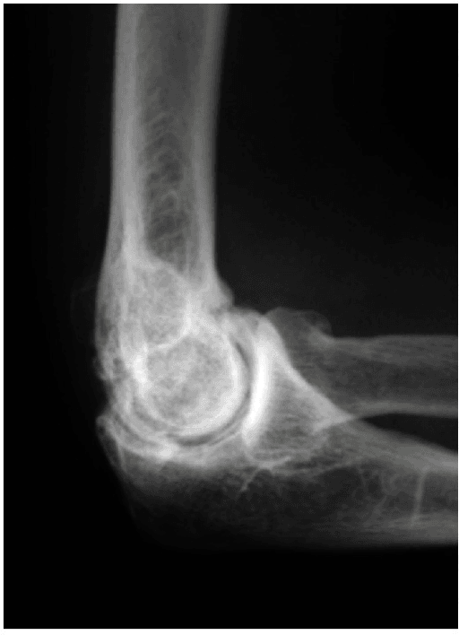
If the ulnohumeral space is globally obliterated, reconsider rheumatoid or post-traumatic arthritis, both of which show global joint-space loss.
Open joint-preserving surgery. These patients are often younger and higher-demand than the ideal TEA candidate, so treatment preserves the joint. The classic open operation is the Outerbridge–Kashiwagi ulnohumeral arthroplasty ("OK procedure"): through a posterior approach a fenestration is created through the floor of the olecranon fossa, and through that single window both the posterior (olecranon) and anterior (coronoid) osteophytes and the loose bodies are removed, with capsular release to restore the arc.
Arthroscopic joint-preserving surgery. Osteophyte excision, loose-body removal and osteocapsular release are achievable arthroscopically in experienced hands. The systematic review in the evidence below (Sochacki) found an average arc gain of roughly 23 degrees and a Mayo Elbow Performance Score improvement of about 24 points. Ulnar decompression is frequently combined with debridement, especially when preoperative flexion is limited.
Recurrence. Osteophytes and motion loss may recur over time, but joint preservation defers arthroplasty in a population for whom lifelong TEA load restrictions are poorly suited.
Haemophilic Arthropathy of the Elbow
The elbow is one of the commonest target joints in haemophilia A and B, alongside the knee and ankle. A target joint is conventionally defined as one that sustains at least three spontaneous bleeds within six months. Both the pathology and the perioperative rules differ fundamentally from the other causes of elbow arthritis.
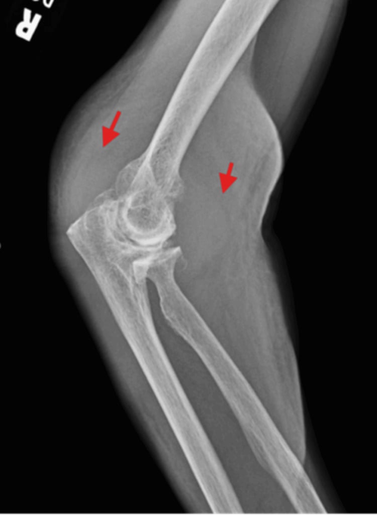
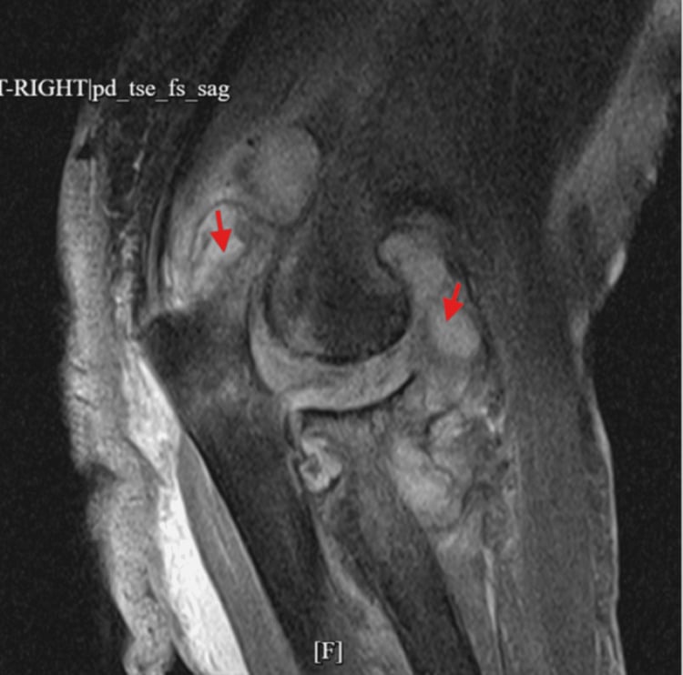
The pathology. Recurrent haemarthrosis deposits iron (haemosiderin) in the synovium, driving a chronic proliferative, hypervascular synovitis. The inflamed synovium releases enzymes and cytokines that degrade cartilage, while the friable neovascular tissue bleeds again, a self-perpetuating cycle. The characteristic changes:
- An enlarged, overgrown radial head
- Widening of the trochlear notch
- Radiocapitellar destruction
- In children, epiphyseal overgrowth from chronic hyperaemia
Haematology first. The foundation is factor replacement or prophylaxis, led by the haematologist, to abolish recurrent bleeds and protect the target joint.
Synovectomy. For persistent bleeding despite prophylaxis, synovectomy reduces bleed frequency. Radiosynovectomy, an intra-articular radioactive isotope such as yttrium-90, rhenium-186 or phosphorus-32, is minimally invasive and first-line in many centres, a way to break the bleed-synovitis cycle before cartilage is lost. Surgical synovectomy, arthroscopic or open, is reserved for its failure.
End-stage disease. In end-stage radiocapitellar disease, radial head excision with synovectomy relieves pain and improves forearm rotation. Total elbow arthroplasty is reserved for end-stage destruction, but carries higher infection and revision risk in these typically younger patients.
Any procedure on a haemophilic elbow, even an aspiration, demands haematologist-guided perioperative factor replacement to safe levels and inhibitor screening beforehand; operating on an uncorrected clotting deficiency risks catastrophic bleeding.
Guidelines, Registries & Global Practice
Global epidemiology:
- Symptomatic elbow arthritis affects under 1% of the population — far rarer than hip or knee disease
- Inflammatory (RA) and post-traumatic causes predominate worldwide; primary OA accounts for only a minority and is overrepresented in male manual workers and athletes
- TEA is a comparatively low-volume procedure globally (a few cases per surgeon per year), concentrated in specialist upper-limb units
- Falling RA disease burden in high-income settings (driven by early DMARD/biologic therapy) has shifted the dominant TEA indication toward acute distal humeral fracture and post-traumatic arthritis
- Focus
- Medical RA control
- Key Position
- Treat-to-target with MTX plus short-course steroids, escalate to biologic/JAK; optimise before surgical referral
- Focus
- Surgical decision-making
- Key Position
- TEA reserved for low-demand patients with end-stage disease; emphasise lifelong load restriction and infection vigilance
- Focus
- Service & referral
- Key Position
- Elbow arthroplasty concentrated in specialist units; primary TEA endorsed for unreconstructible distal humeral fractures in the elderly
- Focus
- Trauma & fracture
- Key Position
- Attempt stable fixation in younger patients; TEA where comminution precludes fixation in low-demand elderly
- Norwegian Arthroplasty Register and the Australian (AOANJRR) and New Zealand joint registries track elbow implants alongside hip/knee
- Registry data consistently show higher revision rates than hip/knee arthroplasty, with aseptic loosening and deep infection the leading reasons
- RA and inflammatory indications historically show better implant survival than post-traumatic OA in pooled registry series
- Low annual volumes limit statistical power, so single-centre cohorts (Mayo, BESS units) remain important evidence sources
- High-resource settings: early biologic RA control, ready access to TEA, fluoroscopy and arthroscopic debridement
- Limited-resource settings: later presentation with advanced destruction; interposition arthroplasty, debridement, and arthrodesis retain a larger role where implants, revision capacity, and infection management are constrained
- Hemophilic arthropathy is more prominent where factor replacement is limited
- Lifelong load restrictions after TEA may be impractical for manual labourers, shifting selection toward joint-preserving options
Controversies and Areas of Uncertainty
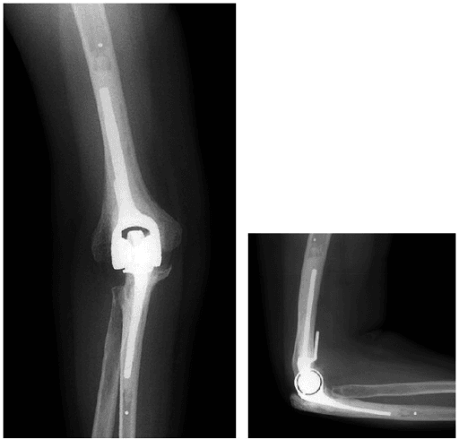
Linked or unlinked TEA. Linked (semiconstrained) designs tolerate ligament deficiency and bone loss but transfer load to the cement-bone interface, risking loosening. Unlinked designs preserve bone but demand competent soft tissues and risk instability. No high-level trial establishes superiority, and the choice is patient- and surgeon-specific.
The young, high-demand patient. No durable solution exists for end-stage arthritis in active patients under 60. Debridement, interposition arthroplasty, arthrodesis, or accepting TEA with strict restrictions all carry significant trade-offs and limited evidence.
Triceps management in TEA. Triceps-reflecting, triceps-splitting and triceps-sparing (paratricipital) approaches each have advocates. Triceps insufficiency remains a recognised complication, and the optimal exposure to minimise it is unsettled.
MCQ Practice Points
Q: What is the most common cause of inflammatory elbow arthritis? A: Rheumatoid arthritis. Unlike hip and knee where primary OA predominates, elbow arthritis is most commonly inflammatory (RA) or post-traumatic. Primary OA of the elbow is rare.
Q: What is the first-line treatment for rheumatoid elbow arthritis? A: Medical management with DMARDs and biologics. Surgery (TEA) is reserved for failed medical management in appropriate patients (age over 60, low demand).
Q: What is the 10-year survival rate of total elbow arthroplasty in rheumatoid arthritis patients? A: 90% 10-year survival in RA patients. This is better than trauma patients (75% 10-year survival). RA is the primary indication with best outcomes.
Q: What is the most common complication after total elbow arthroplasty? A: Significant ulnar nerve complications occurred in 3% of arthroplasties in the series cited here, which used routine in situ release - older reviews quote 10-15% because they count any transient symptom, so state which you mean. Most are neuropraxias that recover, but some may be permanent. The nerve must be identified and protected at the start of surgery, and preoperative flexion under 100 degrees marks the elbow at higher risk.
Q: What are the weight restrictions after total elbow arthroplasty? A: Lifelong restrictions: under 5 kg for single lift, under 2 kg for repetitive activities. These restrictions are essential to prevent aseptic loosening, which is the most common long-term failure (15% at 10 years).
Exam Viva Scenarios
Practise clinical reasoning and management decisions out loud
“A 65-year-old patient presents with bilateral elbow pain and stiffness. Examination shows synovial thickening, limited ROM, and ulnar nerve symptoms. How do you differentiate between rheumatoid arthritis and post-traumatic arthritis, and what is your management approach?”
“A 72-year-old patient with rheumatoid arthritis presents with severe elbow pain and stiffness despite optimal medical management. Walk me through your decision-making for total elbow arthroplasty, including patient selection, surgical approach, and key technical points.”
“A patient presents 3 months after total elbow arthroplasty with numbness and weakness in the ulnar distribution. How do you assess and manage this?”
Key Etiology
- RA: most common inflammatory, bilateral, systemic
- Post-traumatic: after fractures/dislocations, unilateral
- Primary OA: rare, manual laborers
- Hemophilic: bleeding arthropathy
Management Algorithm
- Conservative first: medical management (RA), activity modification, injections
- Surgical: debridement (temporary), TEA (definitive)
- TEA indications: failed conservative, age over 60, low demand
- Weight restrictions: under 5 kg single, under 2 kg repetitive (lifelong)
TEA Outcomes
- RA: 90% 10-year survival, 90% pain relief
- Trauma: 75% 10-year survival, 85% pain relief
- Functional arc: 30-130° flexion for ADLs
- Aseptic loosening: 15% at 10 years (most common failure)
Ulnar Nerve
- Ulnar nerve involvement is common in elbow arthritis - examine and document it
- 3% significant nerve complication after TEA with routine in situ release
- Protect at start of surgery, transposition if symptomatic
Evidence Base and Key Trials
Linked Semiconstrained TEA in RA — 461 Elbows Over Three Decades
- 461 Coonrad-Morrey TEAs in 387 RA patients; median follow-up 10 years
- Survivorship free of revision/removal: 92% at 10 yr, 83% at 15 yr, 68% at 20 yr
- Median Mayo Elbow Performance Score 90 in surviving implants
- Bushing wear seen radiographically in 23%, but revision for isolated wear uncommon (2%)
- Risk factors for revision: male sex, concomitant trauma, PMMA-surfaced ulnar component
EULAR Recommendations for RA Management (2019 Update)
- Start methotrexate plus short-term glucocorticoids as first-line; treat-to-target remission
- Add a bDMARD or JAK inhibitor if poor prognostic factors or csDMARD failure at 3-6 months
- Early, tight disease control limits structural joint destruction including the elbow
- On sustained remission DMARDs may be tapered but not stopped
Arthroscopic Debridement for Primary Elbow OA — Systematic Review
- 9 studies, 213 elbows, mean age 46 yr, mean follow-up 42 months
- Global arc of motion improved by 23 degrees (94.5 to 117.6 degrees)
- Mayo Elbow Performance Score improved by 24 points (61 to 85)
- Low complication rate 2.8% and reoperation rate 4.2%
Open vs Arthroscopic Debridement in Primary Elbow OA — Meta-analysis
- 21 studies, 586 elbows (286 arthroscopic, 300 open)
- Both techniques improved MEPS and range of motion
- Open debridement gave greater flexion gain; arthroscopic improved pain VAS
- Complication rate lower for arthroscopy (6%) than open (12%)
ORIF vs Primary TEA for Distal Humeral Fracture in the Elderly — RCT
- Multicentre RCT, 42 patients over 65 yr with comminuted intra-articular distal humeral fractures
- TEA gave superior Mayo Elbow Performance Scores at 3, 6, 12 and 24 months
- 25% of fractures randomised to ORIF were intraoperatively converted to TEA (unfixable)
- Reoperation rates not significantly different (TEA 12% vs ORIF 27%)
Long-Term Implant Survival of TEA for Fracture (RCT Follow-Up)
- Long-term follow-up of the McKee RCT cohort; mean 12.5 yr for survivors
- Of 25 fracture TEAs, only 1 required revision; 15 died with a well-functioning implant in situ
- No patient required a late revision arthroplasty
- For most elderly fracture patients, a well-performed TEA is the last elbow procedure needed
Ulnar Nerve In Situ Release During TEA
- 83 primary TEAs; routine in situ release, transposition only when nerve tracked abnormally (5%)
- 3% incidence of significant ulnar nerve complications, comparable to systematic reviews
- Preoperative flexion under 100 degrees was associated with postoperative nerve symptoms
- Routine transposition increases nerve handling and operative time without clear benefit
Kudo Type-5 Cementless-Humeral TEA in RA — Minimum 10-Year Follow-Up
- 41 unlinked Kudo type-5 TEAs in RA; mean follow-up 141 months
- Kaplan-Meier survival 87.8% at 5 yr but fell to 70.7% at 10 yr
- Aseptic loosening of the cemented ulnar component was the dominant failure mode
- RA duration under 15 yr and preoperative arc over 85 degrees were revision risk factors