Enneking Staging of Musculoskeletal Tumours
Examiners expect you to state the stage explicitly and then state the surgical margin it demands. A common viva mistake is describing an operation without first naming the stage. Always: (1) grade the tumour (G1 low or G2 high), (2) define the compartment (A intra or B extra), (3) state the stage, and (4) specify the margin. A high-grade (G2) intracompartmental (A) tumour is stage IIA and demands a wide excision — not a marginal one.
Benign Staging (S1, S2, S3)
Benign musculoskeletal tumours are classified into three stages based on their biological behaviour, not histological grade. The stage determines whether the lesion can be observed, curetted, or needs formal excision.
- Behaviour
- Inactive, may regress; no symptoms or minimal pain
- Radiographic Appearance
- Well-defined sclerotic margin; no cortical expansion
- Typical Examples
- Non-ossifying fibroma, simple bone cyst (inactive), lipoma
- Surgical Margin
- Observation; intralesional if intervention needed
- Behaviour
- Progressive growth; symptomatic with pain or swelling
- Radiographic Appearance
- Cortical expansion or thinning; defined but non-sclerotic margin
- Typical Examples
- Aneurysmal bone cyst, giant cell tumour (typical), chondroblastoma
- Surgical Margin
- Intralesional curettage with adjuvants (phenol, cryotherapy, cement)
- Behaviour
- Rapid growth; penetrates natural barriers; may invade soft tissue
- Radiographic Appearance
- Cortical destruction; soft-tissue extension; ill-defined margin
- Typical Examples
- Giant cell tumour (aggressive), aggressive osteoblastoma, desmoplastic fibroma
- Surgical Margin
- Marginal or wide excision; extended curettage with adjuvants may suffice if anatomy allows
LAA — Latent · Active · AggressiveBenign stages — the traffic light
Hook:Behaviour drives the intervention, not the histological name — a 'benign' GCT can be S3 and need a margin.
Giant cell tumour of bone is the classic exam example of a benign lesion that can behave aggressively (S3). It typically occurs around the knee in a skeletally mature patient, involves the metaphysis-epiphysis, and can extend into soft tissue. Examiners often test whether you classify it as benign S3 and choose extended intralesional curettage with adjuvants versus wide excision.
Malignant Staging (Stages I–III)
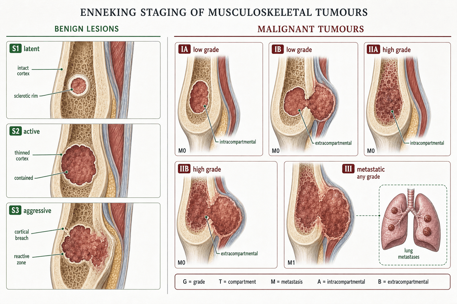
The malignant staging system uses two axes: histological grade (G1 low, G2 high) and anatomical compartment (A intracompartmental, B extracompartmental). A third axis — presence of metastases — defines stage III.
- Grade
- G1 — Low
- Compartment
- A — Intracompartmental
- Metastases
- No
- Typical Examples
- Low-grade chondrosarcoma (intracompartmental), parosteal osteosarcoma
- Grade
- G1 — Low
- Compartment
- B — Extracompartmental
- Metastases
- No
- Typical Examples
- Low-grade chondrosarcoma with soft-tissue extension, desmoid tumour
- Grade
- G2 — High
- Compartment
- A — Intracompartmental
- Metastases
- No
- Typical Examples
- High-grade osteosarcoma confined to bone, high-grade MFH within fascial boundary
- Grade
- G2 — High
- Compartment
- B — Extracompartmental
- Metastases
- No
- Typical Examples
- Conventional osteosarcoma with soft-tissue mass, high-grade leiomyosarcoma beyond compartment
- Grade
- Any G
- Compartment
- Any
- Metastases
- Yes — Distant
- Typical Examples
- Any sarcoma with lung or bone metastases at presentation
Grade × Compartment (III = mets)Malignant stages — the grid
Hook:A 2×2 grid of grade (G1/G2) × compartment (A/B) gives IA–IIB; stage III sits outside the grid — it's metastasis, regardless of grade or compartment.
Surgical Margins — The Stage-Margin Link
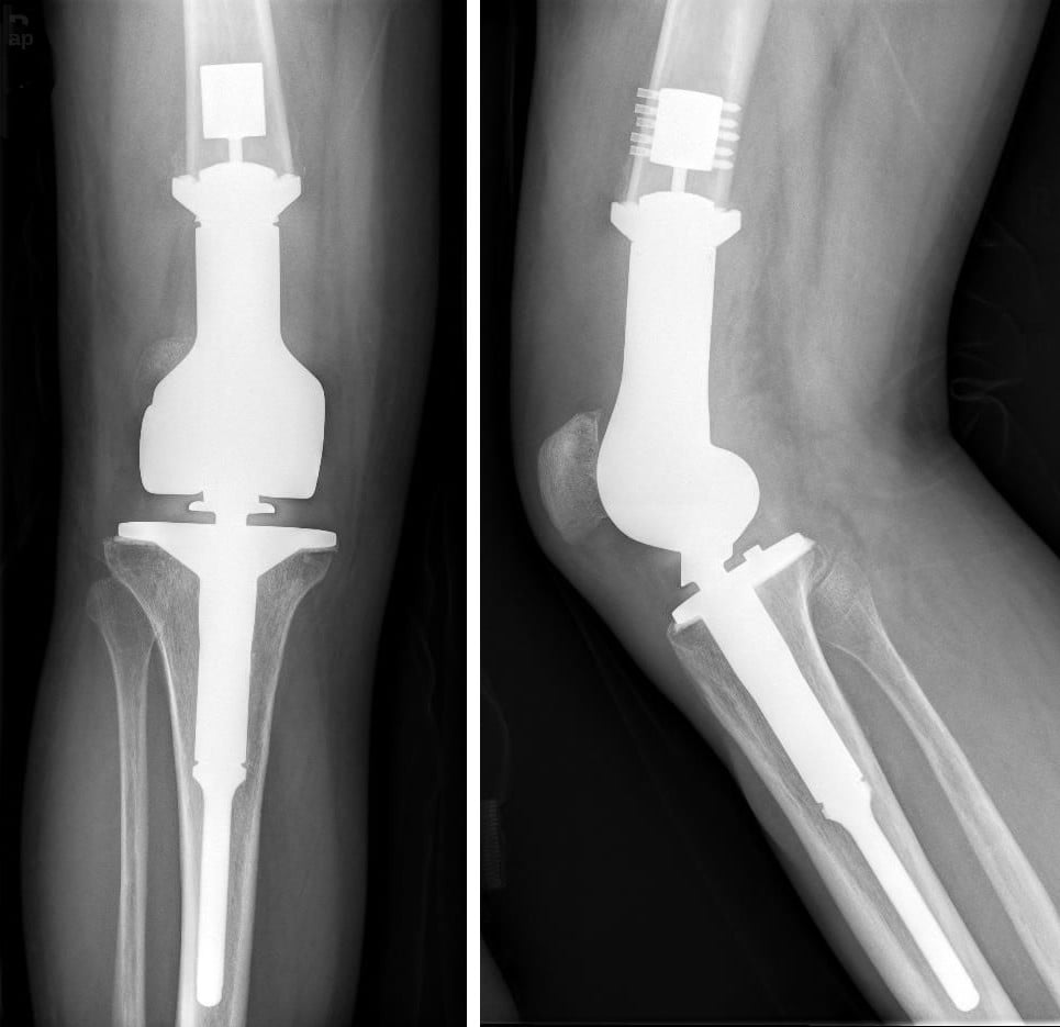
The Enneking stage directly dictates the minimum surgical margin required for local control. A margin is defined by the tissue layer through which the surgeon passes outside the tumour.
- Definition
- Dissection passes through the tumour pseudocapsule and within the lesion
- Tissue at Margin
- Tumour tissue
- Residual Tumour Risk
- High — macroscopic and microscopic residual
- Indicated Stage
- Benign S1, S2 (curettage)
- Definition
- Dissection passes just outside the pseudocapsule through the reactive zone
- Tissue at Margin
- Reactive tissue, possible satellite nodules
- Residual Tumour Risk
- Moderate — microscopic skip lesions possible
- Indicated Stage
- Benign S3; sometimes palliative malignant
- Definition
- Dissection passes through normal tissue beyond the reactive zone but within the compartment
- Tissue at Margin
- Normal tissue ( cuff of healthy tissue around the tumour)
- Residual Tumour Risk
- Low — residual skip metastases within compartment possible
- Indicated Stage
- Stage IA, IB, IIA — standard of care for most sarcomas
- Definition
- Dissection passes outside the entire compartment; the whole compartment is removed
- Tissue at Margin
- Normal tissue outside the compartment boundary
- Residual Tumour Risk
- Very low — removes all skip metastases in the compartment
- Indicated Stage
- Stage IIB with extensive contamination or recurrent disease
A wide margin is the minimum acceptable margin for an intracompartmental sarcoma. Attempting an intralesional or marginal excision for a malignant tumour virtually guarantees local recurrence and is considered inappropriate outside palliative settings. If a wide margin cannot be achieved while preserving a functional limb, amputation providing a radical margin must be discussed.
IMWR — Intralesional · Marginal · Wide · RadicalMargins from inside out
Hook:Concentric rings outward from the tumour: the higher the malignant stage, the further out you must cut — but every malignant stage needs at least a WIDE margin.
Enneking's four margins are qualitative (defined by the plane relative to the reactive zone and compartment). Examiners expect you to know that modern pathology reports the margin quantitatively and in two complementary ways:
- Metric (millimetre) margin — the actual distance from tumour to the inked resection surface. There is no single universal threshold, but a clear margin is reported in millimetres so adequacy can be judged case by case.
- Residual-disease (R) classification — R0 = microscopically negative (no tumour at the inked margin), R1 = microscopically positive margin, R2 = macroscopic residual tumour. R0 is the goal; R1/R2 predict local recurrence and trigger re-excision and/or (neo)adjuvant radiotherapy.
The barrier concept is the nuance that scores marks: a given millimetre distance is not equally protective in every tissue. A thin margin (even 1 to 2 mm) against a robust anatomical barrier — fascia, periosteum, epineurium, vessel adventitia, articular cartilage — can be oncologically adequate, whereas the same distance through fat is inadequate and the same distance through muscle is intermediate. This is why a planned close (marginal) margin against a critical neurovascular structure, combined with adjuvant radiotherapy, is sometimes accepted to preserve a functional limb rather than amputating — the modern refinement of Enneking's all-or-nothing "wide" requirement. It dovetails with the evidence that the margin achieved (not whether the operation was a resection or an amputation) is what drives local control.
Compartments — Defining the Boundary
Understanding what constitutes a compartment is critical because the A/B designation depends on whether the tumour has breached the natural fascial boundary.
- Compartments (Intracompartmental Sites)
- Anterior (quadriceps), Posterior (hamstrings), Medial (adductors)
- Key Boundaries
- Fascial septa and adductor hiatus
- Compartments (Intracompartmental Sites)
- Anterior, Lateral, Deep posterior, Superficial posterior
- Key Boundaries
- Intermuscular septa; interosseous membrane
- Compartments (Intracompartmental Sites)
- Anterior (biceps/brachialis), Posterior (triceps)
- Key Boundaries
- Medial and lateral intermuscular septa
- Compartments (Intracompartmental Sites)
- Intracortical and intramedullary (a bone is its own compartment)
- Key Boundaries
- Cortical bone; periosteum is the boundary
- Compartments (Intracompartmental Sites)
- Intra-articular space
- Key Boundaries
- Joint capsule and synovium
A tumour confined to the medullary canal of the femur is intracompartmental (A) — bone itself is a compartment. Once it breaches the cortex and extends into the soft tissues it becomes extracompartmental (B). This single cortical breach changes the stage from IIA to IIB (if high-grade) and typically enlarges the surgical challenge significantly.
Clinical Application — Staging Workup
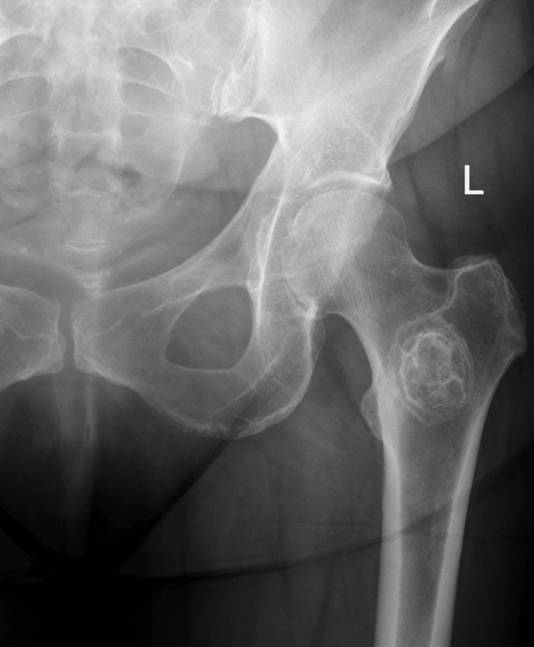
When a patient presents with a suspected musculoskeletal tumour, the staging workup must be completed before any biopsy or surgical intervention. The Enneking stage is determined by combining three assessments:
- Grade (G1 or G2): Determined by histological examination of tissue — mitotic rate, cellular atypia, necrosis, and matrix production. Low grade (G1) is well-differentiated with few mitoses; high grade (G2) shows marked atypia, high mitotic rate, and areas of necrosis. Biopsy should be performed after imaging and planned along the future incision line so the biopsy tract can be excised at definitive surgery.
- Compartment (A or B): Determined by MRI. Assess whether the tumour is confined within its compartment of origin (fascial boundaries, cortical bone, periosteum) or has extended beyond it. The T2-weighted MRI sequence with fat suppression best demonstrates the compartment boundary and soft-tissue extension.
- Metastases (present or absent): Staged with CT chest (the most common site of sarcoma metastasis is the lung) and bone scan or whole-body MRI. PET-CT is increasingly used for high-grade lesions.
- Investigation
- Plain radiographs (two views)
- Purpose
- Lesion characterisation; Enneking does not replace radiographic diagnosis
- Critical Detail
- Lesion in bone: geographic, moth-eaten, or permeative pattern gives the first clue to aggressiveness
- Investigation
- MRI with contrast
- Purpose
- Define compartment status, soft-tissue extension, skip lesions, and neurovascular involvement
- Critical Detail
- T1 for marrow extent, T2 fat-suppressed for soft-tissue plane, gadolinium for tumour vs peritumoral oedema
- Investigation
- CT chest
- Purpose
- Detect pulmonary metastases (defines stage III)
- Critical Detail
- Must be done before biopsy to avoid false-positive nodules from post-biopsy inflammatory changes
- Investigation
- Biopsy (Tru-Cut or open)
- Purpose
- Histological grade (G1 vs G2)
- Critical Detail
- Biopsy tract must be in line with the planned definitive incision and excised en-bloc at resection
The biopsy is the step examiners probe hardest, because a badly-placed biopsy can convert a salvageable limb into an amputation. Mankin's landmark "hazards of biopsy" studies found that diagnostic error occurred in roughly one in five biopsies, and that complications and management-altering problems were several-fold more frequent when the biopsy was performed at the referring hospital rather than the treating sarcoma centre — the evidence behind the rule that suspected sarcomas are referred BEFORE biopsy and biopsied by (or in direct consultation with) the surgeon who will do the definitive resection.
The principles to recite:
- Stage first, biopsy last — complete the imaging before the biopsy so post-biopsy haematoma/oedema does not confound the MRI or create false metastatic nodules.
- Core-needle biopsy is first-line (image-guided where needed) — lower complication and contamination rate than open biopsy; reserve open/incisional biopsy for non-diagnostic cores.
- Longitudinal incision, in line with the planned resection incision — so the entire tract can be excised en bloc. A transverse incision contaminates planes the surgeon cannot then resect.
- Shortest route through a SINGLE compartment — never cross an uninvolved compartment or a neurovascular bundle; every tissue plane the biopsy traverses is contaminated and must be removed.
- Sample the viable periphery, not the necrotic centre; send tissue for histology AND microbiology (infection is the key mimic).
- Meticulous haemostasis — a spreading haematoma seeds tumour cells widely; if a drain is used, bring it out in line with and close to the incision so its tract is excised too.
- Tourniquet by elevation, not Esmarch exsanguination — squeezing the limb can embolise tumour.
Limitations and Modern Context
- The AJCC/UICC eighth edition staging system uses tumour size (greater than or less than 8 cm) rather than compartment status, and incorporates both nodal and distant metastasis. Many centres use AJCC for registry and trial eligibility but apply Enneking to plan the surgical margin. Both systems are valid; know both for the exam.
- Enneking was designed for bone and soft-tissue sarcomas of the extremities. It is less applicable to axial skeletal tumours (spine and pelvis), where clear compartments are harder to define. The Weinstein-Boriani-Biagini (WBB) system is used for spinal tumours.
- Neoadjuvant chemotherapy (for osteosarcoma and Ewing sarcoma) may cause tumour necrosis and shrinkage, but the original Enneking stage determined at presentation guides surgical planning. The tumour bed, not the shrunken mass, must be excised with an adequate margin.
- Post-chemotherapy necrosis mapping (percentage of tumour necrosis in the resected specimen) is an important prognostic factor but does not change the Enneking stage — it informs prognosis and adjuvant therapy decisions.
- Skip metastases (tumour nodules within the same bone but separated from the main lesion by normal marrow) are intracompartmental but can lead to local recurrence if the wide margin does not encompass them. MRI of the entire bone is essential.
Guidelines, Registries and Global Practice
- NCCN Guidelines (US) recommend both Enneking-based surgical margin planning and AJCC eighth edition staging for bone and soft-tissue sarcomas. The guidelines state that the goal of surgery is a wide margin with a cuff of normal tissue.
- ESMO/EURACAN European guidelines endorse the Enneking system for surgical planning of extremity sarcomas and note its value in determining limb salvage versus amputation.
- BOA/BOAST standards (UK) require that all suspected bone and soft-tissue sarcomas be referred to a specialist tumour centre (MTM DT) before biopsy; the Enneking stage is applied at the MDT meeting to plan margins.
- Global variation: In resource-limited settings, MRI may not be available for compartment assessment; in these environments, clinical assessment combined with CT may be used, but the principle of staging before operating remains. The use of Enneking versus AJCC varies by region, but the surgical margin principle is universal.
Viva practice
Exam Viva
Practise clinical reasoning and management decisions out loud
“A 42-year-old man presents with a progressively painful swelling of the distal femur. MRI shows a 10 cm lesion centred in the distal metaphysis with cortical breakthrough and a 4 cm soft-tissue component extending into the anterior compartment. CT chest shows no pulmonary metastases. Biopsy reports a high-grade osteosarcoma. What is the Enneking stage and what surgical margin is required?”
“A 28-year-old woman has a lytic lesion in the proximal tibia involving the metaphysis and epiphysis. It is well-defined with a thin sclerotic border. She has moderate pain and slight swelling. Biopsy shows a giant cell tumour of bone with no atypia or malignancy. An MRI shows cortical thinning but no soft-tissue extension. How would you stage and manage this?”
Exam cheat sheet
Benign stages (S1–S3)
- S1 Latent: asymptomatic, sclerotic border, observe (e.g. NOF, inactive SBC)
- S2 Active: symptomatic, expanding, defined margin — intralesional curettage with adjuvants
- S3 Aggressive: rapid growth, cortical breach or soft-tissue extension — marginal or wide excision
Malignant stages (IA to III)
- IA: G1 low-grade, intracompartmental — wide margin
- IB: G1 low-grade, extracompartmental — wide margin
- IIA: G2 high-grade, intracompartmental — wide margin
- IIB: G2 high-grade, extracompartmental — wide or radical margin
- III: any grade, any compartment, with metastases — systemic treatment plus local control
Surgical margins (inside to outside)
- Intralesional: through tumour — benign S1/S2 only
- Marginal: through reactive zone — benign S3, palliative malignant
- Wide: through normal tissue beyond reactive zone — standard for all malignant stages
- Radical: entire compartment removed — recurrent or contaminated high-grade disease
Key clinical pearls
- Stage BEFORE biopsy: X-ray, MRI entire bone, CT chest, then biopsy along the planned incision line
- A bone is its own compartment — cortical breach changes A to B
- Know both Enneking (surgical planning) and AJCC eighth edition (registry and trials)
- Skip metastases within the same bone are intracompartmental but demand an MRI of the entire bone
Evidence
A system for the surgical staging of musculoskeletal sarcoma
- Proposed the surgical (GTM) staging system stratifying bone and soft-tissue sarcomas by Grade (low/high), anatomic setting (intra- vs extracompartmental) and Metastasis.
- Three stages (I low-grade, II high-grade, III metastatic), each subdivided A (intracompartmental) or B (extracompartmental).
- Defined operative margins (intralesional, marginal, wide, radical) and linked them to the lesion, its reactive zone and compartment.
The effect of the anatomic setting on the results of surgical procedures for soft parts sarcoma of the thigh
- 40 thigh soft-tissue sarcomas: recurrence depended on the MARGIN achieved, not on whether the procedure was a resection or an amputation (marginal 2/4, wide 3/12, radical 1/24).
- Wide margins recurred 0/2 for low-grade but 30% (3/10) for high-grade — the margin REQUIRED for control is dictated by grade.
- Adequate margins were achieved more often for intracompartmental (10/13) than extracompartmental (17/27) lesions — the anatomic setting dictates HOW (resection vs amputation) the margin is obtained.
According to PubMed, the GTM surgical staging system and the margin definitions are from Enneking, Spanier & Goodman 1980 (Clin Orthop Relat Res 1980;(153):106-20; PMID 7449206), with the anatomic-setting validation (margin set by grade, achievability by compartment) from Enneking et al. 1981 (Cancer 1981;47(5):1005-22; PMID 7226034). The modern margin/recurrence outcome data (positive margins worsen survival; local recurrence affects overall but not disease-specific survival) are from Potter et al. 2013 (DOI 10.2106/JBJS.L.01149), and the complementary AJCC 8th-edition framework from Amin et al. 2017 (DOI 10.3322/caac.21388).