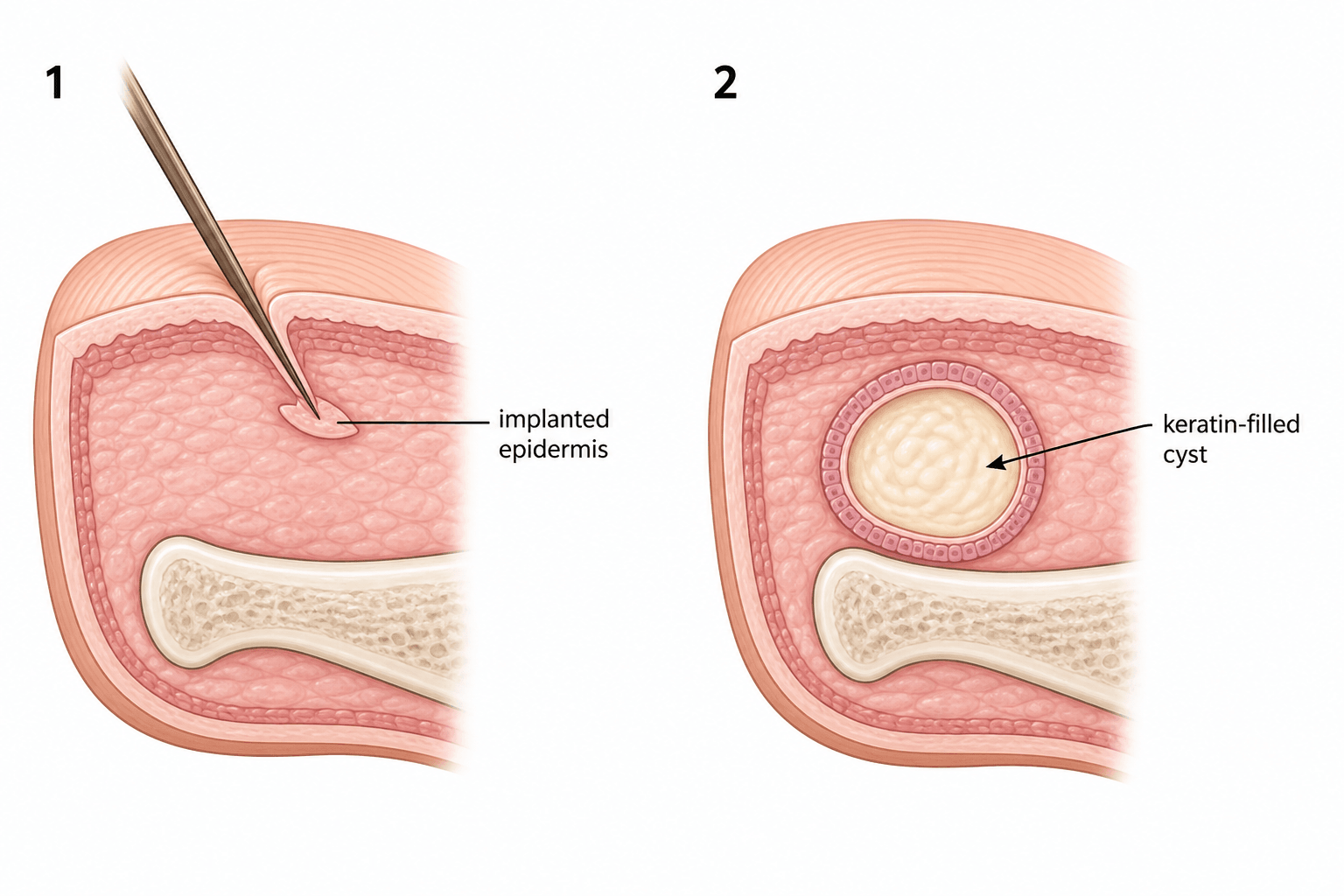
Implanted Epidermis -> a Keratin-Filled Cyst
- An epidermal inclusion cyst (epidermoid/implantation cyst) of the hand forms when keratinising SQUAMOUS EPIDERMIS is implanted into the deeper tissues, usually by PENETRATING TRAUMA or surgery; the implanted epithelium proliferates and produces KERATIN, creating a cyst lined by stratified squamous epithelium and filled with keratin debris.
- It typically presents as a FIRM, slowly enlarging, usually PAINLESS subcutaneous NODULE on the VOLAR fingertip/pulp or palm, often with a HISTORY OF TRAUMA (a puncture, laceration or prior surgery) weeks to years earlier; it can become tender or secondarily infected.
- A distinct INTRAOSSEOUS variant occurs in the DISTAL PHALANX (and the skull), appearing as a well-defined LYTIC lesion that may expand the bone or cause a pathological fracture - it is a recognised benign bone lesion of the hand.
- The DIFFERENTIAL of a hand mass includes a GANGLION (the commonest hand mass, transilluminates, near joint/tendon sheath), GIANT CELL TUMOUR OF THE TENDON SHEATH, foreign-body granuloma, glomus tumour (subungual, painful, cold-sensitive), and - for an intraosseous lytic lesion - ENCHONDROMA (the commonest bone tumour of the hand) and OSTEOMYELITIS (especially with a chronic digital wound, which can coexist with an epidermoid cyst).
- DIAGNOSIS is clinical (a firm nodule with a trauma history) supported by imaging - ultrasound shows a well-defined cyst; radiographs show an intraosseous lytic lesion with a sclerotic margin - but DEFINITIVE diagnosis is HISTOLOGICAL: a cyst lined by stratified squamous epithelium with laminated keratin and NO skin adnexa (which distinguishes it from a true dermoid cyst).
- TREATMENT is COMPLETE SURGICAL EXCISION of the soft-tissue cyst including its entire lining (incomplete removal -> RECURRENCE), or CURETTAGE (+/- bone graft/substitute) for the intraosseous form; where an epidermoid cyst coexists with osteomyelitis, thorough debridement/curettage plus antibiotics treats both.
- “Epidermal inclusion cyst = traumatic implantation of epidermis -> squamous-lined, KERATIN-FILLED cyst (no adnexa).
- “Firm painless volar finger/palm nodule with a trauma history; intraosseous variant = lytic distal phalanx lesion.
- “Differential: ganglion (commonest), GCTTS, glomus, enchondroma, osteomyelitis. Treat by COMPLETE excision/curettage - incomplete removal recurs.
Ganglion (commonest, transilluminates), giant cell tumour of tendon sheath, epidermal inclusion cyst (trauma history, keratin-filled), glomus tumour (subungual, painful).
Enchondroma (commonest hand bone tumour), intraosseous epidermoid cyst, osteomyelitis (chronic wound - can coexist with an epidermoid cyst). Image and biopsy.
Pathogenesis & Presentation
The fingertip and palm are frequently subject to penetrating injuries; if a fragment of surface epidermis is driven into the deeper tissue (or buried at surgery), the trapped keratinising epithelium continues to grow and shed keratin into an enclosing cyst. The result is a firm, well-circumscribed, slowly enlarging subcutaneous nodule, usually on the volar fingertip/pulp or palm, typically painless unless it is large, ruptures or becomes infected. A careful history often reveals a previous puncture, laceration or operation at the site, sometimes years earlier. An intraosseous epidermoid cyst of the distal phalanx presents instead as a localised lytic lesion, with swelling, nail changes or a pathological fracture.
Investigation & Diagnosis
Ultrasound shows a well-defined, often hypoechoic cyst and helps distinguish it from a ganglion or solid tumour; radiographs are essential if an intraosseous lesion is suspected, showing a well-defined lytic lesion with a sclerotic margin in the distal phalanx (and exclude enchondroma/osteomyelitis); MRI can characterise deeper or recurrent lesions. The definitive diagnosis is histological: a cyst lined by stratified squamous epithelium with a granular layer producing laminated (lamellated) keratin, and - importantly - no skin adnexal structures (hair follicles/sebaceous glands), which distinguishes it from a true dermoid cyst. Always send excised tissue for histology, particularly to exclude an unexpected diagnosis.
Management
- Soft-tissue cyst: complete surgical EXCISION including the entire cyst lining/capsule - incomplete removal (or simple rupture/aspiration) leaves epithelium behind and leads to RECURRENCE; marsupialisation is inadequate.
- Intraosseous (distal phalanx): CURETTAGE of the lesion with removal of the lining, filling the cavity with bone graft or substitute as needed; outcomes are generally good with a low recurrence after thorough curettage.
- Coexisting osteomyelitis (chronic digital wound): debridement and curettage plus antibiotics (a bone substitute carrying antibiotic is one option) treats both entities; obtain histology and culture.
- Infected cyst: treat the infection, then excise the cyst electively once settled.
Evidence & Key Studies
Intraosseous epidermoid cyst discovered in the distal phalanx of a thumb: a case report
- Intraosseous epidermoid cyst is a rare benign inclusion cyst found mainly in the skull and phalanges.
- Once differentiated from similar lytic lesions, it can be treated with simple CURETTAGE, seldom needing additional procedures.
- The case was treated successfully without complication, illustrating the favourable outcome of the distal-phalanx variant.
Chronic digital wound: epidermoid cyst, osteomyelitis or both? Two cases and review
- Epidermoid bone cyst and osteomyelitis of the distal phalanx can coexist, and both are linked to previous trauma, nail alterations and chronic inflammatory signs.
- Definitive diagnosis requires histopathology, and good surgical debridement is needed to heal both entities.
- Curettage with filling of the bone defect using an antibiotic-loaded bone substitute is a good treatment option.
According to PubMed, the rarity, distribution (skull/phalanges) and curettage treatment of the intraosseous epidermoid cyst come from the cited Shin case report, and the coexistence with osteomyelitis, the link to prior trauma, and the need for histology and debridement/curettage from the cited Portes-Chiva report. The traumatic-implantation mechanism, the keratin-filled squamous histology and the soft-tissue excision principle are standard hand-surgery teaching. (See also our Ganglion, Giant Cell Tumour of Tendon Sheath, Enchondroma and Hand Infections topics.)
Clinical Decision Scenarios
Practise clinical reasoning and management decisions out loud
“A patient has a firm, painless nodule in the pulp of a finger and recalls a splinter injury there two years ago. What is the likely diagnosis and how would you confirm and treat it?”
“A radiograph shows a well-defined lytic lesion in the distal phalanx of a finger. How do you approach it?”
Mnemonics & Memory Aids
KERATIN
Hook:KERATIN captures the epidermal inclusion cyst story.
GEEO (lytic distal phalanx)
Hook:Lytic distal phalanx? Think GEEO: Glomus, Enchondroma, Epidermoid, Osteomyelitis.
Pathogenesis
- Traumatic/surgical implantation of epidermis into deeper tissue
- Implanted epithelium makes keratin -> squamous-lined, keratin-filled cyst
- No skin adnexa (distinguishes from true dermoid)
Presentation
- Firm, slowly enlarging, usually painless nodule (volar finger/palm)
- History of prior puncture/laceration/surgery
- Intraosseous variant: lytic lesion of the distal phalanx (+/- pathological fracture)
Differential & diagnosis
- Soft tissue: ganglion (commonest), GCTTS, foreign-body granuloma, glomus
- Intraosseous lytic: enchondroma, epidermoid, osteomyelitis (can coexist)
- US/X-ray/MRI; DEFINITIVE = histology (squamous lining + keratin)
Treatment
- Soft tissue: COMPLETE excision of cyst + lining (incomplete -> recurrence)
- Intraosseous: curettage + lining removal +/- bone graft/substitute
- Coexisting osteomyelitis: debridement/curettage + antibiotics; always send histology