Attritional Rupture | Third Compartment | EIP-to-EPL Transfer
- EPL is the sole tendon in the 3rd dorsal compartment, angling sharply around the Lister tubercle pulley
- Most common cause of 'spontaneous' EPL rupture is an apparently minor (often non-displaced) distal radius fracture
- Direct primary repair is almost never possible - tendon ends are frayed, retracted and within an ischaemic 3rd compartment
- EIP-to-EPL tendon transfer is the gold-standard reconstruction; results are reliable and donor deficit is minimal
- Examination hallmark: positive retropulsion (drum) sign - patient cannot lift the thumb off a flat surface palm-down
- “EPL extends IP, MCP and retropulses CMCJ of the thumb (the only retropulsor)
- “After DRF, EPL ruptures weeks to months later even in a cast - mechanism is ischaemic + mechanical at the Lister tubercle
- “Loss of thumb retropulsion with intact EPB strongly suggests EPL rupture rather than PIN palsy
- “EIP is the donor of choice: it is expendable (two tendons to the index), synergistic, and of appropriate length
Overview and Epidemiology
Attritional rupture of the extensor pollicis longus is the commonest closed extensor tendon rupture of the hand and the prototype of attritional rupture at a fibro-osseous pulley. The classic story - sudden, painless loss of thumb extension weeks after an apparently trivial distal radius fracture - is an exam favourite because it tests tendon biomechanics, third-compartment anatomy and the principles of tendon transfer in a single question.
The numbers. Rupture follows about 0.3% of all distal radius fractures but 5% of strictly non-displaced ones, so the benign-looking fracture is the risk group. It declares itself 4-12 weeks after the injury as a rule, with reported cases from one month to a year, and it happens even in a cast.
What the patient loses. The EPL is the only tendon that retropulses the thumb, so its rupture takes thumb IP and MCP extension with it and leaves the thumb lying flat against whatever the hand rests on. Tip pinch, key pinch and grip strength all fall, and patients describe being unable to rest the hand on a table, climb, or pick a coin or other flat object off a surface. The hand also loses its palm-to-fist contour during grip as the thumb collapses into adduction.
Anatomy and Pathophysiology
The tendon and its tunnel. The EPL is the only tendon in the third dorsal compartment, and within it the tendon makes a sharp angular turn of 30-45 degrees around the Lister tubercle, a bony ridge of the distal radius that works as a fixed pulley. It inserts on the dorsal base of the distal phalanx and extends the IP and MCP joints while retropulsing the CMC joint, all in one line of pull. The extensor pollicis brevis, which travels in the first compartment with the abductor pollicis longus, extends the MCP joint only.
- Contents
- APL, EPB
- Site-specific pathology
- De Quervain tenosynovitis; EPB slips in rheumatoid disease
- Contents
- ECRL, ECRB
- Site-specific pathology
- Intersection syndrome; second-compartment syndrome
- Contents
- EPL
- Site-specific pathology
- Attritional rupture: post-DRF, RA, Kienböck, iatrogenic
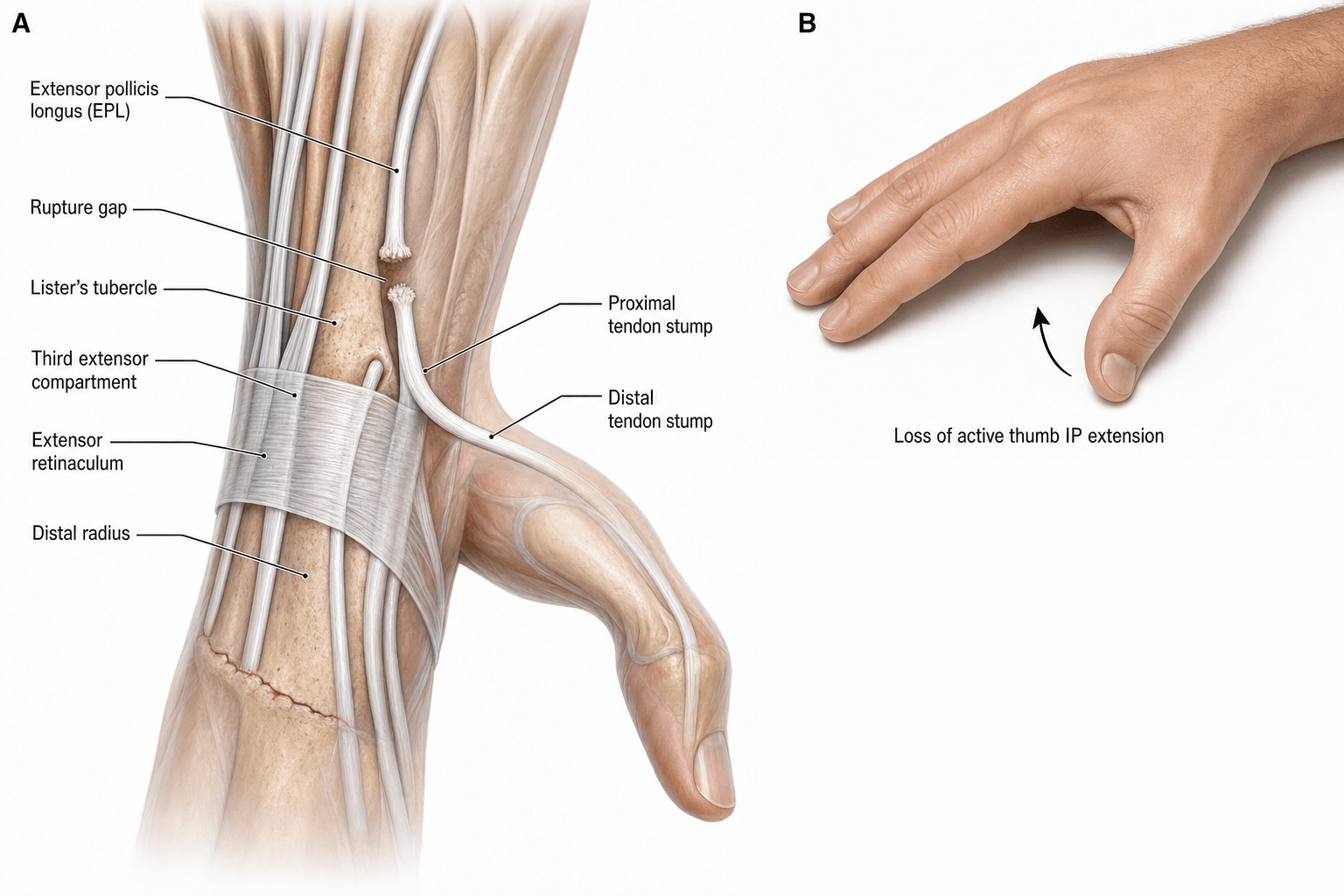
The two hits. The first is ischaemic. The intrinsic blood supply of the tendon is poorest at the level of the tubercle, a watershed zone, and the third compartment is a tight fibro-osseous tunnel in which haematoma, oedema, tenosynovitis, rheumatoid pannus or cast compression raise the pressure further. The second is mechanical: the weakened tendon grinds over an unforgiving bony ridge with every thumb movement. The two summate, and the tendon gives way at or just proximal to the Lister tubercle, often inside a retinaculum that looks intact - a combination unique to the EPL.
Why the undisplaced fracture is the dangerous one. A non-displaced fracture bleeds into an intact third compartment and raises the pressure inside it, and the cast then holds a retinaculum-bound tendon under sustained compression against bone. A radius that heals short sharpens the angle the tendon has to turn through, and the tendon frays and gives way with no fresh injury to explain it.
The volar-plate era version. Volar locking plating changed the mechanism. The plate is fixed from the front and its screws are measured from the front, so every millimetre a locking screw overshoots the dorsal cortex is a millimetre protruding into an extensor compartment - and the third compartment holds the EPL. Perry's cadaveric study passed a wire through every hole of three common volar plates and found that every plate has specific high-risk holes, the central screws most often, whose trajectory points directly at the EPL groove. This is not random bad luck; it is predictable from the plate design and the hole chosen.
Two rules follow. A screw is either long or it is not - there is no "slightly proud" - so check with a dorsal tangential view rather than trusting a lateral; Kiyak found the view detected screw penetration with no false positives in his series. And when an EPL ruptures after plating rather than after a simple fracture, think hardware first and check the plate position and the screw lengths before putting it down to attrition, because the management then includes dealing with the offending screw.
The rheumatoid wrist: caput ulnae. Dorsal synovitis attenuates the ECU subsheath and the ECU subluxates volarly; the DRUJ is destroyed and the ulnar head erodes dorsally into the caput ulnae. The extensors lying over that prominence fail first and the EPL, on the radial side, ruptures later as synovitis reaches the third compartment. The EIP may itself have ruptured, and the FPL may be failing at the scaphoid in the same hand - the Mannerfelt overlap. The EPL is easy to miss in a rheumatoid hand unless it is tested specifically.
Vaughan-Jackson syndrome. The extensor ruptures follow a named, examinable pattern: sequential, progressive rupture of the finger extensors beginning on the ulnar side and marching radially - extensor digiti minimi first, then the EDC to the small finger, then the ring, then the middle. The cause is attrition over the dorsally prominent, eroded distal ulna combined with dorsal tenosynovitis, since the most ulnar tendons sit directly over the offending bone. The clinical clue is a rheumatoid patient who cannot extend the little finger MCP joint, then weeks later the ring: the droop is at the MCP joints and passive tenodesis is lost.
Its flexor mirror. Mannerfelt syndrome is attritional rupture of the FPL over a scaphoid or trapezial osteophyte inside the carpal tunnel - the same disease on the opposite side of the wrist. Finding an EPL rupture in a rheumatoid hand should therefore prompt a search for ulnar extensors that have already gone, and any transfer here is combined with dorsal tenosynovectomy and treatment of the distal ulna (Darrach or Sauvé-Kapandji) to stop further ruptures.
Classification and Types
Two axes matter. The aetiology dictates the reconstruction - transfer for attrition, repair for laceration - and the onset determines whether the bed of the tendon is healthy enough for a repair at all.
Metal belongs on the list too - a scaphoid screw, an external fixator pin, a prominent dorsal plate - and a knife, glass or a saw divides the tendon outright.
- Mechanism
- 3rd compartment ischaemia + Lister attrition
- Timing
- 4-12 weeks typical; range 1-12 months
- Notes
- Most common cause of 'spontaneous' EPL rupture
- Mechanism
- Tenosynovitis, caput ulnae, attritional wear
- Timing
- Insidious, often multiple tendons
- Notes
- Often part of Vaughan-Jackson or Mannerfelt syndrome
- Mechanism
- Carpal collapse alters Lister pulley mechanics
- Timing
- Progressive
- Notes
- Treat the underlying cause; transfer is palliative
- Mechanism
- Scaphoid screw, dorsal plate, pin site
- Timing
- Early post-op
- Notes
- Remove offending hardware + transfer
- Mechanism
- Direct division over the thumb MC or wrist
- Timing
- Acute
- Notes
- Only scenario where direct repair is feasible
Clinical Assessment
History. The rupture is usually described as spontaneous, so the questions that make the diagnosis are about the wrist rather than the thumb: a distal radius fracture, even one three to six months ago, rheumatoid disease, Kienböck disease, previous wrist surgery, scaphoid screws. Sudden, painless loss of thumb extension is the classic account, and the absence of pain at the moment of rupture is itself an important feature.
The retropulsion (drum) test. Lay the palm flat on a hard table with the fingers adducted and the thumb alongside the index, then ask the patient to lift the thumb straight up off the table. An intact EPL lifts it cleanly, retropulsing the CMC joint while extending the MCP and IP joints. After rupture the thumb stays down: the EPB may still produce a small amount of MCP extension, but it cannot retropulse the thumb or extend the IP joint. That is the value of the test - it isolates EPL from EPB, and a patient with a strong EPB and a positive sign has an EPL rupture until proven otherwise.
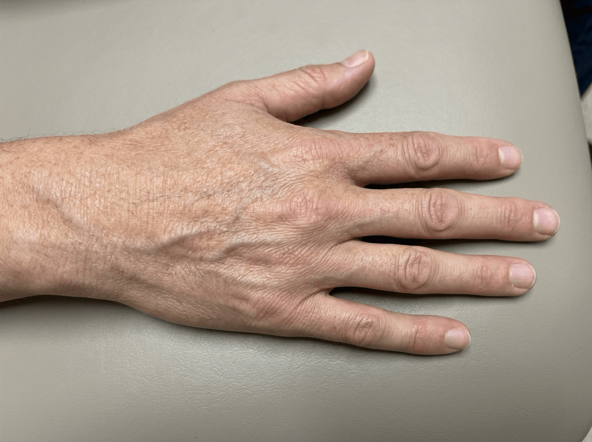
The rest of the examination. Four more checks, every time:
- Tenodesis test - wrist flexion no longer extends the thumb IP joint, because the EPL is discontinuous
- Palpation over the Lister tubercle - the tendon is no longer there, and a gap may be felt
- Every other extensor, and the FPL - the caput ulnae and Mannerfelt spectrum takes more than one tendon
- Finger and wrist extension - intact, which rules out a posterior interosseous nerve palsy
- Thumb posture
- Thumb lies flat, IP flexed, no retropulsion
- Discriminating finding
- Positive retropulsion sign, tenodesis negative
- Key test / imaging
- Clinical; X-ray wrist for DRF callus, US for gap
- Thumb posture
- All fingers and thumb drop, no retropulsion, radial wrist deviation
- Discriminating finding
- Wrist extension preserved (ECRL), no finger or thumb extension at all
- Key test / imaging
- EMG / NCS; MRI for radial tunnel lesion
- Thumb posture
- Flicking, painful snapping over Lister
- Discriminating finding
- Tendon visible and palpable subluxing
- Key test / imaging
- Dynamic US; clinical with active motion
- Thumb posture
- Loss of thumb MCP extension only, IP preserved
- Discriminating finding
- IP extension and retropulsion intact via EPL
- Key test / imaging
- Selective testing of EPB against resistance
- Thumb posture
- Loss of thumb IP flexion in a rheumatoid hand
- Discriminating finding
- Palpable nodule over FPL, attritional rupture at scaphoid
- Key test / imaging
- Active IP flexion lost; EPL intact (the opposite deficit)
- Thumb posture
- Painful radial wrist, weak thumb extension
- Discriminating finding
- Pain limits testing, Finkelstein positive
- Key test / imaging
- Clinical; symptoms predominate over signs
Retropulsion is lost in both, so the sign itself does not separate them. An isolated thumb deficit - a normal-looking hand at rest, normal finger extension, no retropulsion - is an EPL rupture. Loss of thumb and finger MCP extension, with wrist extension preserved through ECRL and deviating radially, is a PIN palsy: the deficit extends well beyond the thumb. Test finger and wrist extension in every patient with a dropped thumb.
Investigations
The diagnosis is clinical. Imaging is for the cause, and it identifies the cause far more often than it confirms the rupture - so in the post-fracture case, do not delay an EIP-to-EPL transfer waiting for an MRI.
Radiographs are mandatory and come first: PA, lateral and oblique views of the wrist and distal radius. Look for the healed fracture and its callus, rheumatoid change (ulnar styloid erosions, caput ulnae, carpal collapse), a sclerotic lunate, a scaphoid nonunion, and any screw or plate lying over the third compartment.
Ultrasound answers the equivocal examination - a palpable gap, a suspected partial rupture, or in-continuity degeneration - by showing discontinuity of the EPL fibres, the retracted stump, fluid in the third compartment and tenosynovial thickening. It is dynamic and cheap, needs no contrast, and can be compared with the other side.
MRI is a planning study rather than a diagnostic one: chronic ruptures with several tendons involved, and assessment of the third-compartment bed. It shows the gap, the scar tissue, marrow oedema in the lunate and active tenosynovitis with enhancement, which is what makes it valuable in a rheumatoid hand, where every ruptured tendon must be mapped before reconstruction.
- Implication
- Confirms attritional post-DRF rupture
- Management shift
- EIP-to-EPL transfer alone
- Implication
- Iatrogenic attrition
- Management shift
- Remove hardware + EIP transfer
- Implication
- RA with multiple extensor ruptures likely
- Management shift
- Dorsal tenosynovectomy + multiple transfers + Darrach or Sauvé-Kapandji
- Implication
- Kienböck disease driving attrition
- Management shift
- Stage the lunate (Lichtman); treat Kienböck + transfer
- Implication
- Carpal collapse changes EPL mechanics
- Management shift
- Scaphoid reconstruction / 4-corner fusion + transfer
Management Algorithm
The operation follows the tendon bed. An attritional rupture leaves frayed, retracted ends in a diseased third compartment and is reconstructed by transfer; a clean laceration in a healthy bed can be repaired directly.
Indication. An attritional rupture with a frayed, retracted stump and a diseased third-compartment bed: the standard operation after a distal radius fracture and for most rheumatoid ruptures. The extensor indicis proprius (EIP) is the donor in over 95% of cases.
Setup. General or regional anaesthesia (axillary block), supine with an arm table and a tourniquet high on the arm, and a single dose of intravenous antibiotic per local policy.
Harvest the EIP. An oblique incision over the index MCP joint. The EIP lies ulnar and deep to the extensor digitorum tendon to the index; confirm it by independent index extension against resistance, then strip it with a distal periosteal tongue to maximise length and deliver it into the wound.
Prepare the EPL. A dorsoradial incision over the thumb metacarpal to find the distal stump. Excise the frayed ends back to healthy tendon, debride the third compartment, and release the proximal stump from the retinaculum and the Lister tunnel, excising the diseased tendon so the transfer runs free.
Route it. Subcutaneously from the index incision, around the radial border of the wrist, to the thumb, superficial to the extensor retinaculum so that the transfer never re-enters the diseased third compartment.
The weave. A Pulvertaft weave of the EIP through the distal EPL stump over the thumb metacarpal, three passes minimum, each pass with 4-0 braided non-absorbable suture (Ticron or similar) and a running 6-0 epitendinous suture.
Tension. Set it with the wrist neutral and the thumb in full retropulsion with the IP and MCP joints extended. Slight overcorrection is right, since the transfer loosens a little in the cast, and the thumb should sit at least level with the index and ideally a few millimetres above it; full passive thumb flexion must still be possible without the transfer falling slack. Under-tensioning is the commonest error in this operation and it reproduces the complaints the patient came in with - an extension lag at the thumb IP joint and weak retropulsion.
Closure and cast. Skin only, with drains rarely needed, then a forearm-based thumb spica cast holding the wrist neutral and the thumb in full retropulsion and extension.
Rehabilitation
- Full-time thumb spica cast
- Finger flexion encouraged within the cast
- Cast off at 4 weeks, protected active thumb motion in a removable splint
- Gentle strengthening and retropulsion retraining from 6 weeks
- Return to most activities at 12 weeks
- Pinch strength returns over 3-6 months; heavy loading after 4-6 months
The choice of EIP is not arbitrary; it satisfies the classic principles of tendon transfer (Boyes/Brand) that examiners expect you to recite for any transfer:
- Supple, mobile joints - the thumb IP/MCP and CMC must be passively correctable first (a stiff joint will not move however good the transfer); release contractures before transferring.
- Expendable donor - taking EIP leaves the index with the EDC, so index extension is preserved (the donor's loss must be tolerable).
- Adequate excursion - the donor's amplitude must match the recipient's need (EPL needs roughly 30 mm; EIP/finger extensors provide it).
- Adequate strength - one grade of power is typically lost after transfer, so the donor must be strong enough.
- Synergy - synergistic donors are easier to re-educate; EIP fires with finger/thumb extension, in phase with the lost EPL.
- Straight line of pull - the transfer should run as directly as possible to its insertion (hence the subcutaneous radial route, not back through the diseased 3rd compartment).
- One tendon, one function - a single transfer should ideally power a single action.
- Soft-tissue equilibrium / supple bed - a healthy, scar-free, well-vascularised bed is needed for gliding (the very reason direct repair of the attritional rupture fails).
Exam point: justify EIP-to-EPL by the transfer principles - expendable, synergistic, correct amplitude and a straight subcutaneous line of pull into a supple thumb - and remember the prerequisite of supple joints and a healthy bed.
Complications
- Incidence
- Common (15-25 percent)
- Risk Factors
- Scarring from prior surgery, prolonged immobilisation
- Management
- Hand therapy; tenolysis if persistent beyond 6 months
- Incidence
- 5-15 percent
- Risk Factors
- Under-tensioning, transfer elongation, adhesions
- Management
- Re-tensioning or tenolysis if functionally limiting
- Incidence
- 2-5 percent
- Risk Factors
- Early return to loading, poor tendon quality
- Management
- Re-transfer (use EDM) or tendon graft
- Incidence
- Up to 20 percent initially, recovers in most
- Risk Factors
- EIP harvest from index finger
- Management
- Re-education; rarely symptomatic beyond 6 months
- Incidence
- 2-5 percent
- Risk Factors
- Severe injury, prolonged immobilisation
- Management
- Early hand therapy, vitamin C prophylaxis in some protocols
- Incidence
- 10-15 percent
- Risk Factors
- Transfer under-tensioned, pre-existing thumb CMCJ OA
- Management
- Splinting, CMCJ stabilisation if arthritic
Outcomes and Prognosis
Who does well. A young patient after a distal radius fracture, with a healthy contralateral hand, reconstructed within 3 months and rehabilitated under supervision.
Who does less well. The elderly rheumatoid hand with several ruptured tendons, a severe caput ulnae, and a late presentation with an established thumb adduction contracture; here the result depends on disease control.
The threshold. Somewhere between 3 and 6 months the outcomes begin to decline steadily, though delayed reconstruction still works. The transfer itself is reliable; the limiting factors are the bed, the disease and the rehabilitation.
- Treatment
- EIP-to-EPL transfer within 3 months
- Expected Outcome
- Good-to-excellent in 85-90 percent
- Long-term Function
- Pinch strength recovers to 80-90 percent of opposite side
- Treatment
- EIP transfer or tendon graft
- Expected Outcome
- Good in 70-80 percent
- Long-term Function
- Some persistent lag, weaker pinch
- Treatment
- EIP transfer + synovectomy
- Expected Outcome
- Good in 75-85 percent, depends on disease activity
- Long-term Function
- Risk of further ruptures from ongoing synovitis
- Treatment
- Multiple transfers, dorsal tenosynovectomy, DRUJ procedure
- Expected Outcome
- Good in 60-75 percent
- Long-term Function
- Functional but disease progression is the main threat
- Treatment
- Direct repair
- Expected Outcome
- Excellent in 90+ percent
- Long-term Function
- Best prognosis of all categories
Guidelines, Registries & Global Practice
- EPL rupture is the most common closed extensor tendon rupture of the hand, with a post-DRF incidence of approximately 0.2-5 percent (higher in non-displaced or minimally displaced fractures)
- Rheumatoid extensor ruptures affect 1-5 percent of RA patients cumulatively, with EPL among the later tendons to fail after the ulnar-sided extensors
- Kienböck and SNAC wrists add a smaller but recognised cohort
- Mechanism (ischaemia plus attrition at Lister) is universal and not country-specific
- High-resource: EIP-to-EPL transfer as a day-case under regional block with immediate hand therapy access
- Limited-resource: cast immobilisation and delayed transfer remain effective; PL or plantaris graft is a low-cost alternative when EIP is unavailable
- Universal principle: the diagnosis is clinical (retropulsion sign); do not delay reconstruction waiting for advanced imaging in clear cases
- Training: EIP harvest is a core hand-surgery skill taught across BSSH/ASSH/EFHS curricula
- Diagnostic emphasis
- Retropulsion sign, isolate EPL from EPB, rule out PIN palsy
- Reconstruction of choice
- EIP-to-EPL transfer as first-line; PL or plantaris graft as alternative
- Special notes
- Strong emphasis on hand-therapy-led rehabilitation
- Diagnostic emphasis
- Clinical diagnosis; X-ray to identify cause, not to confirm rupture
- Reconstruction of choice
- EIP transfer as standard; EDM or tendon graft as salvage
- Special notes
- Caution against under-tensioning; reference to drum sign in teaching
- Diagnostic emphasis
- Bedside retropulsion test; ultrasound if diagnosis unclear
- Reconstruction of choice
- EIP-to-EPL transfer; multi-tendon transfer in RA with DRUJ procedure
- Special notes
- Highlight role of tenosynovectomy and DRUJ management in RA
- Diagnostic emphasis
- Retropulsion sign and clinical examination
- Reconstruction of choice
- EIP transfer; local modifications (e.g. extensor pollicis brevis rerouting) in some units
- Special notes
- Consider regional causes (TB tenosynovitis) in atypical presentations
There is no dedicated registry for EPL rupture because it is a clinical diagnosis treated by an established, low-morbidity tendon transfer. The evidence base is dominated by retrospective case series and a few prospective cohorts (Chung KC et al.). The few randomised comparisons (EIP transfer vs interposition graft; early vs delayed reconstruction) favour EIP transfer as the first-line operation. The single most important practice point - tension the transfer with the thumb in full retropulsion - is consistent across all hand surgery traditions.
Record in every suspected EPL rupture:
- Retropulsion (drum) sign: positive or negative
- Tenodesis test at the thumb IP joint: positive (loss of passive extension with wrist flexion) or negative
- Examination of all other extensor tendons (EDC, EDM, EIP) and the FPL
- A working diagnosis: isolated EPL rupture vs PIN palsy vs caput ulnae / Mannerfelt spectrum
- Cause identified (post-DRF, RA, Kienböck, iatrogenic)
- Reconstruction offered: EIP-to-EPL transfer, with alternatives and donor-site morbidity explained
A missed EPL rupture, or one misattributed to a PIN palsy, is a recurring source of complaints globally. A positive retropulsion sign with preserved finger extension should drive a timely tendon transfer.
Controversies & Areas of Uncertainty
Some surgeons reconstruct within weeks of the diagnosis, others prefer to wait 3 months for soft-tissue consolidation. Functional outcomes appear similar, but earlier reconstruction reliably returns patients to work sooner. There is no high-quality trial that establishes the optimum delay.
A few small comparative series suggest interposition grafts (palmaris longus, plantaris) give comparable retropulsion but with higher rates of adhesions and reoperation. EIP transfer is now universally preferred when the donor is available, but graft reconstruction remains a valid fallback.
Some authors advocate prophylactic retinacular release at the time of DRF fixation or in high-risk rheumatoid hands to prevent EPL rupture. The evidence is mixed: the release may destabilise the tendon and the procedure is not without morbidity. It is not a universal standard.
When a dorsal plate is in place, surgeons vary on whether to remove the hardware, retain it, or stage the procedures. Hardware removal adds morbidity but may improve tendon glide. Decision is individualised by symptoms, fracture healing, and the plate's prominence over the 3rd compartment.
MCQ Practice Points
Q: In which dorsal compartment of the wrist does the extensor pollicis longus tendon travel, and what structure does it angulate around? A: Third dorsal compartment, which the EPL occupies alone. The EPL makes a sharp angular turn around the Lister (dorsal radial) tubercle of the distal radius, which acts as a fixed pulley and is the site of attritional rupture.
Q: What is the retropulsion (drum) sign, and what does it indicate? A: With the palm flat on a hard surface, the patient is asked to lift the thumb dorsally off the table. An intact EPL retropulses the thumb at the CMCJ while extending the MCP and IP joints. A positive sign (the thumb stays on the table) is pathognomonic for EPL rupture, with preserved EPB explaining any residual MCP extension.
Q: Why does the EPL rupture after a non-displaced distal radius fracture? A: A two-hit mechanism at the Lister tubercle: (1) ischaemia of the EPL within a tight, oedematous 3rd compartment (haematoma, cast compression, tenosynovitis), and (2) mechanical attrition as the weakened tendon glides over the bony ridge with every thumb motion. Rupture typically occurs 4-12 weeks after injury and is often within an apparently intact retinaculum.
Q: What is the gold-standard reconstruction for attritional EPL rupture, and why is direct repair usually not possible? A: Extensor indicis proprius (EIP) to EPL tendon transfer, routed subcutaneously around the radial wrist and woven into the distal EPL stump using a Pulvertaft technique. Direct repair is not feasible because the tendon ends are frayed, retracted and within a diseased 3rd-compartment bed; primary suture in this setting fails.
Q: How is the EIP-to-EPL transfer tensioned? A: With the wrist in neutral and the thumb in full retropulsion and extension at the MCP and IP joints, the transfer should hold the thumb at least at the level of the index, ideally a few millimetres higher (slight overcorrection). Full passive thumb flexion must still be possible without the transfer falling slack. Under-tensioning is the most common cause of a poor result.
Q: How do you distinguish EPL rupture from posterior interosseous nerve (PIN) palsy at the bedside? A: In EPL rupture, the deficit is isolated to the thumb (positive retropulsion sign, no IP extension) with normal finger and wrist extension. In PIN palsy, all finger MCP extension and thumb extension are lost, with wrist extension preserved but radially deviated (ECRL only). The retropulsion sign is positive in both, but the global pattern of weakness points to PIN palsy.
Exam Viva Scenarios
Practise clinical reasoning and management decisions out loud
“A 58-year-old solicitor fell on her outstretched hand 10 weeks ago and was treated in a below-elbow cast for a minimally displaced distal radius fracture. The cast was removed at 6 weeks and radiographs showed a healed fracture. Yesterday, while reaching for a coffee cup, she noticed she could no longer lift her right thumb off the table. There is no pain, no wound, and she can move all her other fingers normally. Examination reveals a positive retropulsion (drum) sign, with intact finger extension and wrist extension. What is the diagnosis and how do you manage it?”
“A 62-year-old woman with long-standing seropositive rheumatoid arthritis on methotrexate presents with progressive loss of finger and thumb extension in her dominant right hand over the past 4 months. She can no longer extend her small finger MCP joint, the ring finger MCP, or her thumb IP. On examination, there is a dorsal tenosynovial swelling, the ulnar head is prominent and ballotable dorsally (caput ulnae), and the ECU subluxates volarly on supination. The retropulsion sign is positive, and she cannot extend the small, ring, or thumb IP joints. X-rays show ulnar styloid erosions, ulnar head resorption, and DRUJ destruction with early carpal collapse. How do you approach this?”
Key Anatomy
- EPL sits alone in the 3rd dorsal compartment, angling sharply around the Lister tubercle
- EPL inserts on the dorsal base of the distal phalanx; extends IP, MCP and retropulses CMCJ
- EPL is the only retropulsor of the thumb - EPB extends the MCP only
- Lister tubercle is a fixed pulley and the site of attritional rupture
Diagnosis
- Retropulsion (drum) sign: cannot lift the thumb off a flat surface with the palm down
- Tenodesis test: wrist flexion fails to extend the thumb IP
- Always test all other extensors and wrist extension to rule out PIN palsy
- Plain radiographs identify the cause (DRF callus, RA, Kienböck, screw impingement)
Pathogenesis
- Post-DRF attrition is the most common cause, even after non-displaced fractures
- Two-hit mechanism: ischaemia in the 3rd compartment + attrition over the Lister ridge
- Rheumatoid ruptures occur in the caput ulnae / Mannerfelt spectrum
- Kienböck, SNAC wrist, scaphoid screws and dorsal plates are recognised causes
Reconstruction
- Direct repair is rarely possible - tendon ends are frayed and the bed is diseased
- EIP-to-EPL transfer is the gold standard: harvest EIP, route subcutaneously, Pulvertaft weave
- Tension with the wrist neutral and the thumb in full retropulsion and extension
- Alternatives: EDM, palmaris longus or plantaris graft; FDS transfer as a last resort
Outcomes and Pitfalls
- Good-to-excellent in 85-90 percent of post-DRF EIP transfers
- Donor-site morbidity is minimal: independent index extension recovers in 6-12 weeks
- Under-tensioning the transfer is the most common cause of a poor result
- Always address the underlying cause (tenosynovectomy, DRUJ, hardware, Kienböck)
Evidence Base and Key Trials
Attrition ruptures of flexor tendons in rheumatoid arthritis caused by bony spurs in the carpal tunnel: a clinical and radiological study
- Original description of attritional FPL rupture at the level of the scaphoid in rheumatoid hands
- Established the concept of 'closed' tendon rupture from attrition against a bony or retinacular edge in inflammatory disease
- Coined the term that now describes the same disease spectrum affecting the EPL and other tendons
- Highlighted the importance of dorsal tenosynovectomy as prophylaxis before tendon failure
Incidence of extensor pollicis longus tendon rupture after nondisplaced distal radius fractures
- 3 EPL ruptures among 61 strictly nondisplaced distal radius fractures - an incidence of 5%, far higher than the 0.3% Hove reported across all distal radius fractures
- Ruptures occurred at a mean of 6.6 weeks after the fracture
- Nondisplaced was defined rigorously: within radiographic norms AND confirmed nondisplaced by all four reviewers (two hand surgeons, a musculoskeletal radiologist, a senior resident)
- This is an incidence study only - it reports no treatment outcomes
Delayed rupture of the thumb extensor tendon: a 5-year study of 18 consecutive cases
- 18 consecutive delayed EPL ruptures over 5 years: 4 spontaneous, 14 after distal radius fracture - most of those undisplaced or only slightly displaced
- The 14 post-fracture ruptures came from 4,400 distal radius fractures - an overall incidence of 0.3%
- 15 of 18 treated by tendon transfer, 13 of them extensor indicis proprius to EPL; one ECRL transfer, one later revised with the little-finger extensor as motor
- Reported outcome: nearly complete subjective satisfaction; all regained thumb elevation to the level of the palm with full independent index extension
Rupture of the extensor pollicis longus tendon after Colles fracture and by rheumatoid arthritis
- Compares EPL rupture patterns between Colles fracture and rheumatoid arthritis
- Confirms that EPL rupture is a recognised complication after both aetiology groups
- Supports the unifying concept of attritional rupture at a fibro-osseous pulley
- Provides a direct author link between the FPL Mannerfelt spectrum and EPL rupture
Which Volar Plate Screws Threaten the EPL - a Cadaveric Map
- Three commonly used fixed-angle volar locking plates were positioned in 18 cadaveric arms and a wire passed through every hole to record its dorsal trajectory
- Every plate had specific 'high-risk' holes whose trajectory points directly at the EPL groove
- The central screw holes were most often implicated in EPL injury
- Minor changes in technique - hole selection, shorter screws, checking the dorsal cortex - could plausibly reduce attritional rupture
The Dorsal Tangential View Confirms Screw Penetration
- Twelve patients with extensor tendon complications after volar plating had a dorsal tangential view (DTV) before implant removal, with direct exploration as the reference standard
- Screw penetration into an extensor compartment was confirmed at exploration in 10 of 12
- The DTV detected 7 of the 10 penetrations with NO false positives; breaches were found in the second, third and fourth compartments
- The authors recommend the DTV as a routine intraoperative view to reduce extensor tendon complications