Non-Langerhans Cell Histiocytosis
- ECD is a rare non-Langerhans cell histiocytosis of foamy CD68-positive, CD1a/Langerin-negative histiocytes, on the spectrum of clonal myeloid neoplasms, with a marked male predominance
- BRAF V600E in about half; the rest carry MAP2K1, NRAS/KRAS/ARAF or PIK3CA
- Skeletal hallmark: bilateral, symmetric osteosclerosis of the lower-limb long-bone metadiaphyses, with symmetric uptake on bone scan
- Multisystem and potentially life-threatening: 'coated aorta', 'hairy kidney', CNS, orbit, diabetes insipidus, skin and lung
- Diagnosis is the pattern plus biopsy plus BRAF testing; PET maps the extent
- CNS involvement independently predicts death (HR 2.51); interferon-alpha independently predicted survival (HR 0.32, observational)
- BRAF/MEK inhibitors rest on response data and approval, not a randomised survival trial; the orthopaedic role is recognition and biopsy
- “Symmetric distal long-bone uptake on bone scan is near-pathognomonic
- “Distinguish from LCH: CD1a/Langerin-positive, lytic 'punched-out' lesions rather than symmetric sclerosis
- “Examine the nervous system and image the brain; don't just count bones
- “The disease is now treatable, and no randomised trial shows targeted therapy prolongs survival: both are true
Overview & Epidemiology
Erdheim-Chester disease (ECD) is an extremely rare non-Langerhans cell histiocytosis: a late-diagnosed, male-predominant disease of multisystem infiltration. In the landmark multicentre survival cohort (Arnaud 2011), 53 biopsy-proven patients followed from 1981 to 2010, there were 39 men and 14 women.
Why the orthopaedic surgeon sees it. Skeletal involvement is the hallmark that brings patients to orthopaedic attention. The cardiovascular, retroperitoneal, CNS, orbital, pituitary, cutaneous and pulmonary infiltration is what defines the illness and its dangers.
The historical outlook. The Arnaud cohort predated targeted therapy and described high mortality with noncodified management. That is the backdrop against which BRAF and MEK inhibition changed practice.
What is not known. The literature cited here gives no validated incidence, and no proportion with skeletal involvement drawn from a defined cohort, so none is quoted. The evidence base is case series and referral cohorts.
Pathophysiology & Classification
A clonal myeloid neoplasm. ECD arises from mutated haematopoietic stem/progenitor cells that seed foamy histiocytes into the tissues, and it behaves as an inflammatory myeloid neoplasm. A subset of patients have an associated myeloid malignancy (MDS/CMML).
The MAPK/ERK engine. Activation of the MAPK/ERK pathway drives the disease. BRAF V600E is found in about half of patients; the remainder carry activating MAP2K1 (MEK1), NRAS/KRAS/ARAF or PIK3CA alterations, which is why MEK inhibition works in BRAF-wild-type disease.
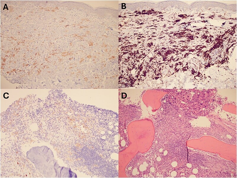
The histology. The lesion is made of foamy histiocytes that are CD68-positive and CD1a/Langerin-negative, often with Touton giant cells and fibrosis. The BRAF V600E mutant protein can be shown directly by immunohistochemistry, as it was in the Al-Abdulmalek case.
Where it sits. The 2016 Revised Classification of Histiocytoses (Emile 2016) divides the histiocytoses into five groups:
- L - Langerhans-related, containing ECD alongside LCH
- C - cutaneous/mucocutaneous
- R - Rosai-Dorfman disease
- M - malignant histiocytoses
- H - haemophagocytic lymphohistiocytosis
The LCH overlap. ECD and LCH share a clonal MAPK-driven myeloid-dendritic origin and can co-occur. A substantial minority of ECD patients, about 15-20%, have concurrent LCH ("mixed histiocytosis"), so the sharp ECD-versus-LCH split is a diagnostic contrast rather than a difference of origin.
Clinical Presentation
The bone. The skeletal hallmark is bilateral, symmetric osteosclerosis of the metadiaphyses of the long bones, particularly of the lower limbs (distal femur, proximal and distal tibia). Lower-limb bone pain is the classic symptom, and the sclerosis is sometimes an incidental radiographic finding.
Beyond the bone. The extraskeletal manifestations are protean and can be life-threatening, and cardiovascular and retroperitoneal disease may be silent until imaged.
- Cardiovascular: periaortic soft-tissue sheathing, the 'coated aorta', and pericardial and myocardial infiltration
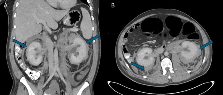
- Retroperitoneal: perinephric infiltration, the 'hairy kidney', with ureteric obstruction
- Neurological: ataxia, dysarthria, and cognitive and personality change. CNS involvement is the dominant prognostic factor, so these symptoms escalate urgency
- Pituitary: central diabetes insipidus from pituitary stalk infiltration, a presenting feature in the Al-Abdulmalek case together with hypogonadism
- Orbital: exophthalmos and retro-orbital infiltration with blurred vision
- Cutaneous and constitutional: xanthelasma/xanthogranuloma, and fatigue
- Pulmonary involvement
Investigations
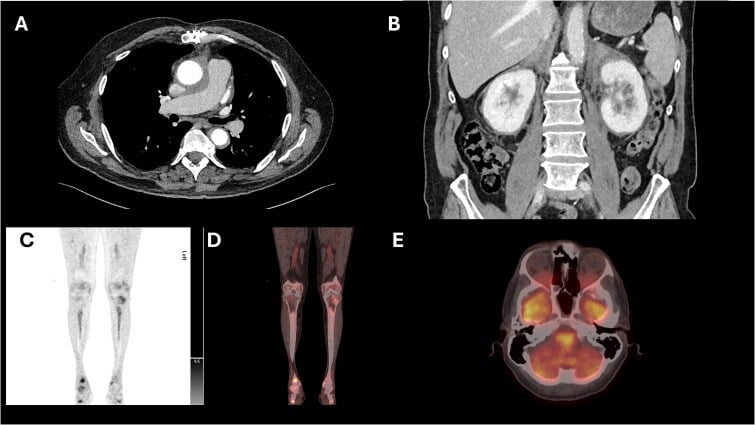
Recognising the pattern. The symmetric distal long-bone osteosclerosis gives matching symmetric increased uptake in the distal long bones on a radionuclide bone scan. This near-pathognomonic pattern often first suggests the diagnosis to the orthopaedic surgeon or radiologist, and it should prompt a search for multisystem involvement.
Mapping the extent. PET is often more informative than CT or MRI, revealing widespread FDG-avid skeletal and soft-tissue lesions. CT and MRI then define the organ involvement: the 'coated aorta', the 'hairy kidney', the CNS and the orbit.
Tissue and mutation. Biopsy confirms the diagnosis with foamy CD68-positive, CD1a-negative histiocytes, and the tissue goes for BRAF V600E and molecular testing. A bone or bone-marrow biopsy can be valuable, and a bone-marrow biopsy can secure the diagnosis even with a normal blood count (Al-Abdulmalek 2026).
Differential diagnosis. ECD must be distinguished from Langerhans cell histiocytosis and from other causes of osteosclerosis. The key is the symmetric distal long-bone sclerosis together with the multisystem features and the immunophenotype.
- Erdheim-Chester disease
- Non-Langerhans (foamy histiocytes)
- Langerhans cell histiocytosis
- Langerhans cells (Birbeck granules)
- Erdheim-Chester disease
- CD68+ , CD1a-/Langerin-
- Langerhans cell histiocytosis
- CD1a+ , Langerin/CD207+ , S100+
- Erdheim-Chester disease
- Bilateral symmetric osteoSCLEROSIS (long-bone metadiaphyses, legs)
- Langerhans cell histiocytosis
- LYTIC 'punched-out' lesions (skull, axial)
- Erdheim-Chester disease
- MAPK pathway (BRAF V600E common)
- Langerhans cell histiocytosis
- BRAF V600E also common
- Erdheim-Chester disease
- 'Coated aorta', 'hairy kidney', CNS, orbit, DI
- Langerhans cell histiocytosis
- Variable (single-system to multisystem)
The other causes of osteosclerosis to exclude:
- Osteoblastic metastases
- Sclerosing dysplasias
- Myelofibrosis
- Renal osteodystrophy
Bilateral, symmetric metadiaphyseal sclerosis of the lower-limb long bones should not be dismissed as nonspecific sclerosis. ECD is multisystem and potentially life-threatening (cardiovascular, retroperitoneal, CNS), and it is now treatable: confirming it on biopsy with BRAF testing and referring to a specialist multidisciplinary team can dramatically change outcome.
Management
The orthopaedic role. Recognise the characteristic bone disease, obtain a planned biopsy, manage the bone pain, and refer. Definitive care sits with a specialist multidisciplinary team addressing the affected organ systems.
The agents. BRAF inhibitors treat BRAF V600E-mutant disease and MEK inhibitors act on the MAPK pathway more broadly; both can produce dramatic responses.
- Vemurafenib, a BRAF inhibitor, was the first agent approved specifically for BRAF V600E-mutant ECD, in 2017
- Dabrafenib, a BRAF inhibitor, is also used, with or without trametinib
- Cobimetinib, a MEK inhibitor, was approved for ECD in 2022, including BRAF-wild-type disease
- Interferon-alpha (or pegylated interferon-alpha) was the historical first-line agent
- Anakinra, sirolimus and others are used in selected cases
Monitoring. Response is assessed by PET. The metabolic response to a BRAF inhibitor is often rapid and dramatic.
Complications
Renal. Perinephric and ureteric infiltration obstructs the ureters, and hydronephrosis and renal impairment follow untreated 'hairy kidney' disease.
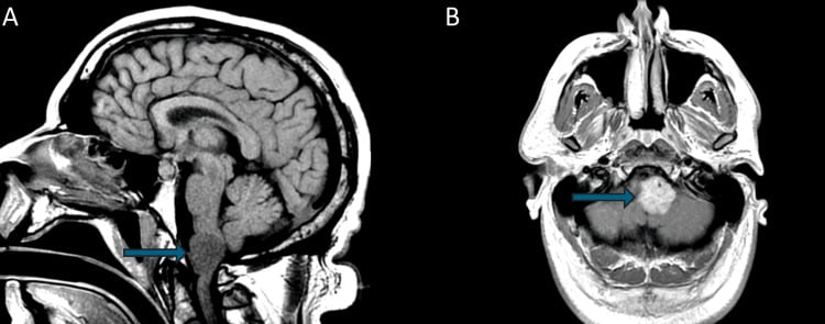
Neurological. The neurological complications are progressive ataxia, cognitive decline and dural disease, such as the foramen-magnum dural mass below.
Endocrine. The endocrine complications are permanent diabetes insipidus and anterior pituitary failure requiring lifelong replacement.
Treatment-related. Vemurafenib brought arthralgia, maculopapular rash, fatigue, alopecia, QT prolongation and cutaneous lesions, with hypertension and dermatological toxicity above melanoma rates (Diamond 2018). Interferon-alpha carries flu-like and marrow toxicity.
Prognosis - and What 'Transformed' Means
What predicts death. The prognosis was historically poor, and the numbers come from the Arnaud cohort. On multivariate Cox analysis, CNS involvement was an independent predictor of death, with a hazard ratio of 2.51 (95% CI 1.28 to 5.52, p=0.006). Cardiovascular involvement is the other major cause of death.
The skeletal pattern is what brings the patient to an orthopaedic clinic, but it is not what determines whether they survive. A patient whose symmetric long-bone sclerosis has just been recognised needs a neurological examination and brain imaging, not only a skeletal survey. The presence or absence of CNS disease, rather than how many bones are involved, is what shapes the conversation about outlook.
Interferon-alpha and survival. In the same analysis, treatment with interferon-alpha independently predicted survival, with a hazard ratio of 0.32 (95% CI 0.14 to 0.70, p=0.006). It was the first time any treatment had been shown to improve survival in ECD, and it is more than the label "historical" implies.
The limits of that finding. It is an observational cohort in which 87% of patients were treated, so confounding by indication cannot be excluded, and the authors said a randomised trial would be needed. It remains a Cox-adjusted observational finding, not a trial result.
What 'transformed' means. The BRAF and MEK inhibitors are the modern standard, and their approval rests on response data: symptom resolution, and metabolic response on PET. In VE-BASKET the objective response rate in ECD was 54.5%, and 80% of PET-assessed patients had a complete metabolic response (Diamond 2018). No randomised trial has shown that a BRAF or MEK inhibitor improves survival, and the only agent with a demonstrated survival benefit remains interferon-alpha. Both statements can be true at once: say the disease is now treatable, and do not say targeted therapy is proven to prolong life.
Mnemonics & Memory Aids
ERDHEIM
Hook:ERDHEIM: Extra-skeletal multisystem, Rare non-Langerhans, Driven by BRAF, Histiocytes CD68+/CD1a-, Evaluate PET+biopsy, Inhibitors (BRAF/MEK), Metadiaphyseal symmetric sclerosis.
FOAMY
Hook:FOAMY histiocytes: CD68-positive, CD1a-negative, MAPK/BRAF-driven - and now treatable.
ARCO
Hook:ARCO arches over the extraskeletal disease: Aorta coated, Retroperitoneal hairy kidney, CNS/pituitary, Orbit.
Clinical Decision Scenarios
Practise clinical reasoning and management decisions out loud
“A bone scan shows symmetric increased uptake in the distal femora and tibiae, with corresponding osteosclerosis. What diagnosis should you consider and how is it managed?”
What it is
- Rare non-Langerhans cell histiocytosis
- Foamy histiocytes CD68+ , CD1a-/Langerin- (vs LCH which is CD1a+)
- MAPK-pathway driven (BRAF V600E common)
Skeletal hallmark
- Bilateral symmetric osteosclerosis of long-bone metadiaphyses (lower limbs)
- Symmetric increased uptake on bone scan (near-pathognomonic)
- Bone pain common
Multisystem features
- Cardiovascular: periaortic 'coated aorta'; pericardial/myocardial
- Retroperitoneal: perinephric 'hairy kidney' (ureteric obstruction)
- CNS, orbit (exophthalmos), pituitary (diabetes insipidus), skin (xanthelasma)
Diagnosis & treatment
- PET (extent) + CT/MRI (organs); biopsy (CD68+/CD1a-) + BRAF testing
- Distinguish from LCH (CD1a+, lytic) and other osteosclerosis causes
- BRAF/MEK inhibitors transformed treatment (response data; no survival RCT); MDT care
- Prognosis: CNS involvement HR 2.51 for death; interferon-alpha HR 0.32 for survival (n=53, observational)
Evidence & Key Studies
From suspected brain malignancy to Erdheim-Chester disease: the role of PET imaging and bone marrow biopsy in diagnosis
- Erdheim-Chester disease is an extremely rare non-Langerhans cell histiocytosis involving MAPK-pathway mutations, notably BRAF V600E.
- PET imaging was more informative than CT or MRI, identifying widespread FDG-avid skeletal and soft-tissue lesions; because of the skeletal lesions, bone-marrow/tissue biopsy was valuable and histopathology confirmed ECD with the BRAF V600E mutation.
- Multidisciplinary evaluation and targeted therapy with a BRAF inhibitor led to symptom resolution.
ECD as a rare non-Langerhans cell histiocytosis involving MAPK-pathway (BRAF V600E) mutations, the superior diagnostic role of PET imaging in revealing widespread FDG-avid skeletal/soft-tissue lesions, the value of biopsy with BRAF confirmation, and the efficacy of targeted BRAF-inhibitor therapy come from the cited Al-Abdulmalek report. The skeletal hallmark (bilateral symmetric long-bone metadiaphyseal osteosclerosis), the CD68+/CD1a- immunophenotype, the multisystem features ('coated aorta', 'hairy kidney', CNS/ orbit, diabetes insipidus), and the distinction from Langerhans cell histiocytosis are standard, well-established teaching. The survival figures - the hazard ratios for CNS involvement and for interferon-alpha, and the sex distribution - come from the Arnaud cohort, which is observational and predates the targeted-therapy era. No randomised trial has shown that a BRAF or MEK inhibitor improves survival in ECD; their approval rests on response data. The L-group classification comes from the Emile 2016 Histiocyte Society revision, and the vemurafenib response figures from the Diamond 2018 VE-BASKET analysis. Nor is there a validated staging system, a surveillance interval, or a figure for what proportion of patients have skeletal involvement drawn from a defined cohort, so none is quoted here.