The Benign Notochordal Lesion (vs Chordoma)
- The BENIGN NOTOCHORDAL CELL TUMOUR (BNCT) is a BENIGN intraosseous lesion arising from NOTOCHORDAL REMNANTS along the axial skeleton - the CLIVUS, the VERTEBRAL bodies and the SACRUM (the embryonic notochord's path) - and is most often an INCIDENTAL, asymptomatic finding.
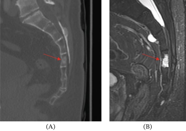
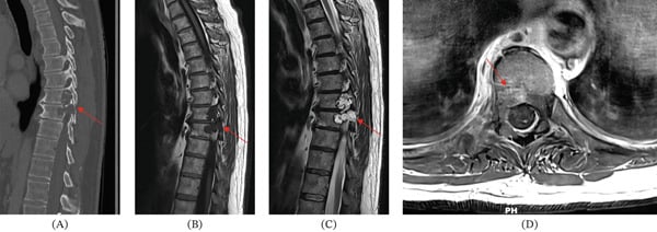
- Its IMAGING is characteristically that of an INTRAOSSEOUS lesion that is SCLEROTIC (or mixed sclerotic) on CT with marrow signal change on MRI, and - critically - shows NO bone DESTRUCTION and NO extraosseous SOFT-TISSUE MASS; this non-aggressive, contained, sclerotic appearance is the key to its benign nature.
- The crucial relationship and DIFFERENTIAL is with CHORDOMA, the malignant notochordal tumour: both arise from notochordal tissue and are BRACHYURY (T)-positive, and BNCT may be a PRECURSOR lesion to chordoma - so distinguishing them matters greatly, and the presence of bone DESTRUCTION, an extraosseous SOFT-TISSUE MASS, a myxoid/lobulated lytic lesion, and clinical symptoms favours CHORDOMA over BNCT.
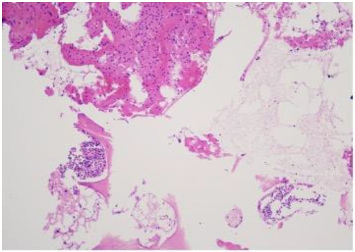
- On HISTOLOGY, BNCT consists of sheets of bland, adipocyte-like vacuolated notochordal cells filling the marrow space WITHOUT the myxoid/chondroid matrix, lobular architecture and bone destruction of chordoma - and it lacks the aggressive features; immunohistochemistry (brachyury, S100, EMA, cytokeratin) confirms notochordal differentiation but does not by itself separate BNCT from chordoma.
- The clinical IMPORTANCE is that recognising a lesion as a benign notochordal cell tumour avoids the morbidity of treating it as a chordoma, while NOT mislabelling a chordoma as benign (which would under-treat a malignant tumour) - the radiological assessment of aggressiveness (destruction/soft-tissue mass) is central, ideally at a specialist spine/tumour centre.
- MANAGEMENT of a benign, asymptomatic BNCT is OBSERVATION with interval imaging - in the largest biopsy-proven series, 16 lesions in 8 patients showed NO radiographic or symptomatic progression at a median 21.6 months, though the authors flag that follow-up as short. NO validated surveillance interval or stopping point exists. A CHORDOMA, in contrast, requires wide/en-bloc oncological RESECTION with consideration of adjuvant RADIOTHERAPY (e.g. proton therapy) - so the diagnosis directly dictates whether the patient is watched or undergoes major surgery.
- The BNCT-versus-chordoma split is not quite binary: a provisional third category, the ATYPICAL NOTOCHORDAL CELL TUMOUR, describes lesions with benign BNCT histology but subtle cortical permeation or minimal soft-tissue extension. In the four reported cases these were followed rather than resected, and the pathologists warned that calling them chordoma precipitates overly aggressive surgery - so minor permeation alone should prompt specialist review rather than sacrectomy.
- “Benign notochordal cell tumour (BNCT) = benign intraosseous lesion from notochordal remnants (clivus/vertebra/sacrum); usually INCIDENTAL; INTRAOSSEOUS, SCLEROTIC, NO destruction, NO soft-tissue mass.
- “Key differential = CHORDOMA (malignant): LYTIC/destructive with an extraosseous SOFT-TISSUE MASS, myxoid/lobulated. Both notochordal + BRACHYURY-positive; BNCT may be a chordoma PRECURSOR.
- “Management: OBSERVE/surveil the benign BNCT; chordoma needs en-bloc oncological RESECTION +/- radiotherapy. Don't under-treat a chordoma or over-treat a BNCT.
Incidental, intraosseous, sclerotic, no bone destruction, no soft-tissue mass. Bland vacuolated notochordal cells. Observe/surveil.
Lytic, destructive, extraosseous soft-tissue mass, myxoid/lobulated. Both brachyury-positive. Needs en-bloc resection +/- radiotherapy.
Overview & Epidemiology
What it is. The benign notochordal cell tumour (BNCT) is a benign intraosseous tumour of notochordal remnant origin, arising along the path of the embryonic notochord - the clivus, the vertebral bodies and the sacrum. It sits within the notochordal tumour spectrum alongside its malignant relative, chordoma, and the two share their brachyury (TBXT) marker.
How common. Notochordal rests are common autopsy findings - BNCTs have been reported in up to about 20% of spines at autopsy (a widely repeated figure for which no primary autopsy series was retrieved here - treat it as an order of magnitude, not a measured prevalence). The vast majority remain stable, asymptomatic and undetected; only a tiny fraction come to clinical attention, usually as an incidental finding on axial imaging.
Who presents, and how. Reported biopsy-proven series are small: the largest (Iorgulescu 2013) describes 16 lesions in 8 adults identified on imaging performed for back pain - so "incidental" describes the lesion's relationship to the symptom, not a screening discovery. There is no established sex or age predilection from cohort data; reported cases are adults.
Why it matters. The entity exists as a clinical problem almost entirely because of its resemblance to - and possible precursor relationship with - chordoma. Recognising it avoids the morbidity of unnecessary oncological resection; missing a chordoma mislabelled as BNCT allows a malignant tumour to progress.
What It Is & The Chordoma Distinction
BNCT is a benign intraosseous lesion from notochordal remnants in the axial skeleton (clivus, vertebrae, sacrum), usually incidental. It is intraosseous and sclerotic with no bone destruction and no soft-tissue mass - the hallmark of its benign nature. It shares notochordal origin and brachyury positivity with chordoma and may be a precursor to it; the presence of bone destruction, an extraosseous soft-tissue mass, a myxoid/lobulated lytic lesion and symptoms favours chordoma. Histologically BNCT is sheets of bland vacuolated notochordal cells filling the marrow without chordoma's myxoid matrix, lobular architecture and destruction.
- Benign notochordal cell tumour
- Benign (often incidental)
- Chordoma
- Malignant
- Benign notochordal cell tumour
- Intraosseous, sclerotic, NO destruction
- Chordoma
- Lytic, destructive
- Benign notochordal cell tumour
- Absent
- Chordoma
- Present (extraosseous)
- Benign notochordal cell tumour
- Sheets of bland vacuolated cells; no myxoid lobules
- Chordoma
- Myxoid/lobulated; physaliphorous cells
- Benign notochordal cell tumour
- Notochordal; brachyury+
- Chordoma
- Notochordal; brachyury+
- Benign notochordal cell tumour
- Observation/surveillance
- Chordoma
- En-bloc resection +/- radiotherapy
Pathophysiology: Notochordal Biology, Ecchordosis Physaliphora, and Brachyury
- The notochord and its remnants. The notochord is the embryonic axial midline structure; it normally regresses, its only adult remnant being the nucleus pulposus of the intervertebral disc. Notochordal cell rests persist in the clivus, vertebral bodies and sacrococcygeal region, and BNCT (also called a "giant notochordal rest/hamartoma") arises from these intraosseous rests.
- The extraosseous relative. Ecchordosis physaliphora is a related benign notochordal remnant that is extraosseous/intradural - classically on the dorsal clivus/prepontine region - the intracranial counterpart of the intraosseous BNCT.
- How common. Notochordal rests are common - BNCTs are found incidentally in up to about 20% of spines at autopsy - and most remain stable; only a small proportion behave as precursors, with some chordomas arising adjacent to a BNCT.
- The gene behind brachyury. Brachyury is the T-box transcription factor T (gene TBXT), the master regulator of notochord development, expressed in all notochordal cells (BNCT, chordoma and nucleus pulposus). Germline TBXT duplication and a common TBXT variant are chordoma susceptibility factors - the molecular basis of the shared marker and the precursor link.
Q: What is the notochordal biology behind BNCT, and what is brachyury?
A: The notochord is the embryonic axial structure that normally regresses, leaving only the nucleus pulposus; notochordal cell rests persist in the clivus/vertebrae/sacrum, and BNCT (a "giant notochordal rest") arises from them. Ecchordosis physaliphora is the extraosseous/intradural (dorsal-clivus) counterpart. Notochordal rests are common (BNCT in up to ~20% of spines at autopsy), mostly stable. Brachyury is the T-box transcription factor T (gene TBXT), the master regulator of notochord development - expressed in BNCT/chordoma/nucleus pulposus; germline TBXT duplication is a chordoma susceptibility factor.
The Grey Zone: When the Lesion Is Neither Clearly Benign Nor Clearly Chordoma
Treating destruction and soft-tissue extension as an absolute switch between observation and en-bloc surgery is clean but incomplete. A recognised intermediate group exists, and knowing it is what separates a safe answer from a confident one.
Four adults (aged 53 to 83) with lumbar or sacral notochordal tumours showed subtle cortical permeation with minimal soft-tissue extension on imaging, yet the histology was that of a benign notochordal cell tumour (two with focal myxoid change). Three were followed with serial imaging for 26 to 120 months: two showed no progression, and one grew 3.7 mm over ten years. The pathologists proposed the provisional label atypical notochordal cell tumour for lesions failing the criteria for either diagnosis, arguing that calling them chordoma "precipitates potentially overly aggressive surgical management". Four patients - a proposal, not an established category.
The grey zone cuts both ways. In a three-case series, two classic lesions were observed without progression, while the third - showing an atypical lytic pattern with contrast enhancement - underwent en-bloc resection with significant associated morbidity, and the specimen contained coexistent foci of incipient chordoma. So an enhancing or lytic notochordal lesion is not over-called when it is treated seriously; the price of getting it wrong in that direction is a missed malignancy, and in the other direction a sacrectomy.
The confident advice to observe rests on modest follow-up. In the largest biopsy-proven series, 16 lesions in 8 patients were followed radiographically for a median of 21.6 months (range 8.5 to 71.2) and none progressed radiographically or symptomatically - the authors state plainly that their conclusion is limited by that short follow-up. Two points follow. First, those patients were identified after presenting with back pain, not purely by chance, so "incidental" describes how the lesion relates to the symptom rather than how it was found. Second, no validated surveillance interval or stopping point exists: the published follow-up ranges from under a year to ten years, and no schedule has been tested. Interval imaging is therefore a reasonable practice justified by the natural history observed so far, not a protocol, and should be set with the specialist centre.
Know the three-way, not the two-way: benign notochordal cell tumour (intraosseous, sclerotic, no destruction, observe), chordoma (lytic, destructive, soft-tissue mass, enhances - resect), and the proposed atypical notochordal cell tumour in between (benign histology with subtle cortical permeation, followed rather than resected in the reported cases). Observation is supported by no progression in 16 lesions at a median 21.6 months - reassuring but short.
Clinical Presentation
The typical story is no story at all - BNCT is characteristically asymptomatic and incidental, found on CT or MRI performed for unrelated reasons. When patients do present, the reported context is non-specific back or axial pain that prompts imaging (all 8 patients in the Iorgulescu series presented this way); the lesion is then an incidental finding rather than the proven pain generator.
Red flags that point away from BNCT and toward chordoma: progressive or night pain, neurological symptoms (radiculopathy, sacral plexus symptoms, bladder/bowel disturbance), a palpable presacral mass on rectal examination, and any imaging evidence of cortical destruction or a soft-tissue component. The Terzi cases all presented with subacute mild pain - including the one that harboured incipient chordoma - so symptoms alone do not separate the two; the imaging aggressiveness assessment does.
Examination is usually normal in BNCT. Any neurological finding or palpable mass should be treated as a chordoma until proved otherwise and referred to a specialist spine/tumour centre.
Imaging in Detail: Signal, Enhancement, and the Sclerotic Differential
- BNCT on CT. Focal sclerosis (or mixed sclerosis), well-defined, intraosseous, with no cortical destruction, no periosteal reaction and no soft-tissue mass.
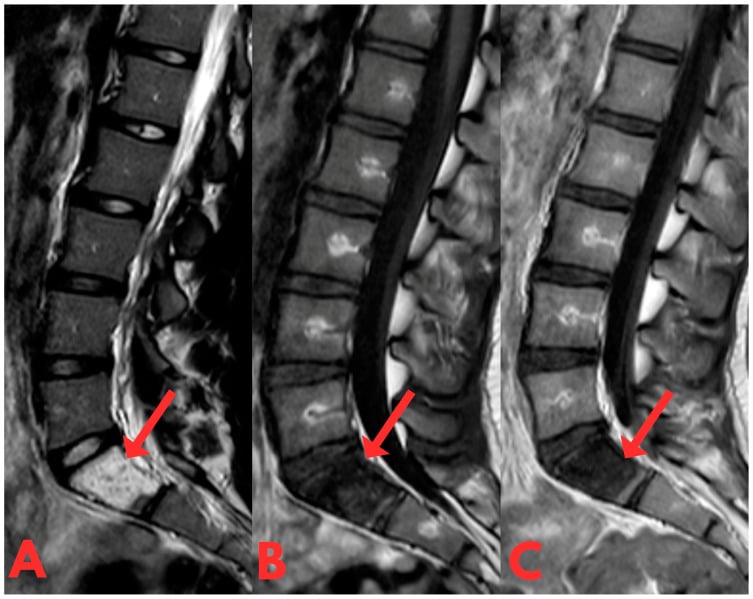
- BNCT on MRI. Low signal on T1 (replacing the normal fatty marrow), high signal on T2, and - a key discriminator - little or no contrast enhancement (unlike chordoma, which enhances heterogeneously).
- The non-chordoma differential.
- Bone island / enostosis: dense homogeneous sclerosis with radiating "thorny" margins that blend into trabeculae, and is low on all MRI sequences (no marrow-replacing T2-high signal).
- Vertebral haemangioma: "corduroy"/"polka-dot", fat-containing (T1-high), enhances.
- Sclerotic metastasis: older patient, known primary, often multiple, may destroy cortex, enhance or be PET-avid.
Q: What are the imaging features of BNCT, and how does it differ from other sclerotic/marrow lesions?
A: BNCT is a focal sclerotic (or mixed) intraosseous lesion on CT with no cortical destruction/soft-tissue mass; on MRI it is low T1, high T2, with little or no enhancement (a key discriminator from the enhancing chordoma). It differs from a bone island (dense with thorny margins, low on all MRI sequences), a vertebral haemangioma (corduroy/polka-dot, fat-containing/T1-high, enhances), and sclerotic metastasis (older, known primary, multiple, may destroy/enhance/be PET-avid).
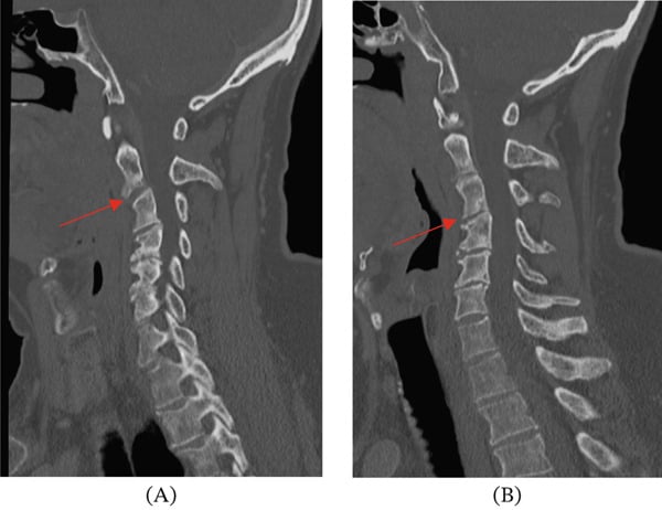
Management
- BNCT (benign, asymptomatic): observation with interval imaging to confirm stability - supported by 16 lesions followed a median 21.6 months without progression. No validated interval, modality or stopping point exists, so agree the schedule with the specialist centre rather than quoting a protocol.
- The lesion that is neither: benign histology with subtle cortical permeation or minimal soft-tissue extension has been designated a provisional atypical notochordal cell tumour and followed rather than resected in the small reported series - so do not let minor permeation alone trigger a sacrectomy without specialist review.
- Distinguish from chordoma: assess for bone destruction, soft-tissue mass, myxoid/lytic features and symptoms (favour chordoma); biopsy/management at a specialist spine/tumour centre.
- Chordoma: wide/en-bloc oncological resection with consideration of adjuvant radiotherapy (e.g. proton therapy).
- Avoid: over-treating a benign BNCT, and under-treating a chordoma mislabelled as benign.
Complications & Prognosis
The lesion itself. In the available follow-up, biopsy-proven BNCTs do not progress: 0 of 16 lesions grew or became symptomatic at a median 21.6 months (Iorgulescu 2013), and the atypical-category cases were similarly indolent (one grew 3.7 mm over ten years; Carter 2017). The prognosis of a genuine BNCT is therefore excellent.
The complication that matters is transformation - or coexistence - with chordoma. BNCT is hypothesised to be a chordoma precursor (shared notochordal origin, shared brachyury), and the Terzi case proves an atypical notochordal lesion can harbour incipient chordoma. What no series provides is a rate: the cohorts are too small and too short to quantify transformation risk, which is precisely why surveillance has no validated stopping point.
The iatrogenic complications are the other half of the story. En-bloc sacral or spinal resection carries significant morbidity (as the Terzi resected case illustrates) - sacral nerve sacrifice, bladder/bowel and sexual dysfunction, wound and reconstruction problems. Over-calling a BNCT as chordoma inflicts that morbidity for a lesion that would never have harmed the patient. Both error directions are complications of the diagnosis, and both are minimised by specialist-centre review.
Surveillance as treatment. Interval imaging is the management and the safety net: it confirms stability, and it catches the rare lesion that declares itself aggressive early enough for curative resection. The schedule is set with the treating centre - no published protocol exists.
Mnemonics & Memory Aids
NOTOCHORD
Hook:NOTOCHORD: Notochordal axial, Often incidental, no exTraosseous mass, nO destruction, CHordoma differential, Origin shared (brachyury), Resect chordoma, Don't over-treat BNCT.
BEDSFeatures That Favour Chordoma
Hook:Chordoma BEDS the patient: Brachyury+ (both), Enhances, Destructive, Soft-tissue mass.
RESTBNCT Biology
Hook:A BNCT is a notochordal REST: Remnant origin, Ecchordosis relative, Sclerotic/contained, TBXT (brachyury) positive.
Clinical Decision Scenarios
Practise clinical reasoning and management decisions out loud
“An incidental sclerotic intraosseous lesion is found in a sacral vertebra. How do you decide whether it is a benign notochordal cell tumour or a chordoma?”
What it is
- Benign intraosseous lesion from notochordal remnants (clivus/vertebra/sacrum)
- Usually incidental and asymptomatic
- Histology: sheets of bland vacuolated notochordal cells filling marrow
Imaging (benign hallmarks)
- Intraosseous, sclerotic (CT), marrow signal change (MRI)
- NO bone destruction; NO extraosseous soft-tissue mass
- Non-aggressive, contained appearance
vs Chordoma
- Chordoma: lytic/destructive + extraosseous soft-tissue mass, myxoid/lobulated
- Both brachyury-positive; BNCT may be a chordoma precursor
- Destruction/soft-tissue mass favours chordoma
Management
- BNCT (asymptomatic): observation + imaging surveillance
- Chordoma: en-bloc oncological resection +/- radiotherapy (proton)
- Equivocal/aggressive axial lesions → specialist spine/tumour centre
Evidence & Key Studies
Sacral tumors: imaging, diagnostic challenges and tumor mimics (notochordal lesions)
- Primary tumours of the sacrum/spine can arise from notochordal remnants, with chordoma recognised for its propensity to occur in the sacrum; imaging is essential in diagnosis, pretreatment evaluation and assessing treatment response.
- Many entities have characteristic clinical, epidemiological and imaging findings that allow a confident diagnosis or a narrow differential, enabling a systematic approach to sacral/axial masses.
- A definitive diagnosis is not always achievable on imaging alone, as some lesions lack specific features - relevant to distinguishing benign notochordal lesions from chordoma.
The origin of axial/sacral tumours from notochordal remnants, the propensity of chordoma for the sacrum, the central role of imaging in diagnosis and pretreatment evaluation, and the fact that imaging alone is not always definitive (relevant to the benign-notochordal-lesion versus chordoma distinction) come from the cited Adin sacral-tumours review. The specific imaging hallmarks of benign notochordal cell tumour (intraosseous, sclerotic, no destruction/soft-tissue mass), the shared brachyury-positive notochordal origin with chordoma, the precursor relationship, and the observation-versus-resection management are standard, well-established teaching. The natural-history figures - 16 lesions in 8 patients, median follow-up 21.6 months, no progression - come from the cited Iorgulescu series, a single retrospective centre whose authors state the short follow-up as a limitation. The atypical notochordal cell tumour category, its imaging and the ten-year 3.7 mm growth come from the cited Carter series of four patients and remain a provisional designation rather than a WHO entity. The observation that an enhancing, lytic notochordal lesion resected with significant morbidity contained incipient chordoma is from Terzi and colleagues (Spine 2012, PMID 22772575), a three-case series. No validated surveillance interval, imaging modality or stopping point for a benign notochordal cell tumour was retrieved, and no cohort large enough to quantify the risk of transformation exists - so the schedule should be set with the treating specialist centre rather than from a published protocol. The figure that notochordal rests are found in up to about 20 percent of spines at autopsy is widely repeated in the literature; no primary autopsy series supporting that exact proportion was retrieved here, so it is given as an order of magnitude. See also chordoma; the library has no dedicated sacral-tumour or spinal-bone-tumour topic, and the sacral lesions most often confused with these on imaging are covered in sacral insufficiency fracture.