The Osteoporotic 'Great Mimicker'
- A sacral insufficiency fracture is a STRESS fracture of the type 'INSUFFICIENCY' - ABNORMAL (weakened) bone failing under NORMAL physiological load - as opposed to a FATIGUE fracture (normal bone, abnormal repetitive load); the great majority are OSTEOPOROTIC fractures in elderly women, often after minimal or no trauma.
- Risk factors beyond osteoporosis include PELVIC RADIOTHERAPY, chronic GLUCOCORTICOIDS, rheumatoid arthritis, renal osteodystrophy, Paget disease, and other causes of weak bone; it is part of the FRAGILITY FRACTURE OF THE PELVIS (FFP) spectrum.
- It is a 'GREAT MIMICKER' - presenting as low back, buttock or groin pain with difficulty weight-bearing, often INSIDIOUS and with NO clear injury - and is frequently MISSED or mistaken for mechanical back pain or, worryingly, for bony METASTASES.
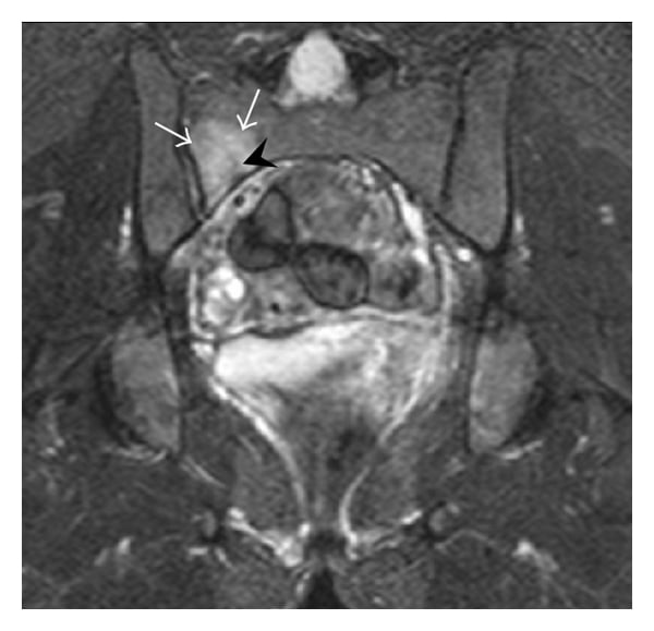
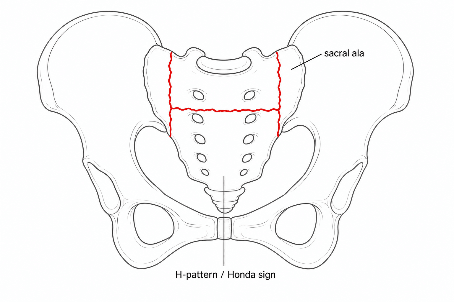
- PLAIN RADIOGRAPHS are insensitive and frequently NORMAL; CT is the gold standard for fracture MORPHOLOGY (and to look for a lytic lesion), while MRI is the MOST SENSITIVE test (~99%) showing BONE-MARROW OEDEMA; a bone scan shows the classic 'HONDA SIGN' (H-shaped uptake) - the typical full H pattern is actually uncommon, so its absence does not exclude the diagnosis.
- It is ESSENTIAL to EXCLUDE MALIGNANCY (metastasis/myeloma) as a cause of the sacral lesion before settling on insufficiency fracture, and to INVESTIGATE AND TREAT the underlying OSTEOPOROSIS - secondary fracture prevention is widely neglected (around 71% of patients do not receive it).
- Most are managed CONSERVATIVELY (analgesia, early mobilisation, osteoporosis treatment); SURGERY/intervention - SACROPLASTY (cement augmentation, for pain) or screw/iliosacral/triangular lumbopelvic FIXATION (for unstable or progressive fractures) - is reserved for refractory pain or instability.
- “Insufficiency = weak bone + normal load (osteoporotic elderly); fatigue = normal bone + abnormal load (athlete/recruit).
- “Radiographs miss it - get MRI (most sensitive, marrow oedema) or CT (morphology); bone scan = Honda/H sign (but the full H is uncommon).
- “Always EXCLUDE malignancy and TREAT the osteoporosis - the fracture is a sentinel for under-treated bone disease.
The pain is often insidious with no injury, and plain radiographs are usually normal - so it is easily dismissed as mechanical back pain. A high index of suspicion in an elderly osteoporotic patient with new buttock/back pain, plus MRI/CT, makes the diagnosis.
A sacral signal abnormality can be read as a metastasis or myeloma - and vice versa. CT/MRI (and the typical fracture morphology) help distinguish them; exclude malignancy before accepting an insufficiency fracture, especially with an atypical pattern or a known primary.
Definition & Risk Factors
A stress fracture is a fracture caused by repetitive sub-maximal load. It has two types:
- Insufficiency fracture - abnormal (weakened) bone fails under normal physiological load. The sacral insufficiency fracture is the classic example: an osteoporotic sacrum in an elderly patient, often after minimal or no trauma.
- Fatigue fracture - normal bone fails under abnormal, repetitive load (e.g. a runner, dancer or military recruit). Beyond osteoporosis, the weakened-bone causes include pelvic radiotherapy, chronic glucocorticoids, rheumatoid arthritis, renal osteodystrophy, Paget disease, hyperparathyroidism and osteomalacia. The sacral insufficiency fracture sits within the fragility fracture of the pelvis (FFP) spectrum and signals under-treated bone fragility.
Presentation - the 'Great Mimicker'
- Low back, buttock or groin pain, sometimes radiating to the hip/thigh
- Difficulty weight-bearing / mobilising; pain worse with standing/walking
- Often insidious with no recalled injury (or trivial trauma)
- An elderly, often osteoporotic patient (or prior pelvic radiotherapy)
- Mimics mechanical low back pain, hip pathology, or metastatic disease
- Examination is often non-specific (sacral tenderness, pain on stressing the pelvis)
- Plain films are commonly normal - the diagnosis needs cross-sectional imaging
Imaging
Plain radiographs are insensitive and frequently normal (overlying bowel gas, osteopenia and the sacral curvature obscure the fracture). CT is excellent for fracture morphology (a sclerotic fracture line/cortical break) and helps exclude a lytic/destructive lesion. MRI is the most sensitive test (around 99%), showing bone-marrow oedema in the sacral ala and the fracture line even when other imaging is normal - and it best distinguishes fracture from tumour. A bone scan classically shows the 'Honda sign' (an H-shaped pattern of increased uptake), though the full H is actually uncommon - unilateral, vertical-only or horizontal-only patterns are frequent, so a missing H does not exclude the diagnosis.
Classification (Fragility Fractures of the Pelvis)
Within the fragility fracture of the pelvis (FFP) classification of Rommens and Hofmann, fractures are graded by anterior and posterior pelvic ring involvement and stability - from FFP I (anterior only) and FFP II (non-displaced posterior, e.g. a sacral ala fracture) to FFP III (displaced unilateral posterior) and FFP IV (bilateral/displaced, highly unstable). Progression from a stable to an unstable pattern is common and is influenced by bone density, pelvic morphology and sarcopenia - which is why follow-up and treating the bone matter. The grade guides treatment (conservative for stable; fixation for unstable/progressive).
Management
The majority of sacral insufficiency fractures are managed conservatively: analgesia, early mobilisation (prolonged bed rest is harmful in the elderly), physiotherapy and fall prevention. Two things are non-negotiable: exclude malignancy, and investigate and treat the underlying osteoporosis (calcium/vitamin D, bone-protective therapy) - secondary fracture prevention is widely neglected (about 71% of patients do not receive it), yet the fracture is a sentinel event for further fragility fractures.
| 0 | 1 |
|---|---|
| Conservative | Most fractures: analgesia, early mobilisation, fall prevention - plus osteoporosis treatment |
| Sacroplasty (cement augmentation) | Refractory pain: gives significant, often rapid PAIN relief and reduced analgesic use; limited stabilising capacity |
| Percutaneous iliosacral / transsacral screw fixation | Unstable or progressive posterior fractures (often with cement augmentation in osteoporotic bone) |
| Triangular lumbopelvic fixation / transsacral bar | Highly unstable patterns (e.g. spinopelvic dissociation), bilateral/displaced fractures |
| Treat osteoporosis (all) | Calcium/vitamin D + bone-protective therapy; secondary fracture prevention |
A fragility fracture of the sacrum carries substantial morbidity in the elderly - loss of independence in 64-89% and mortality of 13-27% in reported series - and most patients are not on adequate bone-protective treatment. Treat it as a sentinel event: arrange bone-health assessment and secondary fracture prevention, orthogeriatric input, and falls assessment, not just symptomatic relief.
Evidence & Key Studies
Chronic pelvic insufficiency fractures and their treatment
- Fragility/insufficiency fractures of the pelvis and sacrum are increasingly prevalent in the elderly; they cause chronic instability, pain and high morbidity (loss of independence 64-89%, mortality 13-27%), and 71% of patients do not receive adequate secondary fracture prevention.
- Conventional radiography often misses sacral fractures; CT is the gold standard for morphology and MRI has the highest sensitivity (~99%) for bone oedema; the Rommens-Hofmann FFP classification guides treatment.
- Conservative treatment is possible, but unstable/progressive fractures are treated surgically (sacroplasty, iliosacral screws, triangular lumbopelvic fixation, transsacral bars; cement augmentation improves fixation in osteoporotic bone), within a multidisciplinary approach including osteoporosis treatment.
Clinical and scintigraphic evaluation of insufficiency fractures in the elderly
- Insufficiency fractures are common in the elderly and generally occur at multiple sites; on bone scintigraphy the typical H-shaped (Honda) sacral pattern was present in only ~4% - its absence should not exclude the diagnosis.
- Most patients with insufficiency fractures who had bone-density measured had osteoporosis/osteopenia.
- These patients had poor survival, underscoring that the diagnosis matters and that specific management (including bone treatment) may improve outcomes.
According to PubMed, the imaging hierarchy (radiograph insensitivity, CT morphology, MRI ~99% sensitivity), the FFP classification, the high morbidity/under-treatment figures and treatment options come from the cited Gewiess review, and the point that the full Honda 'H' is uncommon (and the osteoporosis/poor-survival association) from the cited scintigraphy study. The insufficiency-vs-fatigue definition is standard teaching. (See also our Osteoporosis and Fragility/Geriatric pelvic-fracture material.)
Clinical Decision Scenarios
Practise clinical reasoning and management decisions out loud
“An 78-year-old woman has 3 weeks of low back and buttock pain and difficulty walking, with no injury. Radiographs of the pelvis are normal. What is your differential and how do you investigate?”
“MRI confirms a sacral insufficiency fracture. How would you manage her, and when would you consider intervention or surgery?”
Mnemonics & Memory Aids
SACRUM
Hook:SACRUM: insufficiency stress fracture in the aged - exclude cancer, MRI it, mind the Honda sign, manage the bone.
HONDA
Hook:The HONDA sign: H-pattern, osteoporotic, not always complete, diagnose on MRI/CT, augment/fix if needed.
Definition
- Insufficiency stress fracture: weak (osteoporotic) bone, normal load
- vs fatigue fracture: normal bone, abnormal load
- Risk: osteoporosis, pelvic radiotherapy, steroids, RA, renal osteodystrophy
Presentation & imaging
- Elderly, insidious back/buttock/groin pain, hard to weight-bear, often no injury - the 'great mimicker'
- Radiographs usually normal; MRI most sensitive (~99%, marrow oedema); CT morphology / exclude lytic lesion
- Bone scan Honda/H sign (full H uncommon); EXCLUDE malignancy
Classification
- Pattern: bilateral vertical alar + transverse = H (Honda); variants common
- Rommens-Hofmann FFP (I anterior -> IV bilateral/unstable) guides treatment
- Progression to instability is common
Management
- Most conservative: analgesia, early mobilisation, falls prevention
- ALWAYS: exclude malignancy + treat osteoporosis (secondary prevention) + orthogeriatrics
- Sacroplasty for refractory pain; iliosacral/transsacral screws or triangular lumbopelvic fixation for unstable/progressive