Dancer's Tendinitis | Posteromedial Ankle Pain | Often Coexists With Posterior Ankle Impingement
- FHL is the most posterior of the deep flexors at the ankle ("Tom, Dick, AND Harry") - neurovascular bundle lies ANTERIOR to it
- Coexists with posterior ankle impingement / os trigonum in dancers - assess and treat both (Hamilton)
- Triggering / pseudo-hallux rigidus = nodular tendinosis or adhesions; passive toe motion normal, active restricted
- Steroid injection is controversial - rupture risk; ultrasound-guided peritendinous only, never intratendinous
- Endoscopy reaches Zone 1 only - multi-zone disease (Zone 2/3) needs an open approach
- “FHL stretch test: passive dorsiflexion with hallux extension reproduces posteromedial pain
- “Os trigonum present in 10-25% of the population - reduces tunnel space behind the talus
- “FHL myotendinous junction lies at the ankle joint level - uniquely distal among deep flexors
- “Hindfoot endoscopy (van Dijk, 2-portal prone) is the workhorse minimally-invasive technique
Examiners expect you to differentiate FHL tendinitis from posterior ankle impingement syndrome and recognise the association with os trigonum. Know the anatomical zones and surgical approaches. Be prepared to discuss "dancer's tendinitis" and the unique demands of ballet en pointe.
Overview and Epidemiology
FHL tendinitis is an overuse stenosing tenosynovitis, and later tendinosis, of the flexor hallucis longus where it passes through the fibro-osseous tunnel behind the ankle. The provoking movement is forced or repetitive plantarflexion, so the condition clusters in particular groups rather than in the general public.
Who gets it. Four groups:
- Ballet dancers - the archetypal group, hence "dancer's tendinitis"; en pointe and demi-pointe load the tendon in extreme plantarflexion
- Running and jumping athletes - repetitive push-off in distance runners, gymnasts and triple-jumpers
- Kicking-sport athletes - footballers, with forced plantarflexion
- Non-athletes - after an ankle sprain or from occupational overuse
Not only a dancer's disease. The eponym persists, but in the Corte-Real series none of the patients were dancers or professional athletes, and all related the onset to an ankle sprain. Over-anchoring on the ballet stereotype risks missing the diagnosis in everyone else.
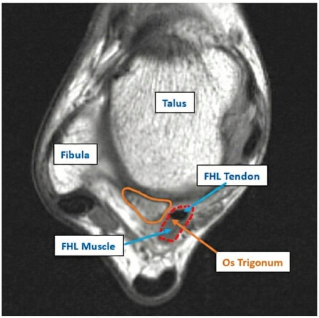
Associations. FHL tendinopathy frequently coexists with posterior ankle impingement syndrome (PAIS), in which a bony impediment sits behind the talus: an os trigonum or a prominent posterolateral talar process. The os trigonum is present in 10-25% of the population and reduces the tunnel space behind the talus.
What else raises the load. Cavus foot, claw or hammer toes and other hallux deformities increase the demand on FHL, and an equinus contracture (a tight Achilles) leads to compensatory FHL overuse. Training errors add to it: a rapid increase in volume or intensity, and the high-level demands of elite performance.
Anatomy and Biomechanics
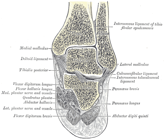
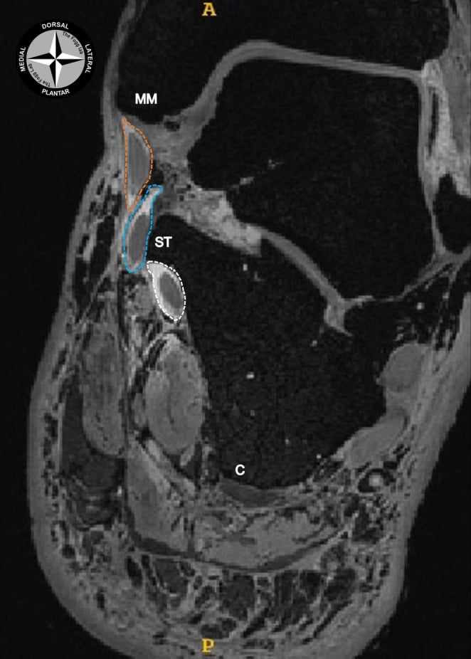
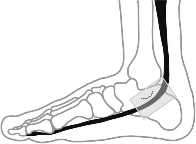
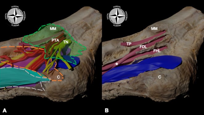
Origin and course. FHL arises from the middle two-thirds of the posterior fibula and the adjacent interosseous membrane. Its muscle belly extends further distally than those of the other deep flexors, and its myotendinous junction lies at the level of the ankle joint, unique among the flexors. The tendon then passes through the fibro-osseous tunnel between the medial and lateral tubercles of the posterior talus, runs beneath the sustentaculum tali, and is joined to FDL at the master knot of Henry in the midfoot, before inserting into the plantar base of the distal phalanx of the hallux.
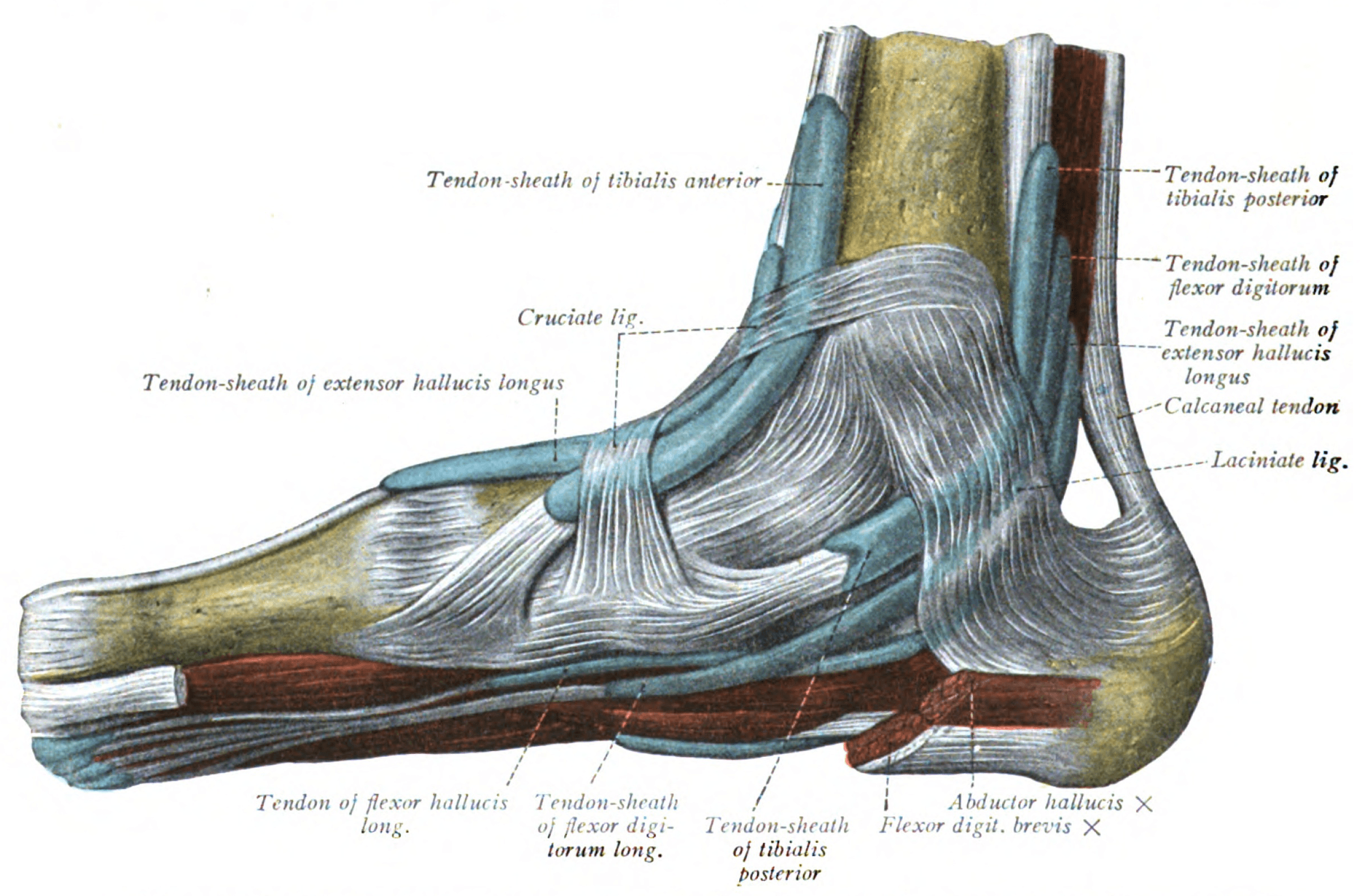
Relations at the ankle. FHL is the most posterior of the three deep flexor tendons (Tom, Dick and Harry), lying posterolateral to FDL. The tibial nerve and posterior tibial vessels lie anterior to it and, at the posterior ankle, just medial to it. That relationship is what makes the tendon the medial safety landmark in hindfoot endoscopy.
The three zones of pathology. Those three sites along the course are where the disease lives, and the zone decides the operation:
- Zone 1 - posterior ankle. The fibro-osseous tunnel between the talar tubercles. The most common site of stenosis, and the only zone endoscopy can reach
- Zone 2 - sustentaculum tali. The second constriction point, under the medial talar process: the "knot of the foot"
- Zone 3 - knot of Henry. The FHL-FDL decussation in the midfoot, where adhesions can cause triggering
The knot of Henry. In the plantar midfoot FHL and FDL cross and are joined by one or more tendinous slips, most often an FHL-to-FDL slip, although the pattern varies between individuals. The two tendons are therefore functionally coupled: FHL contributes to lesser-toe flexion through its slip to FDL, and FDL can contribute to hallux flexion.
Why the coupling matters. After FHL injury or harvest proximal to the knot, some active hallux flexion is retained through FDL. That is why harvesting FHL for an Achilles reconstruction usually leaves useful, though weaker, great-toe flexion, and why a proximal FHL laceration may not fully abolish it. The same coupling makes the FHL-to-FDL transfer work: FDL takes over hallux flexion while the interconnections and quadratus plantae preserve lesser-toe flexion. Adhesions at the knot tether the coupled tendons and cause triggering, and because hindfoot endoscopy cannot reach the plantar midfoot, Zone 3 disease needs an open approach.
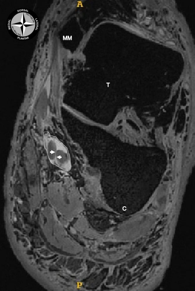
Biomechanics. FHL has the greatest excursion of any tendon in the foot, about 3 cm, and transmits up to 8 times body weight during the push-off phase of gait. It contributes to support of the medial longitudinal arch through the windlass mechanism and can augment FDL for the lesser toes.
Pathophysiology and Mechanisms of Injury
Mechanical impingement. The fibro-osseous tunnel behind the ankle is a potential site of compression. It may be narrowed congenitally or by acquired change between the talar tubercles, and anatomical variants of the tarsal tunnel can affect it. An os trigonum, prominent talar tubercles or spurs from impingement take up the space. Loading closes it further: sustained plantarflexion en pointe (90 degrees or more) in dancers, and the repetitive push-off and toe flexion of running.
From inflammation to degeneration. Repetitive loading drives an inflammatory cascade:
- Peritendinitis - initial inflammation of the paratenon from mechanical irritation
- Tenosynovitis - inflammation of the synovial sheath, with effusion
- Tendinosis - chronic degenerative change with a failed healing response
- Adhesions - scarring to surrounding structures, which limits gliding
- Nodular thickening - focal tendon swelling, which creates triggering
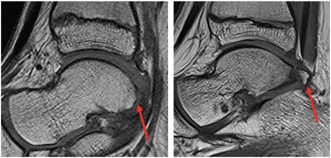
Os trigonum syndrome. An accessory ossicle or an elongated lateral tubercle causes posterior impingement. It may coexist with FHL tendinitis or compress the tendon secondarily.
Clinical Presentation - History
The pain. Deep, aching posteromedial ankle pain with activity, which may radiate to the plantar midfoot and may have a sharp component when the tendon triggers. It is worse with push-off, stair climbing and, in dancers, relevé. Triggering is felt as catching or snapping with great-toe flexion and extension.
Other symptoms. Morning stiffness is common when there is an inflammatory component. Night pain suggests more severe tendinopathy or associated pathology.
Function. Patients describe weakness pushing off or gripping with the great toe, difficulty with stairs and reduced walking tolerance, and tight shoes or heels make it worse. A dancer also loses push-off power. The sport decides the rest:
- keyFeatures
- En pointe pain, relevé weakness, posterior ankle catching
- typicalOnset
- Gradual with increased rehearsal intensity
- functionalLoss
- Cannot maintain en pointe, loss of elevation
- keyFeatures
- Push-off pain, medial ankle tenderness, reduced stride power
- typicalOnset
- After mileage increase or speed work
- functionalLoss
- Reduced pace, altered gait mechanics
- keyFeatures
- Kicking pain, plantarflexion weakness, shooting difficulty
- typicalOnset
- After intensive kicking drills
- functionalLoss
- Reduced shot power, altered technique
- keyFeatures
- Landing pain, vault push-off difficulty, beam work impaired
- typicalOnset
- With increased tumbling volume
- functionalLoss
- Cannot stick landings, reduced elevation
Physical Examination
Look. Posteromedial ankle fullness from tenosynovitis. Muscle atrophy is rare unless the disease is chronic or the tendon has ruptured. Check foot posture for cavus alignment or toe deformity, and watch the gait for an antalgic pattern with reduced push-off.
Feel. Tenderness posterior to the medial malleolus. Follow the tendon: point tenderness under the medial ankle at the sustentaculum, plantar midfoot tenderness if Zone 3 is involved, and posterior ankle tenderness from a coexistent os trigonum. A palpable nodule or thickening with active toe flexion is the trigger point.
Special tests. Four manoeuvres localise the problem to FHL:
- FHL stretch test - passive ankle dorsiflexion with hallux extension reproduces posteromedial ankle pain
- Resisted hallux flexion - active great-toe flexion against resistance is painful; weakness suggests advanced tendinopathy or rupture
- Triggering test - active toe flexion-extension produces palpable catching or an audible snap, indicating nodular thickening or adhesions
- Pseudo-hallux rigidus - restricted active but normal passive toe motion distinguishes FHL adhesions from true MTP joint arthritis
Neurovascular. Look for tarsal tunnel signs of concurrent tibial nerve compression and test sensation in the medial plantar nerve territory to the hallux. Document the dorsalis pedis and posterior tibial pulses, and rule out deep posterior compartment pathology.
The combination of posterior ankle pain with toe flexion activities, positive FHL stretch test, and triggering with great toe motion is highly specific for FHL tendinitis. Always assess for coexistent os trigonum syndrome as management may require addressing both pathologies.
Investigations - Imaging Studies
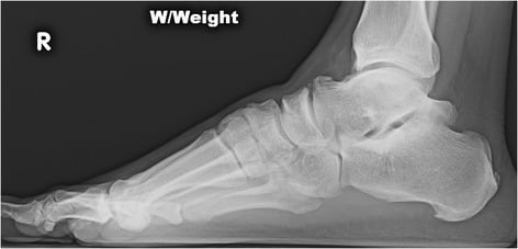
Radiographs. Weight-bearing views are preferred:
- AP and lateral foot - exclude hallux arthritis and sesamoid pathology
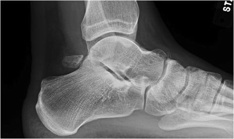
- Lateral ankle - os trigonum, posterior talar spurs, soft-tissue swelling
- Oblique - a better view of the sustentaculum tali
On the films, look for an os trigonum or a prominent lateral tubercle, posterior talar spurring from chronic impingement, abnormality or calcification at the sustentaculum, cavus alignment if it contributes, and posteromedial soft-tissue fullness.
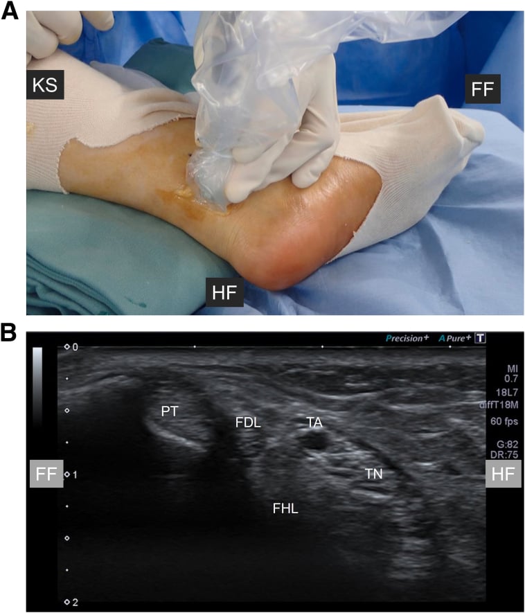
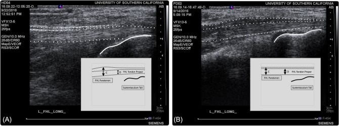
Ultrasound. Dynamic, cost-effective and free of radiation. With the patient in a figure-of-four position, use a high-frequency linear transducer (12-15 MHz), scan the tendon in long and short axis while the patient flexes and extends the toe, and compare with the other side. It can show:
- Tendon thickening (the normal diameter is 3-4 mm)
- Hypoechoic tendinosis or tears
- Peritendinous fluid, indicating tenosynovitis
- Reduced gliding on dynamic assessment
- Triggering at the sites of constriction
- The relationship of an os trigonum to the tendon
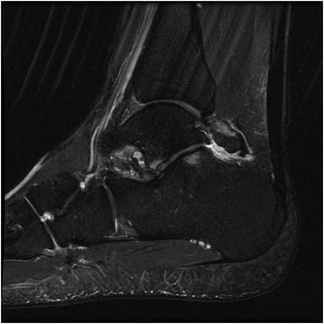
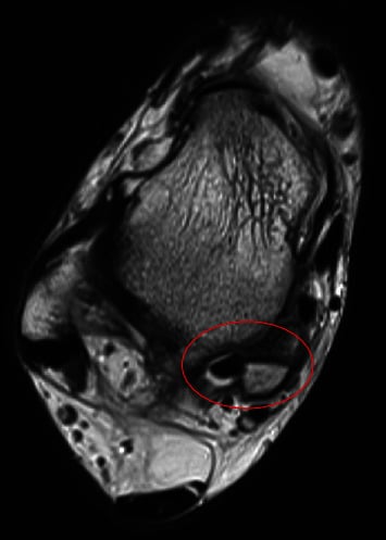
MRI. For an uncertain diagnosis or a suspected rupture, and before surgery. Use an ankle protocol with a foot-ankle coil and T1, T2 and STIR sequences. The findings:
- Tendinosis - increased T2 signal within the tendon substance
- Tenosynovitis - fluid surrounding the tendon in its sheath
- Tear - partial (high signal with intact fibres) or complete (a tendon gap)
- Adhesions - obliteration of the fat planes between tendon and adjacent structures
- Stenosis - a narrowed fibro-osseous tunnel at the involved zone
- Os trigonum oedema - bone marrow oedema if the impingement is symptomatic
- Associated pathology - FDL tendinopathy, tarsal tunnel syndrome
MRI is the gold standard for pre-operative planning when surgery is contemplated. It defines the extent of tendinopathy, identifies the zone(s) of involvement, and reveals associated posterior ankle impingement pathology requiring concurrent treatment.
Diagnostic injection. Ultrasound-guided peritendinous local anaesthetic confirms FHL, rather than other posterior ankle pathology, as the pain generator: significant relief supports the diagnosis. Avoid intratendinous injection because of the rupture risk. Adding steroid may be therapeutic but is controversial for the same reason.
Differential Diagnosis
Several conditions produce posterior ankle or great-toe symptoms like these.
- clinicalFeatures
- Posterior ankle pain with plantarflexion, nutcracker test positive
- examination
- Posterior tenderness, pain with forced plantarflexion
- imaging
- Os trigonum on lateral XR, bone oedema on MRI
- clinicalFeatures
- 1st MTPJ pain and stiffness, osteophyte formation
- examination
- Restricted passive motion, MTPJ tenderness, crepitus
- imaging
- Joint space narrowing, osteophytes on weight-bearing XR
- clinicalFeatures
- Plantar numbness/tingling, night symptoms, Tinel sign
- examination
- Tinel posterior to medial malleolus, sensory changes
- imaging
- MRI may show space-occupying lesion, NCS abnormal
- clinicalFeatures
- Posteromedial ankle pain, flatfoot progression, medial swelling
- examination
- Tenderness posterior to MM, too-many-toes, heel rise weakness
- imaging
- PTT thickening/tear on US/MRI, flatfoot on XR
- clinicalFeatures
- Posterior ankle pain 2-6 cm proximal to insertion
- examination
- Tendon thickening, arc of pain, positive squeeze test
- imaging
- Tendon thickening, intratendinous signal on MRI
- clinicalFeatures
- Exertional posteromedial leg pain, relieved with rest
- examination
- Tenderness over deep flexors, reproduction with exercise
- imaging
- Compartment pressure testing diagnostic
Non-Operative Management
Non-operative care comes first and, applied systematically, succeeds in most patients, although no cited study puts a figure on that success (see Return to Sport and Outcomes).
Load. Relative rest from the aggravating activities for 4-6 weeks, with fitness kept up by cycling or swimming, then a progressive return to loading over 8-12 weeks. Correct the biomechanical error that provoked it, en pointe mechanics in a dancer, and avoid rapid increases in training volume or intensity.
Immobilisation. A CAM boot for 2-4 weeks reduces tendon excursion in severe cases. A night splint holds the foot out of extreme plantarflexion, and kinesiology tape or athletic strapping limits motion. Use the minimum immobilisation necessary, to avoid stiffness.
Drugs and ice. Oral NSAIDs (naproxen 500 mg BD) or topical ones, in 2-4 week courses, monitoring for GI and renal side effects; paracetamol or tramadol if NSAIDs are contraindicated. Ice massage along the course of the tendon for 15-20 minutes.
Physiotherapy. Three phases:
- Phase 1, 0-2 weeks: pain and inflammation. Ice, gentle range-of-motion exercises, soft-tissue mobilisation avoiding direct pressure on the tendon, and intrinsic foot muscle strengthening to reduce FHL demand
- Phase 2, 2-6 weeks: progressive loading. Eccentric strengthening (proven efficacy in tendinopathy), calf stretching for any equinus, tendon-gliding exercises to prevent adhesions, and weight-bearing as tolerated
- Phase 3, 6-12 weeks: return to activity. Sport-specific rehabilitation (en pointe progression for dancers, plyometrics for runners), proprioceptive training on unstable surfaces, and a graded return to full activity with load monitoring
Corticosteroid injection. Methylprednisolone 40 mg with local anaesthetic, placed peritendinously under ultrasound guidance, followed by relative rest for 2 weeks and then a gradual return to loading.
Corticosteroid injections for FHL tendinopathy are controversial. While they may provide short-term symptomatic relief, they carry a risk of tendon rupture and should be used cautiously. Ultrasound guidance is essential to avoid intratendinous injection. Consider no more than 1-2 injections separated by at least 3 months.
Orthoses and footwear. Custom orthoses control excessive pronation or cavus mechanics. A heel lift reduces Achilles tightness and the secondary FHL overload, and a metatarsal pad offloads the great toe if a deformity contributes. Shoes need an adequate toe box, and dancers a rigid sole.
Hamilton, Geppert and Thompson (Landmark - Dancer's Tendinitis)
- Retrospective review of operative treatment of stenosing tenosynovitis of the FHL and/or posterior impingement syndrome in 37 dancers (41 operations), mean 7-year follow-up. 26 operations were for combined tendinitis plus posterior impingement, 9 for isolated tendinitis, and 6 for isolated impingement - underscoring how often the two coexist. 30 of 41 ankles achieved a good or excellent result; a medial incision was used in most. Outcomes were good/excellent in 28 of 34 professional-dancer ankles versus only 2 of 6 amateur ankles.
Operative Management - Indications
Surgery is for symptoms that persist after non-operative care of appropriate duration and compliance:
- Failed non-operative treatment - a minimum of 3-6 months
- Persistent symptoms - pain limiting daily activities or sport
- Triggering - mechanical symptoms suggesting nodular thickening or adhesions
- Professional athletes - earlier surgery for elite performers when the career is affected
- Structural pathology - an MRI-proven tendon tear, severe stenosis, or an os trigonum requiring excision
Before operating. Review the MRI to define the zones involved, the extent of tendinopathy and any associated pathology, because the pathology decides between an open and an endoscopic approach. Agree realistic goals for return to high-level activity, and optimise smoking, diabetes and inflammatory conditions.
Management - Surgical Techniques
Position. Supine, with a bump under the ipsilateral hip to rotate the leg externally and give access to the medial ankle and hindfoot. A thigh tourniquet is controversial, as it may impair visualisation.
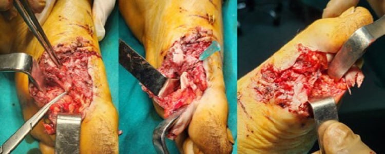
Exposure. An 8-10 cm longitudinal posteromedial incision centred between the medial malleolus and the Achilles tendon. Identify and protect the saphenous vein and nerve, then incise the flexor retinaculum to expose the FHL sheath.
Finding the tendon. FHL is the most posterior of the three tendons; confirm it by passive hallux flexion-extension, which makes it move. Open the sheath along the entire zone of pathology and inspect for tenosynovitis, nodular thickening and partial tears.
The neurovascular bundle (tibial nerve, posterior tibial vessels) lies anterior to the FHL tendon sheath. Use meticulous dissection and avoid excessive retraction. The medial calcaneal branch of tibial nerve is at risk with distal extension. Protect neurovascular structures throughout the procedure.
Decompression. The steps:
- Release the fibro-osseous tunnel at the posterior ankle, between the talar tubercles
- Excise thickened or diseased sheath, with a synovectomy if inflammation is significant
- Release the constriction at the sustentaculum tali if Zone 2 is involved
- Confirm smooth gliding through full passive toe motion
Os trigonum, if indicated. Identify the ossicle or prominent lateral tubercle, protect FHL during the dissection, excise it with a rongeur or osteotome, and smooth the bone to prevent recurrent impingement.
The tendon. Debride partial tears while preserving more than 50% of the width, and tubularise a significant longitudinal split. Repair an acute tear with non-absorbable suture. If more than 50% of the tendon is involved, consider FDL transfer.
Closure. Do not repair the tendon sheath: leaving it open prevents recurrent stenosis. Close the flexor retinaculum loosely and the skin in layers with absorbable sutures, under a soft dressing or a posterior splint in neutral.
Rehabilitation.
- 0-2 weeks - posterior splint, non-weight-bearing, gentle toe range of motion
- 2-6 weeks - CAM boot, progressive weight-bearing, active toe flexion exercises
- 6-12 weeks - wean from the boot, progressive strengthening, gait normalisation
- 3-6 months - return-to-sport protocol and gradual return to full activity
- Professional dancers - 4-6 months before returning to en pointe work
The return-to-sport table below gives a longer range for professional ballet: 6-9 months to en pointe after open release.
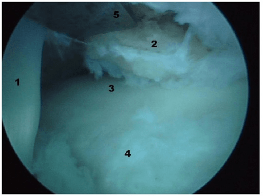
Rietveld and Hagemans - Open vs Endoscopic in Dancers
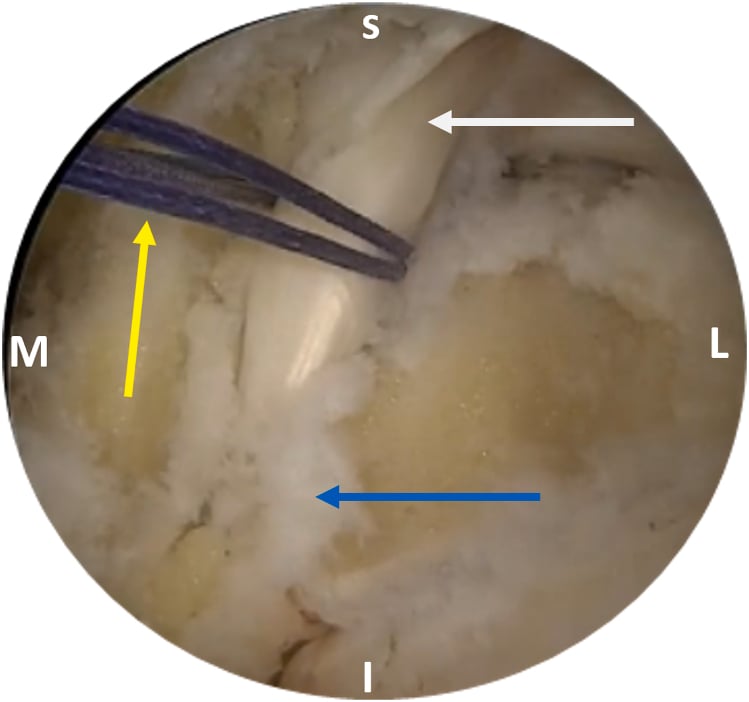
- Comparative series of the first 20 consecutive open versus first 19 consecutive endoscopic operations for posterior ankle impingement syndrome with FHL tendinopathy in dancers, all by one surgeon. Good/excellent results were 90% (18/20) in the open group and 79% (15/19) in the endoscopic group. Both groups returned to barre at a median of 8 weeks. Early postoperative morbidity (haematoma, inflammatory response, deep scar) was less favourable in the endoscopic group. The authors note the small retrospective groups preclude firm statistical conclusions.
Complications
Intraoperative. The risks to name when consenting:
- Neurovascular injury - tibial nerve or posterior tibial vessels (1-2%)
- Medial calcaneal nerve injury - heel numbness
- FHL laceration during the release - repair immediately
- Incomplete release - inadequate decompression and persistent symptoms
- Excessive bone removal - over-aggressive os trigonum excision destabilising the ankle
Early. Infection, haematoma, venous thromboembolism and sural nerve injury:
- incidence
- 2-3% open surgery, less than 1% endoscopic
- management
- Oral antibiotics for superficial, I&D for deep infection
- prevention
- Perioperative antibiotics, sterile technique, careful handling
- incidence
- 3-5%, higher without tourniquet
- management
- Observation if small, evacuation if large or expanding
- prevention
- Meticulous haemostasis, compressive dressing, elevation
- incidence
- Less than 1% with standard prophylaxis
- management
- Anticoagulation per protocol, may require admission
- prevention
- Early mobilisation, chemical prophylaxis if high risk
- incidence
- 1-2% with posterolateral portal
- management
- Observation, most resolve within 3-6 months
- prevention
- Careful portal placement, avoid excessive dissection
Late.
- Persistent pain in 5-10%, which may indicate incomplete decompression or adhesions
- Recurrent stenosis - rare if the sheath is not repaired; may need revision
- Adhesions - loss of excursion requiring repeat release
- FHL weakness - typically improves with rehabilitation, but persists in severe tendinosis
- Stiffness of the ankle or toe from prolonged immobilisation
- Complex regional pain syndrome - rare (less than 1%) but devastating
- Keloid or hypertrophic scar - more common after the open approach
When surgery fails. Repeat the MRI to judge whether the decompression was adequate, use dynamic ultrasound for persistent triggering, reconsider the diagnosis (tarsal tunnel syndrome, posterior impingement), and assess compliance with rehabilitation and its technique. Revision is indicated for:
- Incomplete decompression confirmed on imaging
- Recurrent stenosis with objective evidence
- Persistent triggering from adhesions
- New or missed pathology: an os trigonum, or Zone 2 or 3 stenosis
The revision. A more extensive release of all three zones, thorough debridement of scar and adhesions, attention to any concurrent pathology, and FHL-to-FDL transfer if the tendon is severely damaged. Rehabilitation afterwards is extended.
Corte-Real, Moreira and Guerra-Pinto - Arthroscopic FHL Release
- Series of 27 patients undergoing arthroscopic release for FHL tenosynovitis (mean age 34, mean follow-up 32 months). Notably, none were professional athletes or ballet dancers and all related onset to an ankle sprain - challenging the dogma that FHL tenosynovitis is exclusive to dancers/overuse. Mean postoperative AOFAS score was 89 with 70% excellent/good results; 81% returned to the same work/sport level and 89% would undergo the procedure again. Complication rate was 18% (5 patients) with a 4% (1 patient) reoperation rate.
Return to Sport and Outcomes
After non-operative care. No cited study on this page quantifies the success of conservative treatment, the return to sport after it, or recurrence. The surgical series establish only that operated patients had all failed it: Carreira required a minimum of 3 months of non-operative treatment before surgery, so a trial of at least three months is the standard the literature assumes rather than one it has tested. Significant symptom reduction takes 3-6 months, and the consistent clinical observation is that recurrence follows failure to modify the provoking technique or training load.
After surgery. Good-to-excellent results are reported in roughly 70-90% of appropriately selected patients after open or endoscopic decompression: Hamilton 30 of 41 dancer ankles, Corte-Real 70%, and Rietveld 90% open against 79% endoscopic. Barre work in dancers often resumes at around 8 weeks (Rietveld). After endoscopic surgery for osseous posterior impingement, Ling reported return to previous sport in 49 of 52 at a mean of 5.8 months. A minority have ongoing pain requiring revision or continued conservative care, particularly with missed multi-zone disease.
By sport. Typical timelines and rates of return:
- nonOperative
- 4-6 months to full performance, technique modification essential
- openSurgery
- 6-9 months to en pointe, 9-12 months to full performance
- endoscopic
- 4-6 months to en pointe, 6-9 months to full performance
- prognosis
- In the landmark operative series, 28 of 34 ankles in professional dancers had a good or excellent result - better than the amateur group; the challenge is the 6-12 month timeline, not a worse prognosis
- nonOperative
- 3-4 months to full training volume
- openSurgery
- 4-6 months to competitive racing
- endoscopic
- 3-4 months to competitive racing
- prognosis
- 85% return to pre-injury level
- nonOperative
- 3-4 months to full team training
- openSurgery
- 4-5 months to competitive play
- endoscopic
- 3-4 months to competitive play
- prognosis
- 80% return to pre-injury level
- nonOperative
- 2-3 months to return to activity
- openSurgery
- 3-4 months to full activity
- endoscopic
- 2-3 months to full activity
- prognosis
- Greater than 90% return to desired activity level
Prognostic factors. Better outcomes go with:
- Symptom duration less than 6 months
- No triggering or mechanical symptoms
- Good compliance with rehabilitation
- No associated pathology (os trigonum, tarsal tunnel)
Worse outcomes go with:
- Symptom duration greater than 12 months
- Triggering, indicating nodular thickening
- Multiple zones of involvement
- Previous failed surgery
- Worker's compensation or litigation
Professional status is not on either list. In Hamilton's series professional dancers did better than amateurs (ballet row above); what they face is the longer timeline back to en pointe.
Ling and Walsh - Medium-to-Long-Term Endoscopic Outcomes
- Retrospective case series of 52 patients undergoing 2-portal hindfoot endoscopy for osseous lesions causing posterior ankle impingement syndrome, median follow-up 4.8 years. 49 of 52 (94%) returned to their previous sport/physical activity at a mean of 5.8 months. Mean pain during exercise fell from 7.5 to 0.9, and the Short-Form Revised Foot Function Index improved from 84.4 to 6.7. There were no postoperative infections or other major complications.
van Dijk - Two-Portal Hindfoot Endoscopy (Technique)
- Description and review of the now-standard 2-portal hindfoot endoscopy technique performed prone. Recognised indications include FHL release, os trigonum removal, posterior tibial tenosynovectomy, peroneal tendon work, retrocalcaneal bursitis and Achilles peritendinopathy. The author argues the endoscopic approach offers less morbidity, reduced postoperative pain, outpatient treatment and functional postoperative management compared with open surgery.
Carreira et al. - Arthroscopic Posterior Impingement, ROM Outcomes
- Prospective evaluation of 20 patients (19 competitive athletes) treated with posterior ankle arthroscopy for posterior impingement, mean follow-up 38 months. VAS pain and AOFAS hindfoot scores improved significantly, while Tegner activity scores were maintained, and affected-side range of motion reached statistical similarity to the unaffected side. All 3 professional athletes returned to their previous professional level; 15% reported postoperative neuritis.
Guidelines, Registries and Global Practice
FHL tendinitis is a soft-tissue overuse condition, so it is governed by society consensus and performing-arts/sports-medicine guidance rather than implant registries. Practice converges internationally but resource availability shapes the diagnostic and surgical pathway.
Global Epidemiology
- Concentrated in dancers (especially classical ballet en pointe), running/jumping athletes, gymnasts and kicking-sport athletes worldwide; uncommon in the sedentary general population.
- Os trigonum (a key predisposing factor) is present in approximately 10-25% of the population across studies, with no strong geographic variation.
- Increasingly recognised in non-dancers after ankle sprain or occupational overuse, broadening the at-risk demographic.
Side-by-Side Guidance
- emphasis
- Exhaustive non-operative trial; surgery for refractory cases
- surgicalStance
- Open or arthroscopic decompression both accepted; technique by pathology
- imaging
- MRI for diagnostic doubt and pre-operative planning
- emphasis
- Activity modification and physiotherapy first-line; cautious injection use
- surgicalStance
- Hindfoot endoscopy or open release in specialist foot-ankle units
- imaging
- Ultrasound (dynamic) widely used first; MRI to plan surgery
- emphasis
- Endoscopic posterior ankle techniques strongly developed (van Dijk school)
- surgicalStance
- 2-portal hindfoot endoscopy a reference technique for Zone 1 / os trigonum
- imaging
- MRI and dynamic ultrasound; CT for bony impingement
- emphasis
- Load management, technique correction (en pointe), early return to barre
- surgicalStance
- Combined FHL release and posterior impingement decompression
- imaging
- Clinical diagnosis prioritised; imaging confirmatory
Registry and Resource Notes
- No dedicated registry: Unlike arthroplasty, there is no implant/procedure registry for FHL surgery; evidence is Level III-IV case series and a landmark retrospective cohort (Hamilton).
- High-resource settings: Ready access to MRI, dynamic ultrasound and hindfoot endoscopy enables minimally-invasive Zone 1 surgery and rapid return to sport.
- Limited-resource settings: Diagnosis rests on clinical examination and radiographs; open release through a posteromedial approach remains the dependable, equipment-light option and reaches all three zones.
Controversies and Areas of Uncertainty
Open or endoscopic. The only direct comparison is set out under Endoscopic Release. Technique should follow the pathology (the zone, any tendon tear) and the surgeon's experience rather than a blanket assumption that endoscopy is superior.
Corticosteroid injection. It may give short-term relief but carries a genuine risk of tendon rupture, and no high-quality trial defines a safe frequency. Consensus practice is sparing, ultrasound-guided peritendinous injection, never intratendinous.
Os trigonum: excise or leave? When an os trigonum coexists with FHL tendinopathy and posterior impingement, most evidence supports addressing both. Whether an incidentally imaged, asymptomatic os trigonum should be excised at the time of FHL release is not standardised.
Biologics. PRP and autologous blood injection are alternatives to steroid with limited evidence. For PRP in FHL tendinopathy specifically, the evidence is sparse and extrapolated from other tendinopathies, and it remains investigational rather than standard care.
Clinical Decision Scenarios
Practise clinical reasoning and management decisions out loud
“A 24-year-old professional ballet dancer presents with 6 months of progressive posteromedial ankle pain. She describes pain when en pointe that has worsened despite 3 months of physiotherapy. She occasionally feels a catching sensation when flexing her great toe. She has a major performance in 8 weeks. Examination reveals tenderness posteromedial to the ankle, positive FHL stretch test, and occasional triggering with toe flexion. MRI shows tendinosis of the FHL with synovitis in the fibro-osseous tunnel and a small os trigonum.”
“A 32-year-old distance runner underwent endoscopic FHL release 9 months ago for zone 1 tendinitis. He had initial improvement for 3 months but now has recurrent posterior ankle pain and triggering. He is frustrated as he was told surgery would cure the problem. Examination shows posteromedial ankle tenderness and triggering with great toe flexion. Repeat MRI shows adequate decompression of zone 1 but demonstrates zone 2 stenosis at the sustentaculum tali with tendon thickening and adhesions.”
“A 28-year-old recreational runner presents with 4 months of posteromedial ankle pain that is worse on push-off and climbing stairs. There is no history of an acute injury. On examination there is tenderness posterior to the medial malleolus and pain reproduced by passive ankle dorsiflexion combined with great-toe extension. Active resisted hallux flexion is mildly painful but full strength. How would you approach this patient?”
Must-Know Anatomy
- FHL origin: posterior fibula middle two-thirds
- Three zones: (1) talar tunnel, (2) sustentaculum, (3) knot of Henry
- Greatest tendon excursion in foot: 3 cm
- Myotendinous junction at ankle level (unique among flexors)
- Neurovascular bundle anterior to FHL (tibial nerve, PT vessels)
Classic Presentation
- Professional ballet dancer or distance runner
- Posteromedial ankle pain with push-off activities
- Triggering with great toe flexion-extension
- Positive FHL stretch test (dorsiflexion + hallux extension)
- May have pseudohallux rigidus from adhesions
Key Investigations
- Clinical diagnosis primarily
- XR: lateral ankle for os trigonum (10-25% prevalence)
- Ultrasound: dynamic assessment shows triggering
- MRI: gold standard pre-op - shows tendinosis, synovitis, zones involved
- Os trigonum bone edema suggests concurrent impingement syndrome
Management Algorithm
- Conservative first: 3-6 months activity modification, physio, NSAIDs
- Eccentric strengthening proven effective in tendinopathy
- Steroid injection controversial - rupture risk, max 1-2 injections
- Surgery if failed conservative: endoscopic (zone 1) vs open (multi-zone)
- FHL to FDL transfer salvage for irreparable tears greater than 50%
Surgical Pearls
- Open approach: posteromedial, release all involved zones, DO NOT repair sheath
- Endoscopic: prone position, posterolateral and posteromedial portals
- Protect neurovascular bundle throughout procedure
- Excise os trigonum if symptomatic impingement coexists
- Post-op: early ROM to prevent adhesions, 4-6 months to en pointe work
Viva Traps
- Don't promise quick return to sport - ballet dancers need 6-9 months minimum
- Recognize endoscopic limitations for zone 2/3 pathology
- Multiple steroid injections contraindicated - rupture risk
- Do not claim professionals fare worse - the landmark series found good or excellent results in 28 of 34 professional ankles versus 2 of 6 amateur ankles (small amateur subgroup)
- Failed surgery: assess zone adequacy on MRI, may need open revision
Critical Numbers
- Conservative outcome: not quantified in any cited source; at least 3 months is the trial period the surgical series assume
- Surgical good/excellent: Hamilton 30 of 41 dancer ankles, Rietveld 90% open vs 79% endoscopic, Corte-Real 70% in a non-dancer compensation cohort
- The professional/amateur gap: Hamilton, good or excellent in 28 of 34 professional ankles vs only 2 of 6 amateur
- Return to sport: Ling, 49 of 52 at a mean of 5.8 months after endoscopy for osseous impingement
- Complications are NOT lower after endoscopy in the cited series: Corte-Real 18% after arthroscopic release, Carreira 15% neuritis, and Rietveld found early morbidity worse in the endoscopic arm
Evidence Base
The foundation is Hamilton's landmark 1996 cohort defining dancer's posterior ankle pain and the coexistence of FHL tenosynovitis with posterior impingement. Surgical evidence is predominantly Level III-IV case series: Corte-Real (arthroscopic release, non-dancers), Carreira and Ling (endoscopic posterior impingement outcomes and return to sport), and Rietveld (the only direct open-vs-endoscopic comparison in dancers). The two-portal hindfoot endoscopy technique (van Dijk) underpins the modern minimally-invasive approach. There are no randomised controlled trials specific to FHL tendinitis, which is itself an examinable point: recommendations rest on consistent observational data and expert consensus rather than high-level trial evidence.