The Muscle That Owns the Sesamoids
- Two heads arising from the plantar cuboid, the adjacent lateral cuneiform and a prolongation of the tibialis posterior tendon; each head inserts on its own side of the base of the hallux proximal phalanx.
- EACH TENDON CONTAINS A SESAMOID: the medial head carries the TIBIAL (medial) sesamoid, the lateral head the FIBULAR (lateral) sesamoid. The flexor hallucis longus tendon runs BETWEEN them in the intersesamoid groove.
- Innervated by the MEDIAL plantar nerve (S1, S2) - unlike adductor hallucis, which is a LATERAL plantar nerve muscle.
- The sesamoid complex (both sesamoids, the intersesamoid ligament, the plantar plate, the FHB heads and the abductor and adductor insertions) must glide roughly 1 cm distally as the hallux dorsiflexes - loss of that excursion is the signature of a plantar plate rupture (turf toe).
- The lateral head of FHB fuses with adductor hallucis to form the CONJOINED TENDON on the fibular sesamoid - which is why a bunion lateral release inevitably weakens the lateral head.
- “The sesamoids act as the patellae of the great toe: they elevate the FHB tendons off the metatarsal head, lengthening the plantarflexion moment arm and protecting the flexor hallucis longus.
- “The tibial sesamoid is larger, more medial, bears more load and is bipartite far more often; the fibular sesamoid is smaller and a divided fibular sesamoid should make you suspect a fracture.
- “A bipartite sesamoid has smooth, rounded, corticated edges and the fragments together are LARGER than a normal whole sesamoid; a fracture has sharp, non-corticated, matching edges and no increase in overall size.
- “In hallux rigidus the dorsal osteophyte is the pain generator but the plantar sesamoid complex is the stiffness generator - cheilectomy without mobilising adherent sesamoids under-delivers dorsiflexion.
A complete plantar plate rupture treated as a sprain.
- The Grade 3 injury is a complete disruption of the plantar plate and sesamoid complex, often with sesamoid diastasis or a proximally migrated sesamoid.
- Missed injuries produce progressive hallux valgus or varus, a cock-up hallux, loss of push-off and eventual metatarsophalangeal arthrosis.
- The diagnostic test is comparing sesamoid excursion on a dorsiflexion lateral radiograph with the uninjured side.
Never remove both.
- Excising the tibial and fibular sesamoids destroys the entire FHB insertion and the plantar plate anchor, producing a cock-up hallux (hallux extensus) with loss of push-off.
- Excising one alone unbalances the transverse plane: tibial sesamoidectomy predisposes to hallux valgus, fibular sesamoidectomy to hallux varus.
- Preserve, repair or reconstruct in preference to excision wherever possible.
The nerve of a medial sesamoid approach.
- The medial plantar digital nerve to the hallux runs just plantar and medial to the tibial sesamoid, immediately deep to the medial incision.
- Injury gives a painful neuroma or a numb medial hallux border on a weight-bearing surface, which is far more troublesome than the original complaint.
- Identify and retract it before entering the sheath.
Dorsal bone alone is not the whole operation.
- Resect roughly the dorsal 25 to 30% of the metatarsal head; less and the impingement persists, more and the joint becomes unstable.
- Also release the adherent plantar sesamoid complex and any capsular contracture, then confirm at least 70 to 90 degrees of passive dorsiflexion on the table.
- Persistent stiffness after an adequate bony resection usually means the plantar structures were not mobilised.
PAIRSContents of the Sesamoid Complex
Hook:Every structure that fails in a turf toe is in this list - the plate, the plate's dynamic muscles, and the ligaments that hold the sesamoids.
First Ah Fab OneMedial versus Lateral Plantar Nerve Muscles
Hook:Four muscles only from the medial plantar nerve (S1, S2). Flexor hallucis brevis is one of them; adductor hallucis is NOT.
Overview
Flexor hallucis brevis is a two-headed intrinsic muscle of the third plantar layer running along the plantar surface of the first metatarsal to the base of the hallux proximal phalanx. Its defining feature is that each of its two tendons contains a sesamoid bone, and the flexor hallucis longus tendon passes between them in the intersesamoid groove.
Because of this, flexor hallucis brevis is not simply a hallux flexor. It is the dynamic component of the plantar sesamoid complex, the load-bearing platform beneath the first metatarsal head. Almost every problem on the plantar side of the first metatarsophalangeal joint - sesamoiditis, sesamoid stress fracture, osteonecrosis, turf toe, and the plantar contracture of hallux rigidus - is a disorder of this muscle and the bones inside it.
The mechanical concept the examiner will push on.
- The hallux sesamoids sit in the FHB tendons exactly as the patella sits in the quadriceps tendon. They elevate the tendon away from the joint centre, lengthening the plantarflexion moment arm of flexor hallucis brevis and increasing its mechanical efficiency in terminal stance.
- They protect the flexor hallucis longus tendon, which runs in the intersesamoid groove between them, from direct compression against the metatarsal head.
- They absorb and distribute load, carrying a substantial share of the force under the first metatarsal head. Load through the sesamoids rises steeply as the hallux dorsiflexes in terminal stance, which is why sesamoid pain is always worst at push-off and on an incline or in a heeled shoe.
- They form part of the windlass mechanism. As the hallux dorsiflexes in terminal stance, the plantar aponeurosis tightens around the metatarsal head, the arch rises, the sesamoid complex is drawn distally, and the foot converts from a shock absorber into a rigid lever.
- They are enveloped and articular: each sesamoid has a cartilage-covered dorsal surface articulating with its groove on the plantar metatarsal head, separated by the crista (the median ridge). They are therefore genuine articular structures and can develop degenerative change, osteonecrosis and osteochondral injury.
The sesamoid complex is not fixed to the metatarsal. It is anchored distally to the proximal phalanx through the plantar plate and suspended proximally to the metatarsal neck by the metatarsosesamoid (suspensory) ligaments.
- As the hallux dorsiflexes from neutral to full extension, the whole complex is drawn distally by roughly 1 cm as the plantar plate pulls it forward and it rolls onto the plantar metatarsal head. (That approximate figure is conventional teaching; the number below is the measured one.)
- This distal excursion is reduced or abolished by a plantar plate rupture, because the distal anchor that pulls the complex forward is gone. On a dorsiflexion lateral radiograph the sesamoids fail to migrate distally, and may sit relatively proximal compared with the uninjured foot.
- Put a number on it. Waldrop's cadaveric cutting study measured the distance from the distal margin of the sesamoids to the proximal margin of the proximal phalanx on 45-degree dorsiflexion stress fluoroscopy, dividing the four distal plantar plate ligaments in sequence. The displacement rose with each ligament cut, and an increase of 3 mm or more from the intact state was significant and predicted that at least 3 of the 4 ligaments were torn. Clinically the intact state is the uninjured foot, so stress both sides and compare.
- It is also abolished by adhesion of the complex to the plantar metatarsal head in advanced hallux rigidus, which is why an adequate cheilectomy can still leave a stiff joint if the plantar structures are not mobilised.
- One concept, two very different diseases: no excursion because the tether is torn (turf toe) versus no excursion because the complex is stuck (hallux rigidus). Always compare with the uninjured side.
Attachments, Innervation and Relations
Origin
- Plantar surface of the cuboid (its medial part) and the adjacent plantar surface of the lateral cuneiform.
- A prolongation of the tibialis posterior tendon as it inserts into the plantar midfoot - the reason FHB is functionally coupled to the tibialis posterior, and the anatomical basis for a small contribution to medial arch support.
- Deep fibres from the plantar aspect of the third cuneiform and the long plantar ligament.
- The origin is Y-shaped and tendinous; it lies deep in the midfoot, superficial to the peroneus longus tendon in its groove.
Course and Division
- The muscle runs distally along the plantar surface of the first metatarsal and divides into a medial and a lateral head at about the level of the metatarsal neck.
- The flexor hallucis longus tendon runs forward between the two heads, in the intersesamoid groove, roofed by the intersesamoid ligament.
Insertion
- Medial head: into the medial (plantar-medial) side of the base of the proximal phalanx of the hallux, its tendon containing the TIBIAL (medial) sesamoid. Blends with the insertion of abductor hallucis.
- Lateral head: into the lateral (plantar-lateral) side of the base of the proximal phalanx, its tendon containing the FIBULAR (lateral) sesamoid. Blends with adductor hallucis to form the CONJOINED TENDON.
- Both heads are continuous with the plantar plate of the first metatarsophalangeal joint, so FHB and the plate are functionally inseparable.
The Sesamoids Themselves
- Tibial (medial) sesamoid
- Larger, more oval
- Fibular (lateral) sesamoid
- Smaller, more rounded
- Tibial (medial) sesamoid
- Beneath the medial metatarsal head groove, more distal and more directly under the weight-bearing axis
- Fibular (lateral) sesamoid
- Beneath the lateral groove, closer to the second metatarsal
- Tibial (medial) sesamoid
- Bears the greater share of load - hence the commoner site of sesamoiditis, stress fracture and osteonecrosis
- Fibular (lateral) sesamoid
- Less loaded, but loaded heavily in push-off with the hallux dorsiflexed
- Tibial (medial) sesamoid
- The usual site of a bipartite sesamoid, and frequently bilateral - though less common than often taught: a bipartite hallux sesamoid was seen in only 2.7% of 984 radiographed subjects (Coskun)
- Fibular (lateral) sesamoid
- Distinctly uncommon - a divided fibular sesamoid should raise suspicion of fracture
- Tibial (medial) sesamoid
- Medial head FHB plus abductor hallucis
- Fibular (lateral) sesamoid
- Lateral head FHB plus adductor hallucis (conjoined tendon)
- Tibial (medial) sesamoid
- Predisposes to HALLUX VALGUS - though in Saxena's 16 tibial excisions this occurred in 1 patient, so counsel it as a real risk rather than an expectation
- Fibular (lateral) sesamoid
- Predisposes to HALLUX VARUS - 1 of 10 fibular excisions, and the fibular group also carried the painful-scar and neuroma-like complications in that series
- Ossification begins between about 7 and 10 years of age, from multiple centres, which is one reason bipartition is common. A sesamoid that appears divided in a child may simply be incompletely coalesced.
Action and Biomechanics
Actions
- Plantarflexion of the first metatarsophalangeal joint - its only direct action, and it is the primary metatarsophalangeal plantarflexor. Flexor hallucis longus flexes the interphalangeal joint principally and the metatarsophalangeal joint secondarily.
- Depression and stabilisation of the first metatarsal head in terminal stance, keeping the head loaded and preventing dorsal escape of the first ray.
- Contribution to the windlass mechanism and arch support, through its continuity with the plantar plate, the plantar aponeurosis and (through its origin) the tibialis posterior.
- Medial arch support, indirectly, through the tibialis posterior prolongation at its origin.
- It does not flex the interphalangeal joint - that is flexor hallucis longus. This distinction is the basis of the bedside examination.
Moment Arms and Load
- The sesamoids displace the FHB tendons plantarward by their own thickness, lengthening the plantarflexion moment arm at the metatarsophalangeal joint. Remove a sesamoid and the moment arm shortens.
- Load beneath the sesamoids increases sharply as the hallux dorsiflexes in terminal stance, and is further increased by heeled footwear, a plantarflexed first ray, cavus alignment, and a distally migrated fat pad.
- The sesamoids also share load with the metatarsal head, so sesamoidectomy transfers load to the plantar metatarsal head and can produce transfer metatarsalgia and plantar keratosis.
Length-Tension and What Happens When It Fails
- Mechanism
- Forced hyperdorsiflexion of the MTP joint on a fixed, plantarflexed foot
- Resulting deformity or deficit
- Loss of sesamoid excursion, loss of push-off, progressive hallux malalignment, cock-up hallux, later arthrosis
- Mechanism
- Loss of the medial head insertion and the medial plantar anchor
- Resulting deformity or deficit
- Hallux VALGUS tendency; transfer load to the plantar metatarsal head
- Mechanism
- Loss of the lateral head and conjoined tendon anchor
- Resulting deformity or deficit
- Hallux VARUS tendency; the classic hallux varus of the original McBride
- Mechanism
- Total loss of the FHB insertion and the plantar plate anchor
- Resulting deformity or deficit
- COCK-UP HALLUX (hallux extensus) with no push-off - the deformity to avoid at all costs
- Mechanism
- Denervation of FHB, abductor hallucis, FDB and first lumbrical
- Resulting deformity or deficit
- Weak MTP plantarflexion, hallux valgus tendency (adductor spared), medial plantar sensory loss
- Mechanism
- Sesamoid complex scars to the plantar metatarsal head
- Resulting deformity or deficit
- Loss of sesamoid excursion and therefore of dorsiflexion, independent of the dorsal osteophyte
- Mechanism
- Iatrogenic during sesamoid surgery, or penetrating injury
- Resulting deformity or deficit
- Loss of hallux INTERPHALANGEAL flexion with preserved MTP flexion
Synergists and Antagonists
- Muscle
- Flexor hallucis brevis
- Nerve
- Medial plantar (S1, S2)
- Comment
- Acts through the sesamoids
- Muscle
- Flexor hallucis longus
- Nerve
- Tibial (S1, S2, S3)
- Comment
- Runs between the two FHB heads in the intersesamoid groove
- Muscle
- Abductor hallucis
- Nerve
- Medial plantar (S1, S2)
- Comment
- Shares the tibial sesamoid insertion
- Muscle
- Adductor hallucis
- Nerve
- Deep branch lateral plantar (S2, S3)
- Comment
- Fuses with the lateral head onto the fibular sesamoid
- Muscle
- Extensor hallucis longus and brevis
- Nerve
- Deep peroneal (L5)
- Comment
- Unopposed after loss of both sesamoids - cock-up hallux
Surface Anatomy and Examination
Palpation and Position
- Sit the patient with the ankle in neutral and the hallux relaxed. The sesamoids are palpable through the plantar-medial fat pad beneath the first metatarsal head, and become far easier to localise if the hallux is passively dorsiflexed, which draws them distally and tightens the complex over them.
- Palpate each sesamoid separately. The tibial sesamoid lies beneath the medial third of the metatarsal head; the fibular sesamoid beneath the lateral third, slightly more proximal. Distinguishing which one is tender is a genuine clinical skill and changes the diagnosis.
- The intersesamoid groove and the flexor hallucis longus tendon within it can be palpated between them; crepitus or a painful snap on passive hallux flexion and extension suggests FHL tenosynovitis.
- The medial head of FHB can be felt as a firm longitudinal band deep to abductor hallucis along the plantar-medial first metatarsal.
Isolating Flexor Hallucis Brevis from Flexor Hallucis Longus
This is the examination point that matters, and it mirrors the FDP/FDS logic in the hand.
- How to perform
- Hold the hallux INTERPHALANGEAL joint in full extension to neutralise flexor hallucis longus, then ask the patient to plantarflex the metatarsophalangeal joint against your resistance
- Positive finding
- Weak or absent resisted MTP plantarflexion with the IP joint blocked
- What it means
- Flexor hallucis brevis weakness - medial plantar nerve lesion, plantar plate or sesamoid complex disruption, or post-sesamoidectomy insufficiency
- False positives / pitfalls
- Failure to block the IP joint allows FHL to substitute and masks the deficit; pain inhibition mimics weakness
- How to perform
- Stabilise the proximal phalanx and ask for active flexion of the interphalangeal joint only
- Positive finding
- Weak or absent IP flexion with intact MTP flexion
- What it means
- Flexor hallucis longus lesion - laceration in the intersesamoid groove, rupture, or a proximal tibial nerve problem
- False positives / pitfalls
- Tenodesis effect from ankle position; a stiff IP joint mimics tendon loss
- How to perform
- Stabilise the metatarsal, grasp the proximal phalanx and translate it dorsally relative to the metatarsal head, comparing with the other foot
- Positive finding
- Increased dorsal translation with pain and a soft end-point
- What it means
- Plantar plate and sesamoid complex insufficiency - a positive test in the turf toe setting suggests a Grade 3 injury
- False positives / pitfalls
- Generalised laxity; guarding in an acutely swollen joint; performing it without comparing sides
- How to perform
- Passively dorsiflex the hallux while feeling the sesamoids under the metatarsal head
- Positive finding
- Sesamoids fail to glide distally, or a hard block to dorsiflexion
- What it means
- Loss of sesamoid excursion - a torn plate (turf toe) or an adherent complex (hallux rigidus)
- False positives / pitfalls
- Not comparing with the uninjured side; pain-limited range interpreted as a mechanical block
- How to perform
- Compress the joint axially and rotate through a mid-range arc, avoiding terminal dorsiflexion
- Positive finding
- Pain in mid-range with compression
- What it means
- Intra-articular degeneration - suggests established hallux rigidus rather than isolated dorsal impingement
- False positives / pitfalls
- Pain only at terminal dorsiflexion indicates dorsal impingement, not global arthrosis
- How to perform
- Palpate each sesamoid individually, then ask the patient to rise onto the toes
- Positive finding
- Focal tenderness over one sesamoid, reproduced by push-off
- What it means
- Sesamoiditis, stress fracture or osteonecrosis of that sesamoid
- False positives / pitfalls
- Plantar fat pad atrophy and a plantar keratosis give diffuse rather than focal tenderness
Imaging Views to Request
- Weight-bearing dorsoplantar and lateral radiographs of the foot - alignment, joint space, dorsal osteophyte, sesamoid position.
- Axial (tangential) sesamoid view - the single most useful sesamoid projection: shows each sesamoid in its groove, the crista, degenerative change, fragmentation and position.
- Dorsiflexion (forced dorsiflexion) lateral view, both feet - assesses sesamoid excursion in suspected plantar plate injury; the injured side fails to translate distally.
- MRI - the investigation of choice for turf toe (plate discontinuity, sesamoid marrow oedema, FHL pathology), sesamoid stress fracture and osteonecrosis.
- CT - fracture configuration and fragment position; helpful in distinguishing a fracture line from a bipartite synchondrosis when MRI is equivocal.
- Bone scan or SPECT - occasionally useful in a stress reaction with normal radiographs, though MRI has largely superseded it.
Complications
- Mechanism
- Excision of both sesamoids, or an unrepaired grade 3 turf toe
- Avoidance
- Never excise both; repair the plate; reconstruct with a tendon graft or fuse if both are unsalvageable
- Mechanism
- Loss of the medial head insertion and medial plantar restraint
- Avoidance
- Shell the sesamoid out and repair the tendon defect side to side; consider shaving instead of excision
- Mechanism
- Loss of the conjoined tendon anchor
- Avoidance
- Preserve the sesamoid where possible; repair the conjoined tendon; avoid combining with an aggressive lateral release
- Mechanism
- Incision over weight-bearing plantar skin, or injury to the medial plantar digital nerve
- Avoidance
- Medial mid-axial or dorsal approaches; identify and retract the nerve before deeper dissection
- Mechanism
- Blind division of the intersesamoid ligament
- Avoidance
- Divide it under direct vision only if needed; the tendon lies 2 to 3 mm beneath it
- Mechanism
- Devascularisation of a proximal-pedicle-dependent sesamoid
- Avoidance
- Preserve tendon and plate attachments; do not strip the proximal pole; counsel about slow union
- Mechanism
- Load transferred to the plantar metatarsal head after sesamoidectomy
- Avoidance
- Offload with an orthosis; preserve as much of the sesamoid platform as possible
- Mechanism
- Under-resection, unmobilised plantar sesamoid complex, or unrecognised grade 4 disease
- Avoidance
- Resect 25 to 30% of the head, free the sesamoid complex, confirm 70 to 90 degrees on the table, mobilise early
- Mechanism
- Dorsal or dorsomedial approach to the joint
- Avoidance
- Identify and protect the nerve at the start; a dissatisfied numb hallux after a good cheilectomy is usually this
- Mechanism
- Untreated articular impaction or chronic instability
- Avoidance
- Assess the joint surface on MRI; repair unstable plates; counsel about long-term risk
Clinical Relevance
1. Turf Toe (Plantar Plate and Sesamoid Complex Injury)
The archetypal injury of this muscle.
- Mechanism: forced hyperdorsiflexion of the first metatarsophalangeal joint with the ankle plantarflexed and the heel raised - classically an axial load applied to the heel with the forefoot fixed on a hard surface. Originally described on artificial turf with flexible shoes.
- Pathology: attenuation or rupture of the plantar plate and the sesamoid complex, potentially with sesamoid diastasis (separation of a bipartite sesamoid), a sesamoid fracture, a proximally migrated sesamoid, avulsion of the plate from the phalangeal base, and in severe injuries articular impaction of the dorsal metatarsal head.
- Variants: a medial injury (medial plate and abductor hallucis) leads to progressive hallux valgus; a lateral injury leads to hallux varus; a combined hyperdorsiflexion with valgus is the commonest pattern.
- Do not confuse it with "sand toe" - a forced hyperplantarflexion injury of the first metatarsophalangeal joint, seen in beach volleyball, which injures the dorsal capsule rather than the plantar plate.
- Complete plantar plate rupture with loss of sesamoid excursion
- Sesamoid diastasis or a proximally retracted sesamoid
- Sesamoid fracture with displacement
- Progressive hallux valgus or varus after injury
- Traumatic bunion, a vertical instability that fails to settle, or a retracted plate
- Loose body or osteochondral injury of the metatarsal head
- A high-demand athlete who cannot push off
- Grade I or II injury
- Preserved sesamoid excursion on comparative dorsiflexion lateral views
- No diastasis and no fracture
- Stable joint on vertical Lachman
- Improving symptoms and function through the first weeks
Surgical Relevance
Approaches to the Sesamoid Complex
Medial Approach to the Tibial Sesamoid
- Position: supine, ankle in neutral, hallux held in slight dorsiflexion to draw the sesamoid distally and make it palpable.
- Incision: a medial, mid-axial incision over the first metatarsophalangeal joint, deliberately dorsal to the weight-bearing plantar skin. Never place the incision directly over the plantar sesamoid pad.
- Order encountered: skin; then the medial plantar digital nerve to the hallux running plantar-medial - identify and retract it plantarward before going deeper; then the abductor hallucis insertion; then the medial capsule and the tibial sesamoid within the medial FHB tendon.
- Sesamoid shaving (partial excision): remove the plantar prominence only, preserving the articular dorsal surface and the tendon continuity. Lower morbidity than complete excision.
- Complete excision: shell the sesamoid out of the tendon without dividing the tendon transversely. Repair the resulting longitudinal tendon defect side to side to restore the medial head insertion and prevent progressive hallux valgus.
- Do not divide the intersesamoid ligament unnecessarily - the flexor hallucis longus tendon lies immediately beneath it.
Distances and Safe Zones
- Medial plantar digital nerve to the hallux: runs plantar-medial to the tibial sesamoid, typically within 5 mm of the plantar-medial corner of the sesamoid and immediately deep to the skin. Place the medial incision at the mid-axial line or slightly dorsal to it, never over plantar weight-bearing skin.
- Flexor hallucis longus in the intersesamoid groove: lies immediately deep to the intersesamoid ligament, between the sesamoids. Assume it is within 2 to 3 mm of any blade dividing that ligament.
- Dorsomedial cutaneous nerve to the hallux: crosses the dorsomedial aspect of the first metatarsophalangeal joint subcutaneously, within a few millimetres of the skin - the nerve at risk in every dorsal or dorsomedial approach and the commonest cause of a dissatisfied patient after a technically satisfactory cheilectomy.
- Dorsal digital branches of the deep peroneal nerve: subcutaneous in the dorsal first web space, within about 3 to 5 mm of the skin, if that route is used for the fibular sesamoid.
- Metatarsal head resection depth in cheilectomy: the dorsal 25 to 30%. Resecting beyond a third risks instability and dorsal subluxation.
- Sesamoid excursion: roughly 1 cm distally from neutral to full dorsiflexion. Reproduce it on the table after a turf toe repair, and compare with the uninjured side on imaging.
Excising both the tibial and fibular sesamoids removes the entire insertion of flexor hallucis brevis and the plantar anchor of the plate. The result is a cock-up hallux (hallux extensus) with no push-off, and it is essentially unsalvageable except by arthrodesis.
If both sesamoids are irreparably damaged - typically in severe trauma or sepsis - the honest options are first metatarsophalangeal arthrodesis or reconstruction of the plantar plate with a tendon graft (a flexor hallucis longus or abductor hallucis transfer has been described). Plan for that at the outset rather than discovering it afterwards.
The corollary applies to single sesamoidectomy: repair the tendon defect so the remaining FHB head still has a functional insertion, and warn the patient about the direction of the alignment risk - valgus after tibial, varus after fibular excision.
Sesamoids in Bunion and Arthrodesis Surgery
- Bunion lateral release: dividing the lateral metatarsosesamoid (suspensory) ligament and the conjoined tendon is necessary for the sesamoids to reduce beneath the metatarsal head, but it unavoidably weakens the lateral head of FHB. Preserving the fibular sesamoid itself is what prevents hallux varus.
- First metatarsophalangeal arthrodesis: the sesamoids do not need to be excised. They should be mobilised enough to allow joint preparation and positioning, and are usually left in situ; adherent sesamoids that block positioning may be shelled out of the tendon.
- First metatarsophalangeal replacement and interposition arthroplasty: the plantar complex must be preserved to retain any push-off function, which is one reason these procedures perform less predictably than arthrodesis in a high-demand foot.
Guidelines, Registries & Global Practice
Anatomical Variation Across Populations
- The two-headed pattern with two sesamoids is essentially constant. Absence of a hallux sesamoid is a genuine rarity and, when seen, should prompt a search for prior surgery or trauma.
- Bipartition is reported in a minority of feet, most often affecting the tibial sesamoid, and is frequently bilateral. Reported prevalence varies widely between series because definitions differ; the clinically useful points are the strong tibial predominance and the frequency of bilaterality.
- Multipartite (three or more part) sesamoids are described but uncommon.
- Reported prevalence of a lateral plantar nerve contribution to the lateral head varies between dissection series, consistent with its fusion with adductor hallucis.
- Hallux rigidus prevalence rises with age and is a common cause of forefoot disability in adults over 50; sesamoid disorders are disproportionately seen in dancers, runners, gymnasts and field-sport athletes rather than in the general population.
Side-by-Side Guidance and Practice Differences
- Position on FHB-related pathology
- Aggressive structural assessment of turf toe with comparative dorsiflexion lateral radiographs and MRI in the elite athlete; a relatively low threshold for repairing a complete plantar plate rupture to protect a playing career.
- Position on FHB-related pathology
- Similar diagnostic approach, with proportionally greater emphasis on structured non-operative rehabilitation and a higher threshold for operative repair outside elite sport.
- Position on FHB-related pathology
- Emphasises preservation of the plantar plate and the vascular attachments of the sesamoids, and repair rather than excision wherever technically possible.
- Position on FHB-related pathology
- Broad agreement that cheilectomy suits grades 1 to 2 and selected grade 3 joints with a pain-free mid-range, and that arthrodesis is the reliable operation for grade 4 - with continuing disagreement about the role of implant arthroplasty and interposition.
Areas of Genuine Uncertainty
- Operative versus non-operative Grade 3 turf toe: no randomised evidence exists and there never will be in elite sport. Practice is driven by expert series and the consequences of chronic instability.
- Sesamoid non-union: fixation, grafting or excision? Series are small and heterogeneous; the decision rests on fragment viability, size and the patient's demands.
- Partial (shaving) versus complete sesamoidectomy: partial excision preserves the tendon insertion and alignment but may not relieve pain if the whole bone is diseased.
- Return-to-play timing after turf toe repair: commonly quoted as 3 to 4 months, but based on small series and highly dependent on sport and position.
- First metatarsophalangeal implant arthroplasty and interposition versus arthrodesis: arthrodesis remains the benchmark for pain relief and durability; motion-preserving options continue to be studied without displacing it.
High- versus Limited-Resource Practice
- Well-resourced settings: MRI for turf toe and sesamoid pathology, comparative dynamic radiographs, custom carbon-plate orthoses, suture anchors for plate repair, and access to sports rehabilitation.
- Limited-resource settings: the diagnosis of turf toe and sesamoid disorders is fundamentally clinical and radiographic. Comparative forced dorsiflexion lateral radiographs of both feet cost nothing and answer the key question. A stiff-soled shoe, a simple sesamoid relief pad cut from felt, activity modification and time treat the great majority. First metatarsophalangeal arthrodesis is a reliable, implant-light definitive operation for end-stage hallux rigidus.
- Everywhere: footwear counselling, offloading and clear explanation that the sesamoids are load-bearing bones inside a tendon and therefore heal slowly.
MCQ Practice Points
Q: What is the nerve supply of flexor hallucis brevis? A: The MEDIAL plantar nerve (S1, S2). The medial plantar nerve supplies only four muscles: flexor digitorum brevis, abductor hallucis, flexor hallucis brevis and the first lumbrical.
Q: Which structures contain the hallux sesamoids? A: The two tendons of flexor hallucis brevis - the tibial sesamoid in the medial head, the fibular sesamoid in the lateral head, with the flexor hallucis longus tendon between them in the intersesamoid groove.
Q: How do you test flexor hallucis brevis in isolation? A: Hold the hallux interphalangeal joint in extension to neutralise flexor hallucis longus, then resist metatarsophalangeal plantarflexion. FHB does not flex the interphalangeal joint.
Q: How far do the sesamoids translate as the hallux dorsiflexes, and what abolishes it? A: Roughly 1 cm distally. It is abolished by a plantar plate rupture (the distal anchor is gone) or by adhesion of the complex in advanced hallux rigidus.
Q: What single measurement most reliably distinguishes a bipartite sesamoid from a fracture? A: The combined size of the fragments. A bipartite sesamoid's two fragments together are larger than a normal whole sesamoid; fracture fragments together are the same size. Margins are corticated and rounded in bipartition, sharp and matching in fracture.
Q: What deformity follows excision of the tibial sesamoid, and of the fibular sesamoid? A: Tibial excision predisposes to hallux VALGUS; fibular excision predisposes to hallux VARUS. Excising both produces a cock-up hallux (hallux extensus) with no push-off.
Q: What is the mechanism of a turf toe, and how does sand toe differ? A: Turf toe is forced hyperDORSIflexion of the first metatarsophalangeal joint, injuring the plantar plate and sesamoid complex. Sand toe is forced hyperPLANTARflexion, injuring the dorsal capsule.
Q: How much of the first metatarsal head is resected in a cheilectomy? A: The dorsal 25 to 30%. Confirm 70 to 90 degrees of passive dorsiflexion intraoperatively, and remember to mobilise the plantar sesamoid complex.
Q: What single clinical feature distinguishes Coughlin and Shurnas grade 3 from grade 4 hallux rigidus? A: Pain throughout the MID-RANGE of motion. Grade 3 has pain only at the extremes; grade 4 has mid-range pain and predicts failure of joint-preserving surgery.
Q: Why is osteonecrosis relatively common in hallux sesamoid injuries? A: The dominant blood supply is a proximal pedicle entering through the flexor hallucis brevis tendon and plantar plate, and in a proportion of individuals it is effectively the only supply. Displacement or dissection interrupting it causes non-union and osteonecrosis.
Exam Viva Scenarios
Practise clinical reasoning and management decisions out loud
“A 24-year-old professional footballer sustained a forced dorsiflexion injury of the great toe on a hard pitch three days ago. The first metatarsophalangeal joint is grossly swollen with plantar ecchymosis, he cannot bear weight through the forefoot, and passive dorsiflexion is very painful. How do you assess and grade this, and what would make you operate?”
“A 19-year-old dancer has six months of plantar pain under the great toe, worse on rising onto the toes. A dorsoplantar radiograph shows the tibial sesamoid divided into two pieces. Is this a fracture? How do you proceed and what do you offer?”
“A 58-year-old man had a cheilectomy for grade 2 hallux rigidus four months ago. Radiographs confirm the dorsal osteophyte and about 30% of the dorsal metatarsal head have been resected. He has only 15 degrees of dorsiflexion and pain at push-off. Explain why, and what you would do.”
Anatomy
- Layer 3 of the sole (with adductor hallucis, FDMB)
- Origin: plantar cuboid, lateral cuneiform, tibialis posterior prolongation
- Medial head to medial base of hallux P1 - contains TIBIAL sesamoid
- Lateral head to lateral base of hallux P1 - contains FIBULAR sesamoid
- FHL runs BETWEEN the heads in the intersesamoid groove
Neurovascular
- Nerve: MEDIAL plantar nerve (S1, S2)
- Medial plantar nerve muscles: FDB, abductor hallucis, FHB, 1st lumbrical
- Artery: medial plantar artery, 1st plantar metatarsal, deep plantar
- Sesamoid supply: proximal pedicle through tendon and plate - AVN risk
Sesamoid Complex
- Both sesamoids plus intersesamoid ligament
- Plantar plate distally, metatarsosesamoid ligaments proximally
- Abductor hallucis to tibial, adductor hallucis to fibular
- Excursion roughly 1 cm distally on hallux dorsiflexion
- Acts as the patella of the great toe - lengthens the flexor moment arm
Turf Toe
- Forced hyperDORSIflexion; sand toe is hyperPLANTARflexion
- Grade I stretch, II partial tear, III complete rupture
- Grade III: loss of sesamoid excursion, diastasis, positive vertical Lachman
- Comparative forced dorsiflexion lateral views of BOTH feet
- Repair the plate - never excise both sesamoids
Sesamoid Pearls
- Bipartite: corticated rounded edges, combined size LARGER, usually tibial, often bilateral
- Fracture: sharp non-corticated matching edges, normal combined size
- Tibial excision equals hallux valgus risk
- Fibular excision equals hallux varus risk
- Both excised equals cock-up hallux
Hallux Rigidus
- Coughlin and Shurnas grades 0 to 4
- Grade 4 defined by MID-RANGE pain - fuse it
- Cheilectomy: resect dorsal 25 to 30% of the head
- Mobilise the plantar sesamoid complex and confirm 70 to 90 degrees
- Protect the dorsomedial cutaneous nerve; mobilise early
Evidence Base
Turf Toe Injury
- Review of turf toe injury of the first metatarsophalangeal joint, with emphasis on long-term morbidity
- Long-term morbidity after a previous first metatarsophalangeal injury includes persistent pain with athletic activity and restricted range of motion
- Hallux valgus and early hallux rigidus are described as specific long-term sequelae
- In a review of 20 athletes with a previous turf toe injury followed for more than 5 years, there was a 50% incidence of PERSISTENT SYMPTOMS
- Concluded that turf toe is a significant athletic injury requiring treatment tailored to the severity of the injury
Rehabilitation Following Turf Toe Injury and Plantar Plate Repair
- Reviewed the evaluation, diagnosis and treatment of turf toe injury of the hallux metatarsophalangeal joint
- Emphasised that accurate diagnosis depends on understanding the anatomy of the plantar plate and sesamoid complex and on specific radiographic clues
- Set out the role of surgical repair of the plantar plate where it is indicated by the injury pattern
- Stressed that structured rehabilitation supervised by a physiotherapist or athletic trainer is critical to recovery regardless of the grade of injury
- Reported that with appropriate care athletes can return to their pre-injury level of participation
Radiographic Evaluation of Plantar Plate Injury - an In Vitro Biomechanical Study
- Twenty-four fresh-frozen foot and ankle specimens randomised into four cutting groups; the four main distal ligaments of the plantar plate were sectioned in sequence
- Lateral dorsiflexion stress fluoroscopy at 45 degrees was taken intact and after each cut, measuring the distance from the distal margin of the sesamoids to the proximal margin of the proximal phalanx
- Sesamoid-to-phalanx distance increased with each successive ligament divided - the displacement is graded, not all-or-none
- AN INCREASE OF 3 MM FROM THE INTACT STATE was significant and predictive of severe plantar plate injury, indicating at least 3 of the 4 ligaments had been torn
- This is the first quantitative radiographic grading scale for turf toe; assessment had previously been qualitative
Hallux Rigidus - Grading and Long-Term Results of Operative Treatment
- 110 of 114 patients treated over a 19-year period returned for review; 80 patients (93 feet) had a cheilectomy and 30 patients (34 feet) an arthrodesis
- Mean follow-up 9.6 years after cheilectomy and 6.7 years after arthrodesis
- A NEW FIVE-GRADE clinical and radiographic grading system was introduced and assessed
- Significant improvement in dorsiflexion and total motion after cheilectomy, and in pain and AOFAS scores in both groups
- 97% (107 of 110) had a good or excellent subjective result, and 92% (86 of 93) cheilectomies were successful for pain relief and function
- A good or excellent outcome did NOT correlate with the radiographic appearance of the joint at final follow-up
Dimensions and Arterial Vascular Supply of the Sesamoid Bones of the Human Hallux
- Gross anatomical dissection of the hallux sesamoids in 13 subjects of both sexes
- THE MAJORITY HAVE ONLY ONE ARTERY: a single vessel supplied 63 per cent of medial and 58 per cent of lateral sesamoids - single supply is the RULE, not the exception
- Two sesamoid arteries were found in 26 per cent of medial and 32 per cent of lateral sesamoids
- Three arteries were found in only 11 per cent of medial and 10 per cent of lateral sesamoids, and in left feet only
- The number of arteries corresponded to the size and robusticity of the sesamoid and to anthropometric measures, sex and footedness
Return to Activity After Sesamoidectomy in Athletically Active Individuals
- 26 sesamoidectomies in 24 patients (21 female), mean age 35.4 years, mean follow-up 86.4 months - 16 tibial and 10 fibular sesamoids excised
- Competitive athletes returned to activity at a mean of 7.5 weeks against 12.0 weeks for merely 'active' patients (p less than 0.02) - the fitter group returned sooner
- Complications were few but were CONCENTRATED IN THE FIBULAR GROUP: one hallux varus and two cases of painful scarring with neuroma-like symptoms, all after fibular excision
- One hallux valgus deformity followed tibial sesamoidectomy - so transverse-plane drift did occur, but in 1 of 16 rather than as a rule
The Mechanics of the Foot. II. The Plantar Aponeurosis and the Arch
- NOTE - no abstract is indexed for this 1954 paper; the following is bounded by its title and by the conventional attribution of the windlass concept to this series
- The second in Hicks' series on the mechanics of the foot, addressing the plantar aponeurosis and the arch
- The description of the plantar aponeurosis as a tie-beam whose tensioning by toe dorsiflexion raises the longitudinal arch is conventionally attributed to this work
- It is the origin of the term windlass mechanism as applied to the foot
Incidence of Accessory Ossicles and Sesamoid Bones in the Feet - a Radiographic Study
- Radiographs of 984 subjects reviewed in a two-centre study for accessory ossicles and sesamoid bones of the foot and ankle
- A BIPARTITE HALLUX SESAMOID was seen in 2.7 per cent of radiographs - far lower than the 10 to 30 per cent often quoted in review articles and textbooks
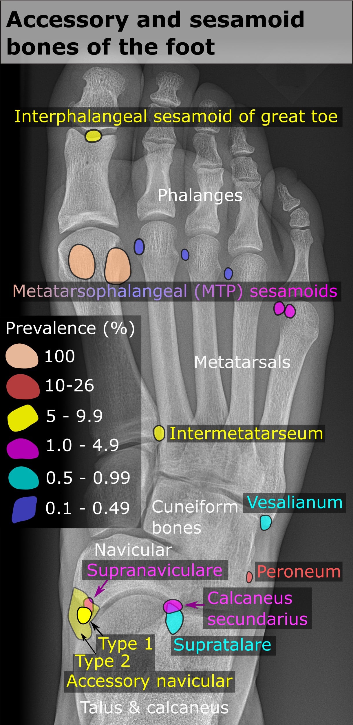
- An interphalangeal sesamoid of the hallux was present in 2 per cent, and lesser metatarsophalangeal sesamoids in 4.3 per cent of fifth digits but under 0.5 per cent of the second to fourth
- Accessory ossicles overall were found in 21.2 per cent and sesamoid bones in 9.6 per cent, the commonest ossicle being the accessory navicular at 11.7 per cent
- Distribution of the common ossicles was similar between men and women
Open Reduction and Screw Fixation of a Diastatic Bipartite Hallux Sesamoid in Turf Toe Injury
- A 25-year-old professional footballer developed severe first metatarsal head pain after opponent contact
- Radiographs showed DIASTASIS between the fragments of a bipartite medial sesamoid - proven acute because radiographs of the same foot taken four years earlier were available for comparison
- CT confirmed the widened interval and additionally showed angulation of the proximal fragment
- Open reduction and screw fixation restored the position of the two fragments and the player returned to the same performance level