Early Active Motion | Zone-Specific Protocols | Preventing Rupture vs Adhesions
- Early mobilisation superior to immobilisation - reduces adhesions without increasing rupture
- Zone 2 (no man's land) requires most careful rehabilitation - FDS and FDP in flexor sheath
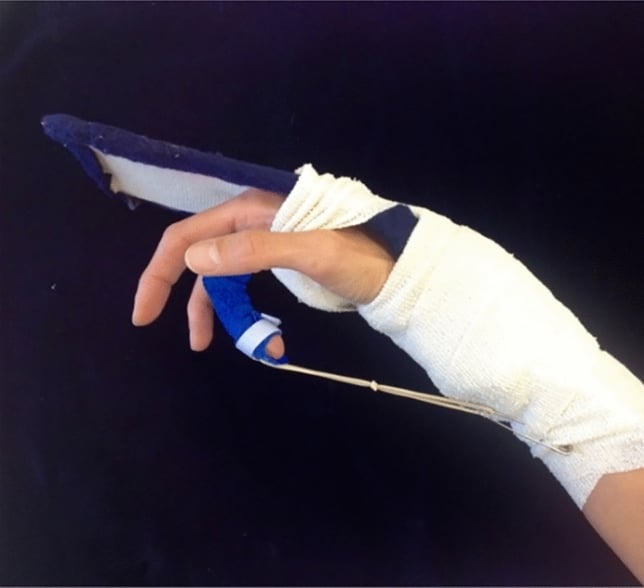
- Kleinert protocol: the rubber band pulls the finger into flexion and the patient actively extends against it
- EAM protocols: Controlled active flexion from day 3-5, superior functional outcomes
- Rupture vs adhesion balance: Too conservative = stiffness, too aggressive = rupture
- “Duran protocol uses passive flexion/extension exercises without rubber band traction
- “Place-and-hold allows passive positioning, then patient actively holds position
- “Strickland criteria: Good result = greater than 70% TAM compared to opposite hand
- “Week 6-8: Transition to unrestricted active motion and light resistance
Overview and Epidemiology
Flexor tendon rehabilitation is one of the most demanding challenges in hand therapy. It balances two competing risks through the critical 4-6 week healing period: too little motion and the tendon forms adhesions, too much and the repair ruptures. Zone 2, where FDS and FDP both run through the restrictive fibro-osseous sheath, is where that balance is hardest to strike.
Who. Working-age adults, mostly male, in manual occupations including construction, manufacturing, food service and agriculture. Power tools, knives and glass are the most common mechanisms. No source cited on this page quantifies the age or sex distribution, the incidence of flexor tendon laceration or the distribution by zone.
Why zone 2 dominates the literature. It is the hardest zone, not necessarily the commonest: both tendons run within the fibro-osseous sheath there, so adhesion and rupture risk are concentrated in it. Every trial on this page recruited zone 2 injuries almost exclusively, and Cochrane records that its included studies focused mainly on zone 2.
The burden. Time away from manual work and the intensity of therapy dominate it, and both run for months rather than weeks. No cited source quantifies the cost, and any figure would be specific to one health system and one labour market. Because treatment by a certified hand therapist produced better motion with smaller contractures in Trumble's trial, access to skilled therapy is an economic determinant of outcome as well as a clinical one.
Immobilisation (before the 1970s). Complete immobilisation for 3-4 weeks carried a low rupture risk (2%) but produced severe adhesions and poor function. Verdan's classic studies demonstrated stiffness rates exceeding 60%, and the approach was abandoned.
Passive motion (1970s-1980s). Kleinert (1967) and Duran (1975) introduced controlled passive motion in place of immobilisation, and the improvement in adhesion-related stiffness was the paradigm shift. No source cited on this page quantifies the outcomes or the rupture rate of that era.
Early active motion (1990s onwards). Multi-strand core sutures of 4-6 strands gave repairs the biomechanical strength for earlier active motion. Each advance in surgical technique, stronger repairs with more core strands, enabled a more aggressive protocol with superior functional outcomes: immobilisation gave way to passive motion, then place-and-hold, then early active motion.
Anatomy
The zones. Verdan divided the flexor tendon system into five zones on structural and functional grounds:
- Zone 1 - FDS insertion to the fingertip; FDP only
- Zone 2 (no man's land) - A1 pulley to the FDS insertion; FDS and FDP within the fibro-osseous sheath
- Zone 3 - the palm at the lumbrical origin, proximal to the A1 pulley
- Zone 4 - the carpal tunnel, with 8 tendons and the median nerve
- Zone 5 - the forearm, at the muscle-tendon junction
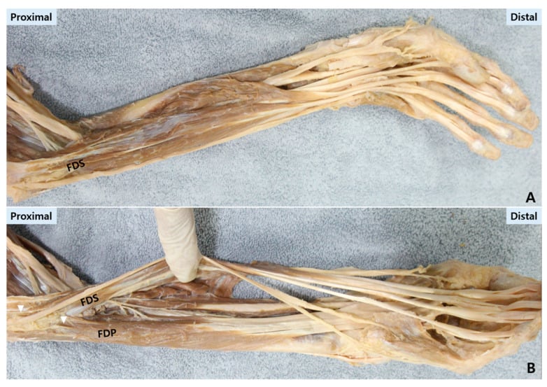
Zone 2 in detail. Both tendons must glide beneath the pulley system, so bulk, oedema and adhesion are especially consequential. The annular pulleys create friction and the synovial sheath, running from A1 to C3, is a site of adhesion. FDS divides around FDP at Camper's chiasm before inserting on the middle phalanx, and FDP continues to the distal phalanx; the FDS decussation lies at the level of the A2 pulley.
The pulleys. The sheath carries annular pulleys A1-A5 and cruciate pulleys C1-C3. A2, over the proximal phalanx, and A4, over the middle phalanx, are biomechanically critical because they prevent bowstringing during flexion, and losing either causes significant mechanical disadvantage and reduced grip strength. How much of them may be vented to let a repair glide is discussed under Controversies.
Nutrition. Flexor tendons are nourished by an intrinsic vascular supply through the longitudinal vincular vessels and by synovial fluid diffusion. Zone 2 has relatively poor vascularity, which contributes to its healing problems.
Pathophysiology
Two kinds of healing. Intrinsic healing comes from tenocytes at the tendon ends, which produce organised collagen fibres that maintain gliding. Extrinsic healing recruits fibroblasts from the synovial sheath and peritendinous tissue and produces disorganised scar that creates adhesions. Early controlled motion promotes intrinsic healing while suppressing excessive extrinsic healing, and stress improves the organisation of the collagen fibres.
The phases of healing. Healing runs through four phases:
- Inflammatory, days 0-5 - haematoma, inflammatory cell infiltration and early fibroblast migration. The repair site is at its weakest and depends entirely on the strength of the suture.
- Fibroplastic, days 5-21 - fibroblast proliferation, type III collagen deposition and neovascularisation. Tensile strength increases but remains low.
- Remodelling, weeks 6-12 - type I collagen replaces type III, fibres align along stress lines and collagen cross-linking matures. Strength increases significantly.
- Maturation, months 3-12 - final collagen reorganisation, with maximum tensile strength of approximately 70-80% of normal tendon.
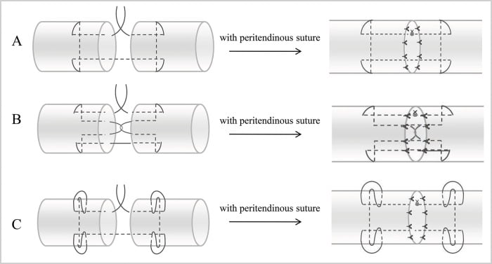
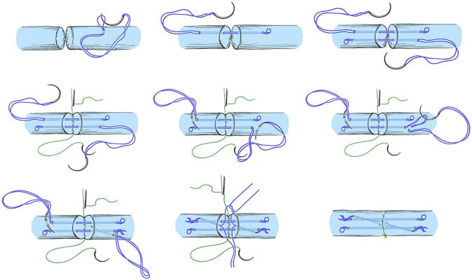
Repair strength. The core suture provides 90% of the strength of the construct and the epitenon suture adds a further 10-20% and smooths the surface. Multi-strand repairs of 4-6 strands are 60% stronger than 2-strand techniques, which is what enables early active motion protocols. Repair strength increases from weeks 3 to 12, and gap formation occurs most commonly in weeks 1-3, when tension exceeds the strength of the healing tissue.
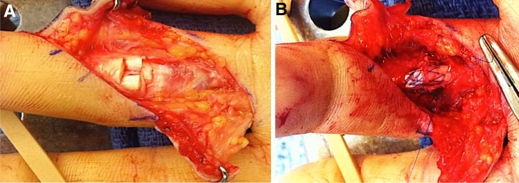
Configuration. Core purchase, locking configuration and strand number determine repair strength and resistance to gap formation, and a smooth circumferential epitendinous suture improves strength and glide. A six-strand locked core distributes load across the repair and supports controlled early active motion when tissue quality, pulley management and patient adherence are favourable; bulky knots or uneven tension increase friction and adhesion. Rehabilitation intensity must match the actual repair, not the skin incision.
Clinical Presentation
What the therapist needs from the surgeon. Repair quality decides which protocol is safe, so the therapist should know the strand count, any pulley venting, any concomitant nerve repair and any concern about tendon quality before selecting a protocol. Injury to both tendons is more complex than injury to FDS or FDP alone.
What the patient brings. Four patient factors shape the choice:
- Compliance - the ability to follow complex instructions decides between a passive and an active protocol
- Cognitive function - elderly or cognitively impaired patients require simpler passive protocols
- Motivation - the return-to-work timeline and functional goals influence rehabilitation intensity
- Manual dexterity - the patient must be able to don and doff the splint and perform the exercises independently
Normal progress. Active flexion should improve by 5-10° a week in the early phase. Passive extension should be maintained to neutral without excessive force, pain with exercises should be minimal (2-3/10 at most), and the repair site should show no sign of infection or inflammation.
Warning signs. Each of these requires immediate assessment:
- Rupture - sudden loss of active flexion, with passive range intact (see Complications)
- Excessive adhesions - a plateau in passive range before week 8, with limited gliding
- Flexion contracture - progressive loss of passive extension
- Complex regional pain syndrome - disproportionate pain, oedema, skin changes and temperature asymmetry
Investigations and Monitoring
Serial measurement. Total active motion (TAM) is measured weekly in weeks 0-6, biweekly in weeks 6-12 and monthly thereafter, and compared with the contralateral hand. Normal composite finger motion is 260°; the formula and the Strickland grades are under Outcome Measurement. Photograph the hand positions at each assessment for the medicolegal record.
At each visit the therapist also records:
- Active and passive range at each joint (PIPJ, DIPJ)
- Pain scores during the exercises
- Oedema, by volumeter or circumference
- Functional grip strength, after week 8
Passive range. The therapist measures maximum passive flexion and extension at each joint. A discrepancy between passive and active range points to adhesions when both are limited or to weakness when only active range is limited, and an isolated limitation suggests a specific anatomical restriction such as an A2 pulley adhesion.
Differential gliding. Reduced differential gliding indicates adhesions between FDS and FDP:
- FDS - block the proximal phalanx and measure isolated PIPJ flexion
- FDP - extend the PIPJ fully and measure isolated DIPJ flexion
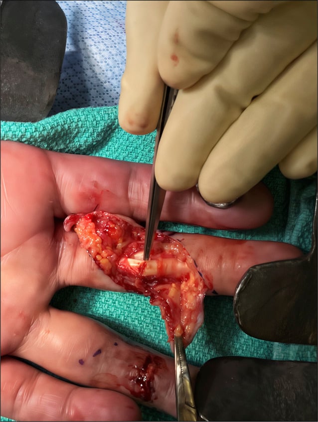
Imaging is rarely required. Dynamic ultrasound can demonstrate tendon gliding, gap formation or adhesions, and is useful when examination is equivocal between rupture and adhesion. MRI is reserved for complex cases with an uncertain diagnosis, where it can show tendon discontinuity, the extent of adhesions or associated pathology such as a ligament injury or occult fracture. Radiographs are taken if an associated fracture is suspected, or to assess joint alignment if a contracture develops.
The partial laceration. A 40% FDP laceration can mimic complete rupture clinically when pain and triggering inhibit motion. Dynamic ultrasound and careful isolated tendon testing help distinguish partial injury, rerupture and adhesion before rehabilitation is changed.
Differential Diagnosis of Poor Motion After Repair
The central diagnostic skill in flexor tendon rehabilitation is identifying why a finger is not moving, and the decisive clinical step is comparing active with passive range.
- Active ROM
- Lost (flexion lag)
- Passive ROM
- Full / preserved
- Discriminating Feature
- Sudden loss of active flexion, possible palpable gap, often minimal pain
- Active ROM
- Reduced
- Passive ROM
- Reduced (both limited)
- Discriminating Feature
- Gradual plateau after week 6-8; gliding lost but no sudden event
- Active ROM
- Reduced
- Passive ROM
- Often near-full
- Discriminating Feature
- Active lag with good passive range; differential gliding deficit on testing
- Active ROM
- Reduced
- Passive ROM
- Reduced extension specifically
- Discriminating Feature
- Fixed loss of passive extension; volar plate / capsular tightness
- Active ROM
- Reduced
- Passive ROM
- Full
- Discriminating Feature
- Active lag improves with strengthening; no mechanical block
- Active ROM
- Reduced in adjacent digits
- Passive ROM
- Full
- Discriminating Feature
- Weak grip and incomplete flexion of NON-injured fingers
- Active ROM
- Reduced efficiency
- Passive ROM
- Often full
- Discriminating Feature
- Visible/palpable bowstringing of tendon on resisted flexion
Active lag with full passive range = rupture (until proven otherwise) or isolated adhesion. Both active and passive limited = dense adhesion or joint contracture. This single comparison directs the entire downstream pathway: urgent surgical referral for suspected rupture versus a therapy-first trial for adhesions and contractures.
Management
The decision. The fundamental management decision is the rehabilitation protocol, chosen on repair strength, patient compliance and functional goals, and it balances functional outcome against rupture risk. Work through it in order:
- Repair strength - a multi-strand repair (4-6 core strands) permits an aggressive protocol; a 2-strand repair requires a conservative one
- Compliance - reliable patients with strong repairs are candidates for early active motion, and non-compliant patients get passive motion only
- Zone - zone 2 requires the strictest adherence; zones 1, 3 and 5 permit faster progression
- Associated injuries - nerve injury, fracture, vascular compromise or multiple-digit involvement may necessitate a modified protocol
Rupture and the protocol. The familiar claim that early active motion trades motion for ruptures is not supported by the randomised evidence. Trumble randomised 119 digits to active place-and-hold or passive motion and found interphalangeal motion of 156° against 128°, with two ruptures in each arm; the authors concluded that active therapy gives greater motion without increasing the risk of rupture. The Cochrane review rated every such comparison very low-certainty and could pool rupture data across only three trials.
Matching protocol to repair. What the evidence does show is that an active protocol must be matched to the repair: Xu found excess rupture specifically where active flexion-extension was paired with a 2-strand core suture. In Trumble's trial smoking, concomitant nerve injury and multiple-digit injury worsened outcomes.
- How it works
- Kleinert: rubber band traction, active extension. Duran: therapist-guided passive range, no rubber band
- Suited to
- Weak (2-strand) repair; non-compliant, elderly or cognitively impaired patient; Duran needs supervised therapy
- What the cited trials show
- Trumble's passive arm: IP motion 128 degrees, 2 ruptures in ~60 digits. Ahmed: 55% excellent by Strickland with early passive mobilisation
- How it works
- Passive placement, active hold
- Suited to
- Moderate compliance or concern for the repair; 3-strand repair
- What the cited trials show
- Trumble's active arm was place-and-hold: IP motion 156 degrees, 2 ruptures in ~60 digits
- How it works
- Controlled active flexion from day 3-5
- Suited to
- Compliant, motivated patient with a strong (4+ strand) repair
- What the cited trials show
- Ahmed: 80% excellent by Strickland with controlled active motion. Xu: excess rupture when active motion was paired with a 2-strand repair
Common to every protocol. Whatever the protocol, the same principles apply:
- Oedema control - elevation, compression wrapping and retrograde massage in the first 2 weeks
- Dorsal blocking splint - wrist 20-30° flexion, MPs 50-70° flexion, IP joints extended only to neutral and never hyperextended
- Progressive loading - tendon stress increased gradually from passive to active to resistive
- Therapist supervision - at least weekly during the protected phase (weeks 0-6)
- Patient education - the signs of rupture, the importance of compliance and realistic expectations of outcome
Returning to activity. Strengthening begins after week 8, once collagen remodelling provides adequate repair strength. The general timeline:
- Weeks 6-8 - light activities of daily living (ADLs): eating, writing, grooming
- Weeks 8-12 - unrestricted ADLs, light work
- Weeks 12-16 - progressive strengthening, return to light manual work
- Months 4-6 - full return to manual labour and contact sport
The early active motion timeline under Rehabilitation Protocols brings light ADLs (weeks 5-6) and contact sport (months 3-4) forward.
Complications and Problem-Solving
Rupture. Prevention rests on protocol adherence, patient education and appropriate protocol selection. A suspected rupture needs immediate referral to the surgeon to consider re-repair or reconstruction, on the timescale set out under Management.
- Sudden loss of active flexion - the patient cannot flex the DIPJ (FDP) or the PIPJ (FDS)
- Flexion lag - passive flexion is possible, but active flexion cannot maintain the position
- Palpable gap - tendon discontinuity on palpation, not always present
- Pain - often minimal despite the rupture
- Recognition
- Limited passive ROM, lacks final 10-20° flexion
- Prevention
- Early motion protocols, differential gliding exercises
- Management
- Weeks 8-12: Aggressive therapy, consider tenolysis if plateau after 3 months
- Recognition
- PIPJ cannot extend to neutral passively
- Prevention
- Splint compliance, extension exercises
- Management
- Static progressive extension splinting, night extension splints
- Recognition
- Loss of independent finger flexion, all fingers flex together
- Prevention
- Proper FDP tendon tensioning at surgery
- Management
- Surgical revision if severe, therapy for mild cases
Rehabilitation Protocols
Dose and position. Exercise dose is adjusted to swelling, glide, lag and repair strength rather than to a rigid calendar alone. Mild wrist and MCP flexion reduces tension across the repair, while active and passive PIP and DIP extension prevents flexion contracture.
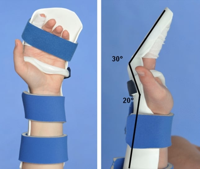
The splint. The controlled-active-motion splint illustrated blocks wrist and MCP extension while leaving the IP joints free for controlled extension. It sets the wrist at 20° and the MCPs at 30°, lower than the 50-70° MCP flexion given under Management, and protects the repair without holding the hand in excessive flexion.
EAM Timeline
Dorsal blocking splint with elevation, ice and oedema control. No exercises, to allow initial healing.
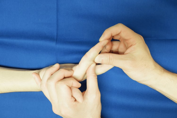
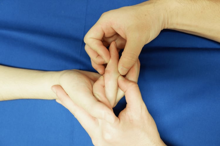
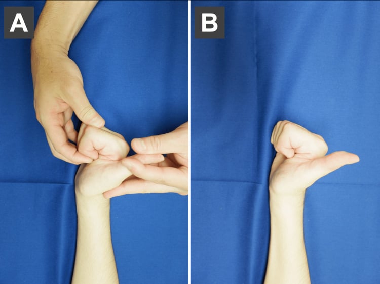
Active differential gliding, 10-12 repetitions every waking hour, with the splint removed for exercises and replaced between sessions:
- FDS blocking - hold the proximal phalanx and flex the PIPJ
- FDP isolated - flex the DIPJ with the PIPJ extended
- Composite fist - all joints flexed together
Continue active flexion and increase the hold to 5 seconds. Add passive extension, the therapist extending the fingers to neutral, and tenodesis exercises (passive wrist motion with finger flexion and extension). Check for flexion lag, the sign of rupture.
Allow gentle passive IP extension beyond neutral. Wean the splint to daytime only and continue night splinting. Light ADLs: eating, writing, grooming.
Full active range at all joints with no restriction on extension, gentle blocking exercises to isolate FDS and FDP, and light resistance with therapy putty and light grip. Discontinue the splint at week 8.
Progressive resistance with hand grippers and weighted exercises. Return to work on light duty first, then full duty; non-contact sport first, contact sport at months 3-4.
Zone-Specific Considerations
- Rehabilitation Challenge
- Single tendon; good prognosis, usually good outcomes
- Protocol Modification
- Standard EAM or passive, simpler than Zone 2
- Rehabilitation Challenge
- Both tendons in the sheath; highest adhesion risk, most challenging
- Protocol Modification
- Strict protocol adherence critical, consider passive if non-compliant
- Rehabilitation Challenge
- Good gliding, larger space
- Protocol Modification
- EAM typically safe, faster progression
- Rehabilitation Challenge
- Nerve injury concern, adhesions to transverse ligament
- Protocol Modification
- Nerve gliding exercises added, standard tendon protocol
- Rehabilitation Challenge
- Best healing, ample space
- Protocol Modification
- Faster progression, strengthening earlier (week 6)
- Strict splint compliance is mandatory
- Consider a passive protocol if there is any doubt about compliance
- Monitor for flexion lag, the sign of rupture, at every session
Synergistic Wrist-Finger Motion: the Basis of Tenodesis Protocols
The principle. During normal grip the wrist extends as the fingers flex and flexes as they extend, a coupled or synergistic pattern. Used deliberately in therapy, extending the wrist while the fingers flex produces a large flexor tendon excursion for very little active muscle force: wrist extension pre-tensions the flexors (the tenodesis effect) and slackens the antagonist extensors, so the finger flexors barely have to contract to close the hand. Flexing the wrist while extending the fingers glides the tendon the other way, again at low tension.
Why it protects the repair. Tendon healing needs glide, which prevents adhesions, but not tension, which risks gap formation and rupture. Synergistic motion maximises glide while minimising the active force, and therefore the tension, on the repair. It is the rationale behind tenodesis exercises, place-and-hold and controlled active motion.
The corollary: avoid a fixed, markedly flexed wrist. The original Kleinert splint held the wrist in marked flexion, but a flexed wrist increases the work of flexion, because the flexors must overcome stretched extensors and a tightened sheath, and so raises repair tension when the patient does move. Modern dorsal blocking orthoses therefore keep the wrist closer to neutral or slight extension, and several protocols allow controlled synergistic wrist motion within the splint.
Oedema Control and the Work of Flexion
Oedema control and gliding resistance are usually taught separately, but they are the same problem: swelling is one of the biggest determinants of whether early active motion is safe.
Work of flexion. The total resistance the flexor tendon must overcome to glide: friction in the sheath and under the pulleys, the bulk of the repair and, critically and modifiably, post-operative oedema and inflammation. The higher the work of flexion, the more active muscle force, and therefore repair-site tension, is needed to move the finger. That raises rupture risk and, when the tendon cannot glide, promotes adhesions.
Why oedema control is biomechanical. A swollen hand has a high work of flexion, so the same active-flexion exercise loads the repair far more than in a decongested hand. The oedema measures of the first two weeks lower the work of flexion, making active motion both safer, with less tension, and more effective, producing real glide rather than muscle effort against a stiff, swollen finger. Controlling oedema is therefore a prerequisite for an active protocol, not an afterthought.
In practice. If the hand is very swollen, or the repair feels tight and high-friction when tested on the table, the surgeon and therapist should decongest first and lean toward a lower-tension protocol, place-and-hold or passive, until the work of flexion falls. The protocol is matched to the achievable gliding resistance, not to the strand count alone.
Outcome Measurement
Total active motion. TAM = (active PIPJ flexion + active DIPJ flexion) - (PIPJ extension lag + DIPJ extension lag). Strickland's criteria grade it against normal:
- Excellent - greater than 85% of normal (greater than 220°)
- Good - 70-84% of normal (180-219°)
- Fair - 50-69% of normal (130-179°)
- Poor - under 50% of normal (under 130°)
What the grade means. Good or excellent outcomes, greater than 70% of TAM, correlate with high patient satisfaction and functional independence. Fair or poor outcomes often require tenolysis or reconstruction.
Guidelines, Registries & Global Practice
Global Epidemiology
Flexor tendon lacerations predominantly affect working-age males (roughly 18-45 years, around three-quarters of cases) in manual occupations - construction, manufacturing, food service and agriculture - with knives, glass and power tools the leading mechanisms. Reported incidence is broadly 15-20 per 100,000 population per year in higher-income settings, with zone 2 accounting for approximately 40% of injuries. In lower-resource and agrarian regions, the absolute burden is often higher and presentation later, but population-level registry data are sparse - unlike arthroplasty, there is no large international flexor-tendon outcome registry, so most evidence comes from single-centre series and small RCTs.
Side-by-Side Guidance and Consensus
- Emphasis
- Multi-strand repair plus early controlled mobilization
- Practical position
- Four-strand minimum core repair; early active or place-and-hold within a dorsal blocking orthosis
- Emphasis
- Repair strength dictates rehab intensity
- Practical position
- Active protocols reserved for robust (4-6 strand) repairs in adherent patients
- Emphasis
- Pulley venting plus early active motion
- Practical position
- Judicious A2/A4 venting accepted to permit gliding; active motion favoured
- Emphasis
- Evidence appraisal
- Practical position
- No protocol proven superior - individualize; calls for powered RCTs
These bodies converge far more than they differ: a strong multi-strand repair, judicious pulley management, and early controlled motion under hand-therapy supervision is the shared global standard. Disagreement is largely about the degree of activity (full active vs place-and-hold vs passive) and how aggressively to vent pulleys.
High- vs Limited-Resource Practice Variation
- Well-resourced setting
- Frequent certified hand therapist (CHT) supervision; active/place-and-hold protocols feasible
- Limited-resource setting
- Sparse CHT access; passive (Kleinert/Duran) or simpler home programmes safer
- Well-resourced setting
- In-person weekly review; telerehabilitation as adjunct
- Limited-resource setting
- Reliance on patient self-management and infrequent review
- Well-resourced setting
- Multi-strand repair, microscope/loupes, WALANT available
- Limited-resource setting
- Variable suture material and magnification; emphasis on a secure simpler construct
- Well-resourced setting
- Early, primary repair
- Limited-resource setting
- Often delayed - higher rate of secondary reconstruction/graft
The guiding principle worldwide is to match the rehabilitation protocol to the achievable level of supervision: an active protocol without reliable therapy is more dangerous than a well-run passive one. Telerehabilitation can extend monitoring but cannot fully replace in-person assessment of differential gliding and contracture.
Counsel and record, regardless of health system:
- Rupture risk discussed and quantified relative to the chosen protocol and repair strength
- Rationale for protocol selection (repair strength, zone, adherence, therapy access)
- Therapy attendance and serial active/passive range at each visit
- Prompt action if rupture is suspected, given the narrow re-repair window
Controversies and Areas of Uncertainty
Despite decades of practice, flexor tendon rehabilitation rests on a strikingly thin evidence base. The 2021 Cochrane review graded all comparisons as very low-certainty, so most "rules" are convention rather than proof.
Active versus passive. Newer RCTs (Trumble 2010, Ahmed 2025) favour active or place-and-hold therapy for motion, but the Cochrane synthesis found no protocol definitively superior. The honest answer is that a strong repair plus any early controlled motion matters more than the specific named protocol.
Strand number. Four-strand repair is the accepted minimum for active motion. Whether 6-strand repairs add clinical rather than only biomechanical benefit, against their increased bulk and gliding resistance, remains debated.
Pulley venting. The historical dogma of preserving A2 and A4 at all costs has shifted. Judicious venting (partial A2, full A4) to allow a repaired tendon to glide is now considered safe, but the safe limit of release is not precisely defined.
WALANT and intra-operative testing. Wide-awake repair lets the surgeon confirm active gliding on the table, but Douwes (2025) found it not demonstrably superior in outcome. Its main value may be patient selection and gap detection rather than the anaesthetic itself.
Relative motion flexion orthoses. They promise a smaller, more functional orthosis, but the evidence is limited to small case series with an RCT still pending, and the technique remains investigational.
Therapy dose and telerehabilitation. The minimum effective dose of hand therapy is unknown. Supervised therapy improves outcomes, yet how much can be safely shifted to home programmes or telerehabilitation, critical in limited-resource settings, is unresolved.
MCQ Practice Points
Q: Zone 2 of the flexor tendon system extends from which landmarks? A: A1 pulley to FDS insertion. Zone 2 (no man's land) encompasses the area where both FDS and FDP tendons run within the restrictive fibro-osseous sheath. This zone has the highest risk of adhesions and poorest outcomes historically.
Q: In the Kleinert protocol, what motion does the patient actively perform? A: Active extension against rubber band traction. The rubber band maintains flexion passively; the patient actively extends the finger to neutral against the band resistance. This protects the repair from active flexion forces while maintaining gliding.
Q: How is Total Active Motion (TAM) calculated for flexor tendon outcomes? A: TAM equals (Active PIPJ flex plus Active DIPJ flex) minus (PIPJ extension lag plus DIPJ extension lag). Good outcome is 70-84% of normal, excellent is greater than 85%. This standardized measurement allows comparison across studies.
Q: What is the approximate rupture rate for early active motion protocols compared to passive motion? A: The premise of the question is not supported by the randomised evidence. Trumble randomised 119 digits to active place-and-hold or passive motion and recorded two ruptures in each arm, concluding that active therapy gives greater motion without increasing rupture risk; the Cochrane review of 17 studies could pool rupture data across only three trials and rated every comparison very low-certainty. What is established is conditional: Xu's meta-analysis found excess rupture in the active flexion-and-extension group specifically where the repair was a 2-strand core suture. So the risk lies in the mismatch, not in the protocol - an active regimen after a 4-strand or stronger repair is not the high-rupture option it is often described as.
Q: During which period is the flexor tendon repair weakest and most vulnerable to rupture? A: Weeks 1-3 post-repair. During this inflammatory phase, the repair has minimal intrinsic strength and depends entirely on suture holding power. Tensile strength increases significantly during the fibroplasia phase (weeks 3-8).
Exam Viva Scenarios
Practise clinical reasoning and management decisions out loud
“A 35-year-old tradesman sustained a Zone 2 FDP and FDS laceration to his index finger. You performed a 4-strand core suture repair with running epitenon suture. He is motivated to return to work quickly. What rehabilitation protocol would you recommend and why?”
“A patient returns to clinic at 3 weeks post Zone 2 FDP repair on an EAM protocol. The therapist notes a new flexion lag at the DIPJ - the patient can passively flex to full range but cannot actively maintain the position. What is your assessment and management?”
“A patient is 12 weeks post Zone 2 flexor tendon repair with strict adherence to an EAM protocol. TAM is 120° (45% of normal) with significant limitations in both active and passive motion. No rupture occurred. What is the likely problem and management approach?”
Protocol Selection
- Passive Motion (Kleinert/Duran) = for the weak repair or the non-adherent patient; no cited source quantifies its rupture rate separately
- Place-and-Hold = intermediate tension; not separately quantified in any cited source
- Early Active Motion (EAM) = best motion in the randomised trials; rupture risk not shown to be higher when the repair is 4-strand or stronger
- Zone 2 (no man's land) = both FDS and FDP in sheath, highest adhesion risk
Critical Timelines
- Days 0-2: Splint immobilization, no exercises
- Days 3-5: Begin EAM or passive protocol
- Weeks 0-6: Protected motion phase, strict splint compliance
- Weeks 6-8: Transition to unrestricted active motion
- Weeks 8-12: Progressive strengthening, return to work
- Weeks 1-3: Weakest repair, highest rupture risk
Splint Positioning
- Wrist: 20-30° flexion
- MPs: 50-70° flexion
- IPs: Neutral to slight flexion
- Kleinert: Add rubber band from nail to volar forearm
Exercise Components
- Differential gliding: Isolate FDS (PIPJ flex) vs FDP (DIPJ flex)
- Tenodesis: Passive wrist motion with finger motion
- Composite fist: All joints flexed together
- Place-and-hold: Passive positioning, active maintenance
Complications
- Rupture: Flexion lag, loss of active flexion, palpable gap
- Adhesions 20-30%: Limited passive ROM, consider tenolysis after 3 months therapy
- Flexion contracture: Cannot extend passively, extension splinting
- Re-repair window: Under 2 weeks best, after 6 weeks need reconstruction
Outcome Measurement
- TAM = (PIPJ flex + DIPJ flex) - (PIPJ lag + DIPJ lag)
- Excellent = over 85% (over 220°)
- Good = 70-84% (180-219°)
- Fair = 50-69% (130-179°)
- Poor = under 50% (under 130°)
Evidence Base and Key Trials
Active Place-and-Hold vs Passive Motion after Zone-II Repair (Landmark RCT)
- Prospective RCT: 103 patients (119 digits) with zone-II repairs randomized to active place-and-hold vs passive motion
- Active group had greater IP joint motion at every time point: final mean 156° vs 128° (p less than 0.05)
- Active group had smaller flexion contractures and higher satisfaction scores
- Only 2 ruptures occurred in each group - active motion did NOT increase rupture risk
- Smoking, concomitant nerve injury and multiple-digit injury independently worsened outcomes; CHT-supervised therapy improved them
Cochrane Review: Rehabilitation after Flexor Tendon Surgery
- 16 RCTs plus 1 quasi-RCT, 1108 participants, predominantly zone-II repairs
- Very low-certainty evidence across all 14 comparisons (GRADE) - no protocol proven superior
- Early active flexion plus controlled passive vs modified Kleinert: no clinically important difference in function or motion
- Place-and-hold vs rubber-band traction: very low-certainty signal toward greater active motion at 12 months with place-and-hold
- Identifies an urgent need for adequately powered, standardized RCTs
Controlled Active Motion vs Early Passive Mobilization (Zone II RCT)
- RCT of 40 patients with complete zone-II FDP and FDS lacerations: CAM vs early passive mobilization (EPM)
- Both protocols improved TAM, grip strength and DASH over 12 weeks (p less than 0.001)
- CAM superior to EPM at 6 and 12 weeks for TAM, grip strength and DASH (p less than 0.05)
- At 12 weeks, 80% of CAM patients achieved 'excellent' by Strickland criteria vs 55% with EPM
Evidence-Based Management of Zone II Flexor Tendon Injury
- Systematic review addressing 8 key questions on diagnosis, repair and rehabilitation of zone-II injury
- Repair requires a four-strand or multi-strand core suture, with or without an epitendinous suture
- Judicious pulley venting (including partial A2/A4 release) is safe and effective
- WALANT (wide-awake) technique is not demonstrably superior to other anaesthesia
- Early controlled mobilization (passive or active, matched to repair strength and adherence) is the cornerstone of rehabilitation
Indications, Methods and Outcomes of Primary Zone-2 Repair (Tang concepts)
- Influential practical framework for predictable zone-2 outcomes from a high-volume unit
- Advocates strong multi-strand core repair combined with judicious sheath-pulley venting (release of part of A2/whole A4 as needed)
- Describes a postoperative active motion regimen tailored to repair strength
- Emphasizes outcome evaluation that separates true active gliding from passive range
Relative Motion Flexion Orthoses after Zone I-III Repair
- Narrative review plus case series (18 patients) of relative motion flexion (RMF) orthoses as an early active strategy
- Positioning the injured digit in relatively greater MCP flexion is hypothesized to offload FDP tension via the quadriga effect
- Permits earlier functional hand use within a smaller, less restrictive orthosis
- Authors stress evidence is still limited and a pragmatic RCT is underway