The Arch, the Windlass and the Flexible-to-Rigid Lever
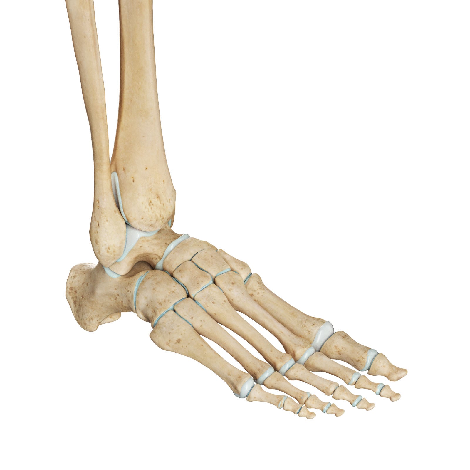
- The foot is divided into HINDFOOT (talus, calcaneus), MIDFOOT (navicular, cuboid, three cuneiforms - linked to the hindfoot at the transverse tarsal/CHOPART joint and to the forefoot at the tarsometatarsal/LISFRANC joint) and FOREFOOT (five metatarsals and the phalanges); it is arranged into a MEDIAL and LATERAL longitudinal arch and a TRANSVERSE arch.
- Arch support is provided in three layers: BONY/static (the wedge-shaped bones; the talar head/navicular acts as the medial-arch 'keystone'), LIGAMENTOUS (the PLANTAR FASCIA, the SPRING/plantar calcaneonavicular ligament, and the long and short plantar ligaments - the principal passive supports), and MUSCULAR/dynamic (intrinsics, TIBIALIS POSTERIOR, peroneus longus, long toe flexors).
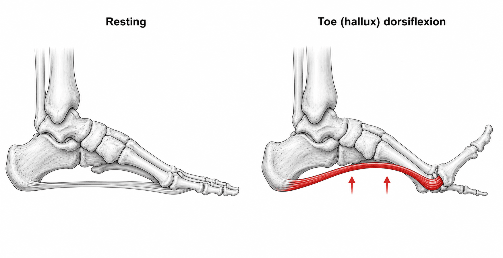
- The WINDLASS MECHANISM: the plantar fascia runs from the calcaneus to the bases of the proximal phalanges, wrapping around the metatarsal heads. DORSIFLEXION of the metatarsophalangeal joints (especially the HALLUX) at push-off winds the fascia around the metatarsal heads like a cable on a drum, SHORTENING the calcaneus-to-metatarsal distance, RAISING the arch and INVERTING the hindfoot - converting a flexible foot into a rigid lever. JACK'S (Hubscher) test - passive hallux dorsiflexion that raises the arch - demonstrates a working windlass.
- The arch also behaves as an elastic ARCH-SPRING: the plantar fascia and other arch tissues STRETCH and store energy in early-mid stance and RECOIL to return energy at push-off - so the fascia is not perfectly isometric but combines an arch-spring with a near-isometric windlass in propulsion.
- During gait the SUBTALAR joint controls a flexible-to-rigid transition: at heel strike the hindfoot PRONATES (everts), which makes the transverse tarsal (talonavicular + calcaneocuboid) joint axes PARALLEL and UNLOCKS the midfoot for a flexible, shock-absorbing foot; in terminal stance the hindfoot SUPINATES (inverts), the axes become NON-PARALLEL, the midfoot LOCKS, and (with the windlass) the foot becomes a rigid lever for push-off.
- The GAIT cycle is ~60% STANCE and ~40% SWING; stance progresses through initial contact (heel strike), loading response, midstance, terminal stance (heel off) and pre-swing (toe off), advancing the body over three ROCKERS - the heel rocker, the ankle rocker and the forefoot (metatarsophalangeal) rocker. This functional anatomy underlies plantar fasciitis, flatfoot, cavus, hallux rigidus and the goals of deformity-correcting surgery.
- “Arch supports in 3 layers: bony (keystone = talar head/navicular), ligamentous (plantar fascia, SPRING ligament, long/short plantar), muscular (tib post is key).
- “Windlass: hallux/MTP dorsiflexion tightens the plantar fascia -> raises arch + inverts hindfoot -> rigid lever; Jack's test demonstrates it.
- “Subtalar pronation unlocks the transverse tarsal (Chopart) joint (flexible); supination locks it (rigid) - the foot is a shock absorber at heel strike and a rigid lever at push-off.
Subtalar pronation -> transverse-tarsal axes parallel -> midfoot unlocked -> a flexible, mobile foot that absorbs shock and adapts to the ground.
Windlass (toe dorsiflexion) + subtalar supination -> transverse-tarsal axes non-parallel -> midfoot locked -> a rigid lever for efficient propulsion.
Functional Anatomy & the Arches
The hindfoot (talus, calcaneus) meets the midfoot (navicular, cuboid, three cuneiforms) at the transverse tarsal (Chopart) joint, and the midfoot meets the forefoot (metatarsals, phalanges) at the tarsometatarsal (Lisfranc) joint. The bones form a medial longitudinal arch (calcaneus-talus-navicular -cuneiforms-medial three metatarsals; higher, more mobile), a lateral longitudinal arch (calcaneus-cuboid-lateral two metatarsals; lower, flatter, contacts the ground) and a transverse arch (across the cuneiforms/metatarsal bases). The arch is held by three layers: bony (wedge-shaped bones, with the talar head/navicular the medial keystone); ligamentous - the plantar fascia, the spring (plantar calcaneonavicular) ligament supporting the talar head, and the long and short plantar ligaments; and muscular/dynamic - the intrinsics and especially tibialis posterior (whose failure causes adult acquired flatfoot), with peroneus longus and the long toe flexors.
The Windlass Mechanism & Arch-Spring
The plantar fascia (aponeurosis) runs from the medial calcaneal tubercle to the bases of the proximal phalanges, passing beneath the metatarsal heads. When the metatarsophalangeal joints dorsiflex - as the heel rises and the toes extend at push-off, the hallux being most important - the fascia is wound around the metatarsal heads like a cable around a drum (a 'windlass'), which shortens the distance between the calcaneus and the metatarsal heads, elevates the medial longitudinal arch, and inverts the hindfoot, stiffening the foot into a rigid propulsive lever. Clinically, passively dorsiflexing the hallux and watching the arch rise is Jack's (Hubscher) test, used to assess a flexible flatfoot and the integrity of the windlass.
The arch is not only a windlass but an elastic arch-spring: the plantar fascia and the other arch tissues stretch and store strain energy as the arch flattens under load in early-to-mid stance, then recoil and return that energy at push-off, improving the efficiency of walking and running. High-speed imaging shows the plantar fascia is not perfectly isometric - it strains through stance - yet still behaves as a near-ideal windlass during propulsion, with its shortening timed to enhance arch recoil at push-off. This energy-storage role also explains why the fascia is highly loaded (and prone to overload/plantar fasciitis) at the medial calcaneal tubercle.
The Gait Cycle & Joint Coupling
A gait cycle (one heel strike to the next on the same foot) is about 60% STANCE and 40% SWING. Stance proceeds through initial contact (heel strike), loading response, midstance, terminal stance (heel off) and pre-swing (toe off); swing has initial, mid and terminal phases. The body advances over three rockers: the heel (1st) rocker at heel strike (ankle dorsiflexors control plantarflexion), the ankle (2nd) rocker in midstance (the tibia rolls forward over the fixed foot), and the forefoot/MTP (3rd) rocker at push-off (the body pivots over the metatarsal heads, engaging the windlass).
The foot must be flexible to absorb load at heel strike and rigid to push off. The subtalar joint controls this:
- Heel strike -> subtalar PRONATION (eversion): the talonavicular and calcaneocuboid joint axes of the transverse tarsal (Chopart) joint become PARALLEL, so the midfoot is UNLOCKED and the foot is a flexible, mobile shock absorber that adapts to the terrain.
- Terminal stance -> subtalar SUPINATION (inversion): the axes become NON-PARALLEL, the midfoot LOCKS, and - together with the windlass - the foot becomes a rigid lever for efficient propulsion. Loss of this coupling underlies many deformities: a foot stuck in pronation (e.g. tibialis posterior dysfunction) cannot lock and re-supinate, giving an inefficient flatfoot gait.
Clinical Relevance
- Plantar fasciitis: overload of the highly stressed plantar fascia at the medial calcaneal tubercle (modelling shows peak fascia tension late in stance, increased substantially by the windlass).
- Pes planus (flatfoot): failure of arch supports - tibialis posterior dysfunction (dynamic) and/or spring-ligament failure (static) - the hindfoot stays pronated and the arch collapses; Jack's test and the double/single heel-rise test assess flexibility and tibialis posterior.
- Pes cavus: a high, rigid arch (often neuromuscular) overloads the heel and metatarsal heads.
- Hallux rigidus / first-ray problems: loss of MTP dorsiflexion impairs the windlass and the forefoot rocker.
- Surgery: arch-restoring procedures (e.g. medialising calcaneal osteotomy, tendon transfers, lateral-column lengthening) and fusions are designed around restoring this bony-ligamentous-muscular balance and the flexible-to-rigid mechanics. (See our Plantar Fasciitis, Adult Acquired Flatfoot / PTTD, Pes Cavus and Hallux Rigidus topics.)
Evidence & Key Studies
The extensibility of the plantar fascia influences the windlass mechanism during human running
- Using high-speed X-ray during running, the plantar fascia was shown to both store/release elastic energy (arch-spring) AND act as a windlass coupling toe dorsiflexion to arch shape.
- Toe plantarflexion at foot strike delays plantar-fascia stretch, redistributing load through other arch tissues.
- In propulsion a quasi-isometric (near-ideal windlass) fascia shortens later in stance, enhancing arch recoil at push-off - reconciling the arch-spring and windlass models.
Finite element analysis of plantar fascia during walking: a quasi-static simulation
- In a 3-D finite-element model, plantar-fascia tension peaked at pre-swing (about 83% of stance) at roughly 493 N (about 0.7 body weight).
- Stress concentrated near the MEDIAL CALCANEAL TUBERCLE - the typical site of plantar fasciitis - and the windlass mechanism increased fascia tension by about 66%.
- Reducing heel rise and Achilles tendon force lowers fascia loading, supporting calf-stretching and gait modification in plantar fasciitis.
According to PubMed, the combined arch-spring/windlass behaviour of the plantar fascia comes from the cited Welte study, and the peak fascia tension, the medial-calcaneal-tubercle stress concentration and the ~66% windlass contribution from the cited Chen finite-element analysis. The arch anatomy, the three layers of arch support and the subtalar/transverse-tarsal coupling are standard, well-established functional anatomy. (See also our Plantar Fasciitis and Adult Acquired Flatfoot topics.)
Clinical Decision Scenarios
Practise clinical reasoning and management decisions out loud
“Describe the arches of the foot and how they are supported, and explain the windlass mechanism.”
“How does the foot change from a flexible structure at heel strike to a rigid lever at push-off during gait?”
Mnemonics & Memory Aids
BLM (arch support)
Hook:Arch support = Bony, Ligamentous, Muscular (B-L-M).
PURL
Hook:PURL: Pronation Unlocks, Re-supination Locks.
Anatomy
- Hindfoot (talus, calcaneus) | Midfoot (navicular, cuboid, 3 cuneiforms) | Forefoot (MTs, phalanges)
- Chopart (transverse tarsal) and Lisfranc (TMT) joints
- Medial (higher, keystone talar head/navicular) + lateral longitudinal + transverse arches
Arch support (3 layers)
- Bony/static: bone shape; keystone = talar head/navicular
- Ligamentous: plantar fascia, SPRING (calcaneonavicular) ligament, long/short plantar
- Muscular/dynamic: intrinsics, tibialis posterior (key), peroneus longus, toe flexors
Windlass & arch-spring
- MTP/hallux dorsiflexion winds plantar fascia round met heads -> raises arch + inverts hindfoot -> rigid lever
- Jack's (Hubscher) test demonstrates it
- Arch-spring stores/returns energy; peak fascia stress at medial calcaneal tubercle
Gait & coupling
- ~60% stance / ~40% swing; heel, ankle, forefoot rockers
- Heel strike: subtalar pronation -> Chopart axes parallel -> flexible (shock absorber)
- Push-off: subtalar supination -> axes non-parallel -> midfoot locked + windlass -> rigid lever