Clostridium perfringens | Alpha Toxin | Myonecrosis | Radical Debridement
- Pain out of proportion to clinical findings is the earliest and most important sign
- Bronze/bronze-brown skin with haemorrhagic bullae is pathognomonic
- Crepitus present in only 50% - absence does NOT exclude diagnosis
- Dishwater exudate - thin, serosanguinous, foul-smelling discharge
- Radical surgical debridement is life-saving - antibiotics alone are inadequate
- “Alpha toxin (lecithinase/phospholipase C) destroys cell membranes - key virulence factor
- “X-ray shows gas in soft tissues - feathery pattern tracking fascial planes
- “Penicillin G + clindamycin is the antibiotic regimen (clindamycin inhibits toxin production)
- “Hyperbaric oxygen is ADJUNCTIVE only - never delays surgery
Overview and Epidemiology
Gas gangrene (clostridial myonecrosis) is a rapidly progressive, life-threatening infection of skeletal muscle caused by toxin-producing Clostridium species. Its hallmark is myonecrosis, the destruction of muscle with gas production, which leads to systemic toxicity and, if untreated, to death. Mortality remains 25-40% even with treatment and approaches 100% without surgery.
How it arises. Gas gangrene is uncommon but highly lethal, with approximately 1,000-3,000 cases a year in developed countries. It classically follows contaminated trauma (military wounds, agricultural injuries, compound fractures), but it can also occur after surgery or spontaneously. The spontaneous form, typically caused by C. septicum, is strongly associated with occult gastrointestinal malignancy, especially colorectal cancer, and should prompt investigation with colonoscopy once the patient is stabilised.
The organisms. Clostridium perfringens, most commonly Type A, is the dominant organism in traumatic and postoperative gas gangrene. The 80% given in the summary above is the figure quoted in textbooks, and it derives from case series rather than from surveillance; no source cited on this page gives a proportion. The other species:
- C. septicum - spontaneous cases
- C. novyi - historically seen in users of contaminated heroin
- C. histolyticum - rare, with aggressive tissue destruction
- C. bifermentans and C. fallax - less common
Risk factors. These are the settings in which it arises:
- Contaminated wounds: soil, faeces, foreign material
- Muscle ischaemia: vascular compromise, tourniquet injury
- Open fractures, especially agricultural and combat injuries
- Immunocompromise: diabetes, malignancy, steroids
- Gastrointestinal pathology: colorectal cancer (C. septicum)
- Intravenous drug use: contaminated injections
Gas gangrene can spread through muscle at 2 cm per hour, and every hour of delay to surgical debridement increases mortality. This is a true surgical emergency: arrange theatre while making the diagnosis, not after it.
Pathophysiology
Alpha toxin. The principal toxin, and the key virulence factor, is alpha toxin: a phospholipase C (lecithinase) and zinc metalloenzyme that hydrolyses phosphatidylcholine and sphingomyelin in cell membranes. It disrupts the membranes of muscle cells, erythrocytes, endothelium and leukocytes, which explains the rapid progression and the systemic toxicity.
In muscle. Destruction of muscle cell membranes produces massive myonecrosis and tissue liquefaction. Damaged muscle releases myoglobin, potassium and creatine kinase, and the tissue becomes non-contractile and non-bleeding.
In the blood. Lysis of erythrocyte membranes causes intravascular haemolysis, with haemoglobinuria, jaundice and renal failure. Haemolysis indicates severe systemic toxin absorption, and patients with it have significantly higher mortality. Look for jaundice, haemoglobinuria (dark urine without red cells on microscopy), a falling haemoglobin and an unconjugated hyperbilirubinaemia.
In the case in the figure, from fulminant C. perfringens sepsis, the blood film showed severe haemolytic change and bacillary forms phagocytosed within a neutrophil. Sudden anaemia, haemoglobinuria, a very high LDH and visible bacilli constitute a critical toxin-mediated emergency even before culture confirmation.
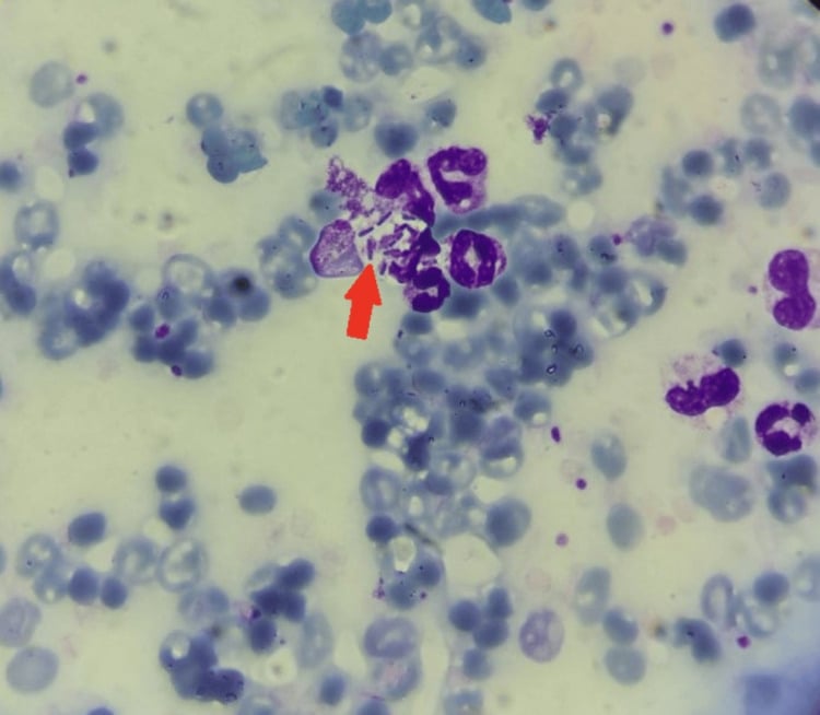
In the vessels. Endothelial damage increases vascular permeability and causes oedema and platelet aggregation. The result is a microenvironment of ischaemia and anaerobiosis that favours further clostridial growth.
The other toxins. Perfringolysin O (theta toxin) is pore-forming and synergises with alpha toxin. Collagenase and hyaluronidase are tissue-spreading enzymes that facilitate rapid invasion.
Why the wound looks bland. Alpha and theta toxin act on the microvasculature to trigger massive leukocyte-endothelial aggregation and adhesion at the vessel margins, leukostasis. The neutrophils pile up inside and around the vessels instead of migrating into the infected muscle, and alpha toxin destroys leukocytes as well, so the tissue and the Gram stain show few or no white cells despite overwhelming infection.
The adherent leukocyte-platelet aggregates, together with toxin-mediated vascular occlusion and endothelial injury, throttle the local blood supply. The regional ischaemia and anaerobiosis that follow propagate the infection, extending the anaerobic environment the organism needs, and account for its relentless spread through apparently healthy tissue.
Because the necrosis begins deep in the muscle and the overlying skin is initially spared, the surface can look unremarkable while the muscle is already dead. The pain is ischaemic, from the toxin-driven vascular shutdown rather than from surface inflammation, and this is the mismatch that "pain out of proportion to the wound" describes.
The anaerobic wound. Infection needs all of the following:
- An anaerobic environment, with low tissue oxygen tension
- Devitalised tissue: ischaemic or necrotic muscle
- Inoculation with spores or vegetative forms
- Nutrients, the damaged tissue providing substrate
- A low redox potential, optimally below -150 mV
Wounds become anaerobic through vascular injury, which reduces oxygen delivery; tissue compression from crush injury or compartment syndrome; foreign bodies, which create microenvironments; oedema, which increases the diffusion distance for oxygen; and aerobic bacteria, which consume the oxygen that remains.
Gas. Clostridia produce gas by fermenting muscle glucose and amino acids, mainly hydrogen, carbon dioxide and nitrogen. The gas dissects along fascial planes and gives the characteristic crepitus.
Systemic toxicity. Toxin and sepsis act on distant organs:
- Effect
- Shock, myocardial depression
- Mechanism
- Alpha toxin cardiotoxicity, sepsis
- Clinical Finding
- Hypotension, tachycardia, reduced cardiac output
- Effect
- Haemolysis, DIC
- Mechanism
- Red cell membrane lysis, coagulopathy
- Clinical Finding
- Haemoglobinuria, jaundice, bleeding
- Effect
- Acute kidney injury
- Mechanism
- Myoglobinuria, haemoglobinuria, hypoperfusion
- Clinical Finding
- Oliguria, rising creatinine
- Effect
- Liver dysfunction
- Mechanism
- Toxin-mediated injury, hypoperfusion
- Clinical Finding
- Jaundice, elevated transaminases
- Effect
- Encephalopathy
- Mechanism
- Toxaemia, metabolic derangement
- Clinical Finding
- Confusion, delirium, obtundation
Clinical Presentation
History. Ask about recent trauma or surgery: open fractures, contaminated wounds, bowel surgery. The incubation period is 6-72 hours, usually 12-24 hours after injury. Pain comes on suddenly and severely, disproportionate to the wound; progression is over hours, not days; and constitutional symptoms follow, with fever, malaise and altered mental status.
Pain out of proportion. Severe pain that exceeds what the wound would suggest is the earliest and most important sign. The patient may look toxic, with tachycardia, fever and confusion, before the skin changes are obvious.
The classic signs. They follow the pain:
- Tense oedema, the limb swollen and woody-hard
- Bronze or bronze-brown skin, a pathognomonic colour change that indicates myonecrosis
- Haemorrhagic bullae filled with dark fluid, a late sign
- Crepitus, present in only approximately 50%, so its absence does not exclude the diagnosis
- Dishwater exudate: thin, serosanguinous and foul-smelling
- A sweet, sickly, mousy odour from the products of anaerobic metabolism
Pain out of proportion, bronze discolouration and systemic toxicity are more reliable early signs than crepitus. By the time crepitus is obvious, extensive myonecrosis has occurred.
Beyond the skin. Diminished pulses and sensory loss may indicate compartment syndrome or vascular compromise. Fever, tachycardia, hypotension and confusion mark septic shock and toxaemia. Dishwater exudate and non-contractile grey muscle found on exploring the wound confirm myonecrosis, and the patient goes to theatre.
Clinical Evolution
Severe pain at the wound, often described as bursting or tearing, while the wound itself may appear unremarkable. The patient becomes restless and anxious, with tachycardia out of proportion to fever. This is the window for intervention.
The skin becomes tense and oedematous and turns bronze or bronze-brown, haemorrhagic bullae may appear, and thin, foul-smelling dishwater discharge leaks from the wound. The pain remains severe and systemic signs of sepsis develop.
Extensive skin necrosis with dusky or black discolouration, and crepitus may now be palpable. The patient is profoundly toxic, with shock, confusion and jaundice, and the muscle at the wound is non-contractile and non-bleeding.
Without intervention, progressive shock, DIC, renal failure and respiratory failure.
In the case in the figure, a small traumatic wound progressed within 48 hours to extensive tense swelling, dusky skin and ruptured bullae. Deep-tissue Gram stain showed abundant Gram-positive bacilli and anaerobic culture confirmed the organism: an initially bland wound can become destructive clostridial myonecrosis within hours.
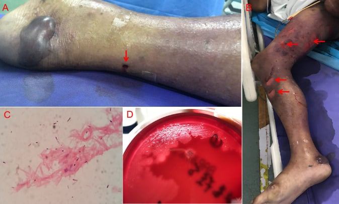
If you wait for bronze skin, crepitus and dishwater discharge, you have waited too long. Pain out of proportion in a contaminated wound is gas gangrene until proven otherwise. Take the patient to theatre for exploration.
Investigations
A clinical diagnosis. Gas gangrene is diagnosed at the bedside. Laboratory tests support the diagnosis and guide resuscitation but should never delay surgical exploration; if clinical suspicion is high, go to theatre immediately.
Blood tests. Expect evidence of myonecrosis, haemolysis, renal injury and DIC:
- Expected Finding
- Leukocytosis (or leukopenia in severe sepsis), anaemia from haemolysis
- Clinical Significance
- Haemolysis is a poor prognostic sign
- Expected Finding
- Markedly elevated (often greater than 10,000 U/L)
- Clinical Significance
- Reflects extent of myonecrosis
- Expected Finding
- Elevated and rising
- Clinical Significance
- AKI from myoglobinuria, haemoglobinuria, shock
- Expected Finding
- Very elevated
- Clinical Significance
- Haemolysis and tissue destruction
- Expected Finding
- Elevated (unconjugated)
- Clinical Significance
- Haemolysis
- Expected Finding
- Metabolic acidosis, elevated lactate
- Clinical Significance
- Tissue hypoperfusion, anaerobic metabolism
- Expected Finding
- Prolonged PT/APTT, low fibrinogen, elevated D-dimer
- Clinical Significance
- DIC developing
- Expected Finding
- May grow Clostridium (10-20%)
- Clinical Significance
- Bacteraemia indicates severe disease
Microbiology. Send deep tissue rather than a superficial swab and request anaerobic culture, but never wait for microbiological confirmation before debridement. The Gram stain shows large Gram-positive rods with blunt ends and few or absent white cells, a combination that is highly suggestive of clostridial infection and an important diagnostic clue. Spores may or may not be visible, since C. perfringens rarely sporulates in vivo, and mixed flora suggests a polymicrobial infection.
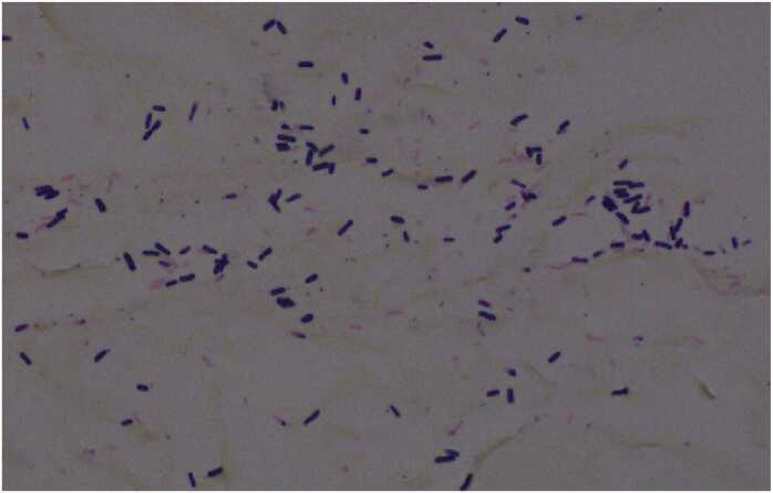
Culture. What the laboratory needs and reports:
- Anaerobic culture is essential; routine aerobic culture will miss the organism
- C. perfringens grows rapidly, in 8-12 hours
- Double-zone haemolysis on blood agar (Nagler reaction)
- Lecithinase-positive on egg yolk agar
- Speciation by biochemical tests or PCR
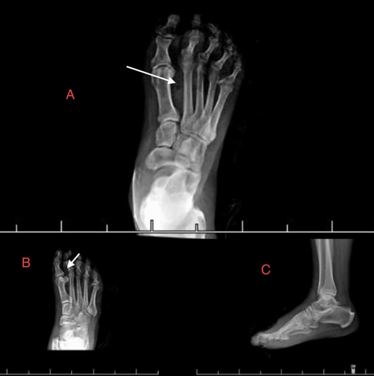
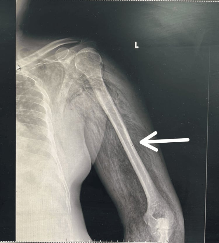
Plain radiographs. Gas gangrene produces a characteristic feathery pattern of gas dissecting deep along muscle and fascial planes, and the gas may extend beyond the clinical margins of the disease. Subcutaneous emphysema from wound contamination has a more linear pattern, and superficial traumatic emphysema may be no more than a small rim.
In the foot case shown, the gas was most conspicuous over the forefoot on the AP and oblique films, and the lateral projection was less sensitive.
A film showing gas is useful for confirming clinical suspicion and supports immediate source control; with systemic toxicity, deep gas mandates immediate surgical exposure. Imaging lags behind the clinical disease, though: a normal film does not exclude the diagnosis, cannot exclude early myonecrosis and must not delay exploration. Trust the clinical examination.
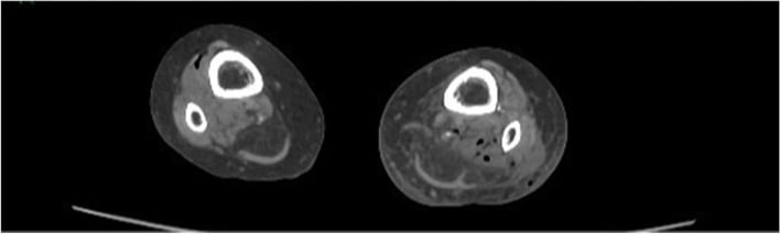
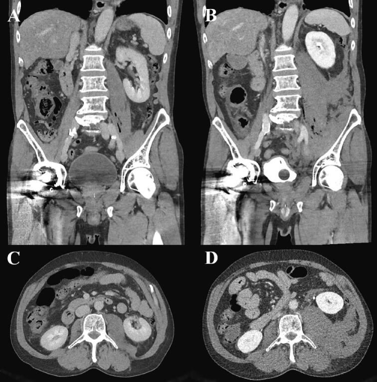
CT. CT is more sensitive for gas than plain film, shows the extent of gas tracking and muscle involvement, and helps surgical planning. Do not delay surgery for CT if the diagnosis is clear. When the patient is stable, image the full clinically threatened region: spontaneous clostridial myonecrosis can produce a skip distribution, with gas in separated muscle compartments. CT maps the extent of disease; it does not decide whether surgery is required.
The tempo. In one case of Clostridium septicum myonecrosis, serial CT only 2.5 hours apart showed progression from minor gas around the iliopsoas to extensive intramuscular gas and swelling. An apparently limited abnormality can become anatomically extensive during a routine diagnostic interval.
Differential Diagnosis: Clostridial Cellulitis and Necrotising Fasciitis
The first hour is the same. Clostridial myonecrosis and necrotising fasciitis are different diseases in different tissue planes with different organisms, and examiners will ask you to separate them. Stevens's review of necrotising soft tissue infections states the practical position plainly: despite subtle nuances that may distinguish one entity from another, the clinical approaches to diagnosis and treatment are highly similar. Resuscitate, give broad empirical cover including a protein-synthesis inhibitor, and get to theatre.
- Gas gangrene (clostridial myonecrosis)
- C. perfringens after trauma or surgery, C. septicum when spontaneous; also C. novyi
- Necrotising fasciitis
- Type I polymicrobial (aerobes plus anaerobes) or Type II group A Streptococcus, sometimes with S. aureus
- Clostridial cellulitis
- Clostridium species (non-invasive)
- Gas gangrene (clostridial myonecrosis)
- MUSCLE, the defining difference; fascia is involved secondarily
- Necrotising fasciitis
- Fascia and subcutaneous tissue, with muscle characteristically spared until late
- Clostridial cellulitis
- Devitalised subcutaneous tissue and fascial planes; muscle spared
- Gas gangrene (clostridial myonecrosis)
- Extremely rapid, over hours; among the fastest of all surgical infections
- Necrotising fasciitis
- Rapid (1-2 cm/hour), usually over hours to a few days
- Clostridial cellulitis
- Slower, more indolent
- Gas gangrene (clostridial myonecrosis)
- Gas typical and often abundant, in muscle planes; crepitus unreliable
- Necrotising fasciitis
- Gas in fascial planes in Type I polymicrobial disease, characteristically absent in Type II streptococcal; crepitus variable
- Clostridial cellulitis
- Abundant superficial gas; crepitus common and often more obvious than in myonecrosis
- Gas gangrene (clostridial myonecrosis)
- Bland early, out of proportion to the pain
- Necrotising fasciitis
- Skin changes often more obvious, with bullae, dusky discolouration and anaesthesia
- Clostridial cellulitis
- Florid gas and crepitus with little pain
- Gas gangrene (clostridial myonecrosis)
- Severe: haemolysis, shock, MODS
- Necrotising fasciitis
- Severe
- Clostridial cellulitis
- Mild
- Gas gangrene (clostridial myonecrosis)
- 25-40%
- Necrotising fasciitis
- 20-30%
- Clostridial cellulitis
- Low (less than 5%)
- Gas gangrene (clostridial myonecrosis)
- Muscle is dull brick-red to grey-green, does not contract to diathermy and does not bleed
- Necrotising fasciitis
- Fascia is grey and friable and dissects off with a finger, with dishwater fluid and no frank pus
- Clostridial cellulitis
- Muscle viable: red, contractile, bleeding
- Gas gangrene (clostridial myonecrosis)
- RADICAL debridement following muscle to bleeding, contracting tissue, which is why amputation is more often necessary
- Necrotising fasciitis
- Wide debridement following the fascial plane, which can often preserve muscle
- Clostridial cellulitis
- Limited debridement
Clostridial cellulitis is the trap. It is a separate entity: a clostridial infection of devitalised subcutaneous tissue and fascial planes that spares the underlying muscle, with comparatively little systemic toxicity and little pain.
Its gas and crepitus can be florid, so it can look more alarming on examination and X-ray than early myonecrosis. The trap runs both ways, over-calling it gas gangrene or being reassured by "only cellulitis" when muscle is in fact involved, and the distinction decides whether the patient needs a limb-sacrificing radical debridement or a much more limited one.
The muscle decides. Imaging cannot reliably separate the two. The only reliable way is surgical exploration and direct inspection of the muscle by the 4 Cs (see Management), which is why the safe response to any crepitant clostridial infection is to explore rather than guess from imaging.
Confirmed cellulitis needs debridement of the necrotic subcutaneous tissue, opening and drainage of the gas, and antibiotics, but not radical muscle excision or amputation. If muscle involvement is found, it is managed as full clostridial myonecrosis.
Neither imaging nor a scoring system should delay theatre in a patient with pain out of proportion, systemic toxicity and a suspicious wound. Gas on plain film supports the diagnosis but its absence excludes nothing, particularly in streptococcal disease. The diagnosis is made by looking at the muscle and the fascia, and the same operation - urgent, wide and repeated - is the answer to both.
Management
- Surgical debridement - the only definitive treatment; arrange theatre immediately
- Resuscitation - IV fluids, blood products, ICU care
- Antibiotics - penicillin G plus clindamycin, started immediately but not instead of surgery
- Hyperbaric oxygen - adjunctive only, never delaying surgery
- Repeat debridement - a planned second look in 6-24 hours
Surgery cannot wait. Radical debridement is life-saving and antibiotics alone are inadequate. Do not delay for imaging, cultures or hyperbaric oxygen, or wait for antibiotics or HBO to work.
Preparation. Notify theatre of the emergency. The patient may need intubation for an unstable airway or shock, invasive monitoring with arterial and central lines, and blood products on standby.
Exposure. Long incisions expose all the affected muscle compartments and extend beyond the clinical margins of the disease. Skin flaps are not a priority; the muscle is the target.
Excision. Remove all non-viable muscle, back to bleeding, contractile muscle. Viable muscle has the 4 Cs:
- Colour - red or pink
- Contractility - twitches with diathermy
- Consistency - firm, not mushy
- Capacity to bleed - bleeds when cut
Gas gangrene muscle is grey-green, non-contractile, mushy and non-bleeding, and all of it must be excised. All infected tissue must be removed or the patient will die.
The most common surgical error is inadequate debridement. If in doubt, remove more tissue. A patient can survive without a muscle group; they cannot survive with residual gas gangrene. Amputation is preferable to death from under-debridement.
The wound. Leave it open for a planned second look, packed loosely with saline-soaked gauze or under VAC therapy. Primary closure traps infection.
The return. Go back at 6-24 hours for reassessment. Multiple debridements are usually required, an average of 3-4, continuing until no further necrotic tissue is identified.
Amputation. Amputation is a definitive source-control procedure, not a failure, and a timely one can convert a dying patient into a survivor. Do not persist with repeated debridements if the patient continues to deteriorate; consider amputation earlier.
- Description
- Uncontrolled shock despite resuscitation and debridement
- Timing
- Emergency amputation
- Description
- Greater than 50% of limb musculature involved
- Timing
- After initial debridement assessment
- Description
- Non-reconstructible vascular damage with ischaemia
- Timing
- Emergency amputation
- Description
- Ongoing sepsis despite 2-3 aggressive debridements
- Timing
- Salvage amputation
- Description
- Osteomyelitis, septic joints beyond salvage
- Timing
- After initial assessment
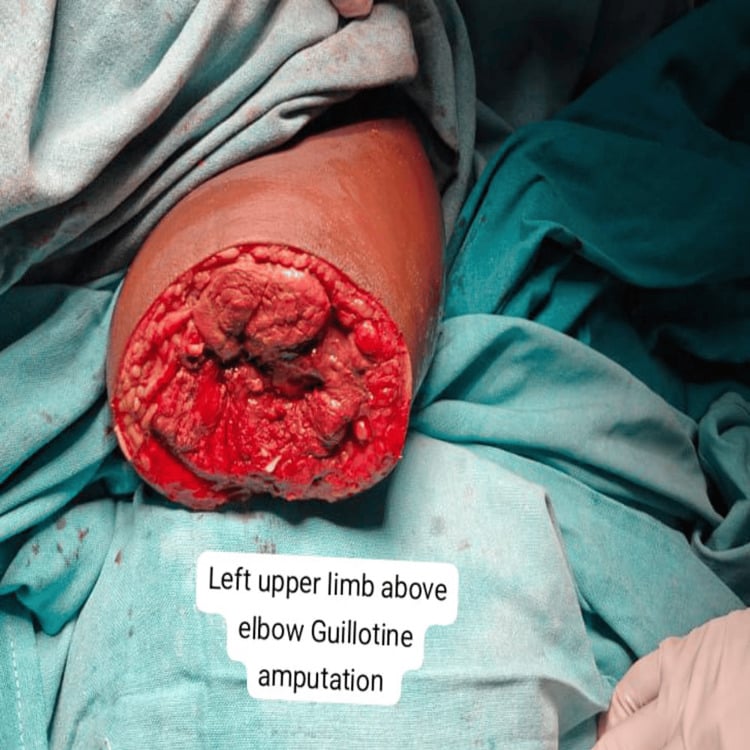
In the upper limb, an above-elbow guillotine amputation provides rapid proximal source control when myonecrosis is too extensive for serial compartment debridement. The stump is deliberately left open, and perfusion and muscle viability are reassessed at a planned second look before definitive revision and closure.
Antibiotics. Start immediately, alongside surgery and never instead of it.
- Dose
- 4 million units IV every 4 hours
- Rationale
- Bactericidal - kills clostridia, first-line agent
- Dose
- 900mg IV every 8 hours
- Rationale
- Inhibits toxin production at ribosomal level
- Dose
- 500mg IV every 8 hours
- Rationale
- If penicillin allergic - covers anaerobes
- Dose
- 1g IV every 8 hours
- Rationale
- Broad Gram-negative and anaerobic cover
Why penicillin and clindamycin. Penicillin provides rapid bactericidal activity against clostridia. Clindamycin inhibits toxin production: by blocking protein synthesis at the ribosome, it reduces alpha-toxin release even from dying bacteria, which matters because a cell wall-active antibiotic such as penicillin can transiently increase toxin release during bacterial lysis.
Clindamycin is also unaffected by inoculum size, penetrates necrotic tissue better and remains effective against stationary-phase bacteria. The combination is superior to either agent alone.
Hyperbaric oxygen. The proposed benefits are that HBO:
- Inhibits clostridial growth, the organisms being obligate anaerobes
- Inhibits alpha-toxin production
- Enhances neutrophil bacterial killing
- Improves tissue oxygen for wound healing
- May demarcate viable from non-viable tissue
There are no randomised controlled trials in humans, and the evidence is limited to case series and animal studies. HBO is not available at most centres, and the chamber is a difficult environment for an unstable patient.
Hyperbaric oxygen is never a substitute for surgery and never delays debridement. If available, it may be used between debridements or after operation. Transfer to an HBO facility should occur only after adequate surgical debridement, and only if the patient is stable for transport.
Critical care. Monitoring and organ support by system:
- Monitoring
- Invasive BP, CVP, cardiac output if available
- Intervention
- Vasopressors, inotropes, fluid resuscitation
- Monitoring
- Hourly urine output, creatinine
- Intervention
- Fluids for myoglobinuria, early RRT if needed
- Monitoring
- Haemoglobin, coagulation, fibrinogen
- Intervention
- Blood transfusion, FFP, platelets, cryoprecipitate for DIC
- Monitoring
- Ventilatory support as needed
- Intervention
- Lung protective ventilation if ARDS develops
- Monitoring
- Early enteral nutrition when feasible
- Intervention
- High protein, high calorie requirements
Complications
Death. Death, at the rate given in the Overview, comes from multi-organ failure and toxaemia. Its management is early radical surgery and ICU support.
- Incidence
- 20-30%
- Mechanism
- Extensive myonecrosis, source control
- Management
- Life-saving procedure when indicated
- Incidence
- 30-50%
- Mechanism
- Myoglobinuria, haemoglobinuria, shock
- Management
- Fluids, renal replacement therapy
- Incidence
- 20-30%
- Mechanism
- Sepsis, toxin-mediated coagulopathy
- Management
- Treat underlying cause, blood products
- Incidence
- 60-80%
- Mechanism
- Systemic toxaemia, bacteraemia
- Management
- Vasopressors, fluids, source control
- Incidence
- 10-20%
- Mechanism
- Sepsis, fluid resuscitation
- Management
- Lung protective ventilation
- Incidence
- Common
- Mechanism
- Radical debridement required
- Management
- Staged reconstruction, skin grafting
- Incidence
- Variable
- Mechanism
- Alpha toxin lysis of red cells
- Management
- Transfusion
Poor prognostic factors include trunk involvement, spontaneous (non-traumatic) onset, leukopenia, haemolysis with jaundice, shock at presentation, renal failure and delayed surgical intervention. Mortality can exceed 50% with multiple risk factors.
Guidelines, Registries & Global Practice
Global Epidemiology
Clostridial myonecrosis is rare but globally distributed. In high-income settings it follows contaminated trauma (agricultural, road traffic, crush/compartment injuries), open fractures, and post-operative or injection-drug-use exposures; spontaneous cases (typically C. septicum) signal occult GI or haematological malignancy. In conflict zones, low-resource settings, and after natural disasters the incidence rises sharply due to delayed wound care, soil contamination, and limited surgical access. Late presentation — common where distances to surgical care are large — is the dominant driver of higher mortality everywhere.
Side-by-Side Guidance
- Antibiotic Position
- Penicillin G + clindamycin for clostridial myonecrosis
- Surgery / Adjuncts
- Urgent surgical debridement; HBO not recommended (may delay surgery)
- Antibiotic Position
- Benzylpenicillin + clindamycin; metronidazole if penicillin-allergic
- Surgery / Adjuncts
- Emergency debridement and planned re-look; amputation for source control
- Antibiotic Position
- Early empirical cover, then targeted
- Surgery / Adjuncts
- Aggressive debridement to viable muscle (4 Cs); staged soft-tissue reconstruction
- Antibiotic Position
- Adjunctive only
- Surgery / Adjuncts
- HBO listed as accepted adjunct AFTER, never instead of, debridement
Guidance converges on the essentials: clinical diagnosis, immediate radical debridement, penicillin + clindamycin, and resuscitation. The main point of difference is HBO — endorsed as an accepted adjunct by hyperbaric bodies but explicitly cautioned against by IDSA where it risks delaying surgery.
Resource-Setting Variation
- Well-resourced centres: rapid theatre access, ICU organ support, repeated re-look debridements, selective limb salvage, and HBO at a minority of sites.
- Limited-resource / remote settings: source control must not wait for transfer — a guillotine amputation performed locally is frequently life-saving, with retrieval to a higher-level ICU only once the patient is stabilised. Telemedicine support and early empirical penicillin + clindamycin (or metronidazole) reduce delay-related mortality.
There is no dedicated international registry for gas gangrene; outcome data derive from single-centre series, the spontaneous-C. septicum literature, and trauma databases.
Controversies & Areas of Uncertainty
Hyperbaric oxygen. This is the most contested issue. Hyperbaric medicine bodies list clostridial myonecrosis as an accepted indication, citing in-vitro toxin suppression and historical series. IDSA and many trauma surgeons argue that there are no randomised data, that transport destabilises critically ill patients and that HBO can delay definitive surgery. The working consensus is the one set out under Management: adjunct only.
Limb salvage or early amputation. When persisting with serial debridement becomes futile has no validated threshold. The indications tabulated under Management favour early amputation as source control, but the exact tipping point is a clinical judgement made under pressure.
Antibiotic nuances. Penicillin plus clindamycin is standard, but rising clindamycin resistance in some Clostridium isolates and uncertainty over the optimal duration are unresolved. Whether to add broad-spectrum cover empirically, for a possible polymicrobial necrotising infection, before the organism is confirmed varies by centre.
Adjunctive IVIG. Intravenous immunoglobulin is debated for streptococcal toxic-shock-associated necrotising infection. Its role in clostridial myonecrosis is not established, and it should never divert resources from surgery.
Exam Viva Scenarios
Practise clinical reasoning and management decisions out loud
“A 65-year-old diabetic man is 36 hours post ORIF of his open right tibial fracture (Gustilo IIIB). He develops severe pain in his leg despite adequate analgesia. His wound is tense and there is bronze discoloration spreading proximally. Temperature 39.2, HR 120, BP 90/60. What is your assessment and management?”
“A 72-year-old woman presents with 12 hours of severe right thigh pain. There is no history of trauma. She has lost 8kg over the past 3 months. Her thigh is swollen, tense, with mottled discoloration. Crepitus is palpable. She is confused with a BP of 80/50. What is your diagnosis and what additional investigation is mandatory after stabilization?”
“You are called to assist at a rural hospital 4 hours from the nearest major center. A 45-year-old farmer was injured 18 hours ago when his leg was trapped under a tractor. He was self-rescued and drove himself to hospital. He now has an obviously necrotic right lower leg with extensive gas gangrene extending to the thigh. He is in septic shock requiring high-dose vasopressors. The hospital has no vascular surgery, limited blood products, and no ICU. What are your options?”
Organism and Toxin
- Clostridium perfringens (80%), C. septicum (spontaneous = GI malignancy)
- Alpha toxin = lecithinase/phospholipase C - destroys cell membranes
- Causes myonecrosis, hemolysis, shock
- Gram-positive rods, few WBCs on Gram stain
Clinical Diagnosis
- Pain OUT OF PROPORTION is earliest sign
- Bronze/bronze-brown skin discoloration
- Crepitus in only 50% - absence does NOT exclude
- Dishwater exudate - thin, serosanguinous, foul smell
- Systemic toxicity - fever, tachycardia, shock, confusion
Investigations
- Clinical diagnosis - do NOT delay surgery for tests
- X-ray: feathery gas pattern in muscle planes
- Bloods: elevated CK, hemolysis markers, acidosis, DIC
- Gram stain: large gram-positive rods, paucity of WBCs
Antibiotics
- Penicillin G 4MU IV 4-hourly (bactericidal)
- PLUS Clindamycin 900mg IV 8-hourly (inhibits toxin production)
- Alternative: metronidazole if penicillin allergic
- Start immediately but NOT instead of surgery
Surgical Management
- EMERGENCY radical debridement - cannot wait
- Excise ALL necrotic muscle (4 Cs: color, contractility, consistency, capacity to bleed)
- Leave wounds OPEN - no primary closure
- Return to OR every 6-24 hours for repeat debridement
- Average 3-4 debridements required
Amputation Indications
- Life-threatening sepsis uncontrolled by debridement
- Greater than 50% limb muscle involvement
- Non-reconstructible vascular injury
- Failed multiple debridements with ongoing sepsis
Hyperbaric Oxygen
- ADJUNCTIVE only - never delays surgery
- No RCT evidence in humans
- Inhibits clostridial growth and toxin production
- Use between debridements if available and patient stable
Key Exam Points
- Spontaneous gas gangrene (C. septicum) = investigate for GI malignancy
- Under-debridement is the most common surgical error
- Mortality 25-40% with treatment, approaches 100% without surgery
- Pain out of proportion + contaminated wound = OR immediately
Evidence Base
Spontaneous C. septicum Gas Gangrene: A Literature Review
- Systematic review of 94 published cases of spontaneous (non-traumatic) C. septicum gas gangrene
- Known or occult malignancy present in 71% of patients
- Overall mortality 67% despite treatment
- Colorectal/GI malignancy the dominant association; haematological malignancy and neutropenia also implicated
Genetic Evidence for the Essential Role of Alpha-Toxin in Clostridial Myonecrosis
- Allelic-exchange plc (alpha-toxin) mutants showed markedly reduced virulence in a mouse myonecrosis model
- theta-toxin (perfringolysin O) mutants were comparatively less attenuated
- Provides definitive genetic proof that alpha-toxin (phospholipase C) is essential for gas gangrene
- Underpins the rationale for toxin-suppressing antibiotic therapy
Hyperbaric-Oxygen Therapy
- Authoritative review of accepted indications for hyperbaric oxygen, including clostridial myonecrosis
- Evidence for gas gangrene rests on animal data and uncontrolled clinical series, not randomised trials
- Proposed mechanisms: bacteriostasis of anaerobes, suppression of alpha-toxin, and improved neutrophil killing
- HBO positioned as an adjunct to surgery and antibiotics, not a substitute
Clindamycin vs Penicillin for Experimental Gas Gangrene
- In a mouse C. perfringens myonecrosis model, clindamycin, metronidazole, rifampin and tetracycline were all more efficacious than penicillin
- Penicillin-treated survival was not significantly better than untreated controls despite very high serum levels
- Clindamycin was effective across a broad dose range; efficacy of all agents fell with treatment delay or larger inoculum
- Companion work showed penicillin permits persistent alpha-toxin activity whereas clindamycin suppresses it
Necrotizing Soft Tissue Infections
- Contemporary review of necrotizing soft tissue infections including clostridial myonecrosis
- Early radical debridement is the single most important determinant of survival; delay increases mortality
- Antibiotics and resuscitation are adjuncts to, not substitutes for, source control
- Diagnosis is primarily clinical — imaging must not delay operative exploration
IDSA Practice Guidelines for Skin and Soft Tissue Infections (2014)
- Recommends urgent surgical inspection/debridement for suspected necrotizing infection or clostridial myonecrosis (strong recommendation)
- Penicillin plus clindamycin is the recommended regimen for clostridial myonecrosis
- Empirical broad-spectrum cover until clostridial aetiology and source are confirmed
- Hyperbaric oxygen not recommended as it may delay resuscitation and surgical debridement