Gastrocnemius (Equinus) Contracture & the Silfverskiöld Test
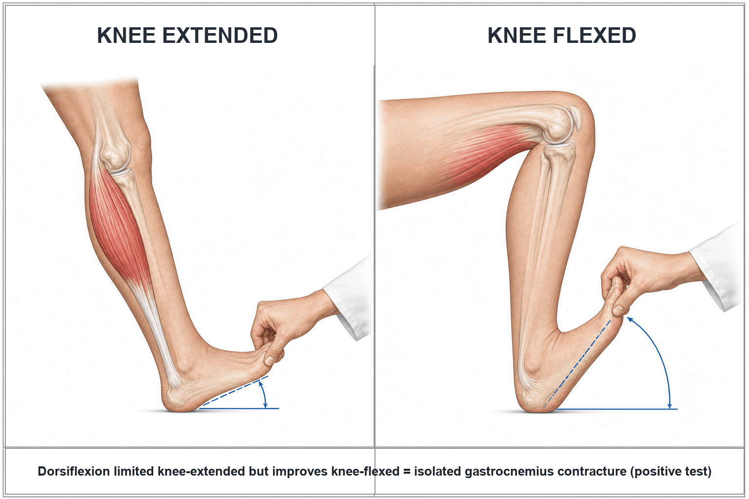
Ankle dorsiflexion is limited with the knee extended but improves with the knee flexed. Flexing the knee slackens the biarticular gastrocnemius (which crosses the knee), so the remaining restriction is gastrocnemius-driven. Treat with gastrocnemius recession.
Dorsiflexion is limited regardless of knee position - the soleus/Achilles (which do NOT cross the knee) are also tight. This is a combined gastroc-soleus contracture, where tendo-Achilles lengthening is considered (accepting some push-off weakness).
MAP-PD — what a tight gastrocnemius causesEquinus drives forefoot/hindfoot pathology
Hook:A tight gastrocnemius is an under-diagnosed root cause across the foot: Metatarsalgia, Achilles tendinopathy, Plantar fasciitis, PTTD, Diabetic ulcer (MAP-PD).
Overview & Epidemiology
Isolated gastrocnemius contracture is increasingly recognised as an under-appreciated, correctable root cause of common foot and ankle complaints, rather than an incidental finding. Some degree of gastrocnemius tightness is highly prevalent in the general adult population and rises with age, sedentary posture and (in women) sustained high-heel wear; it is frequently present in patients presenting with metatarsalgia, plantar fasciitis, Achilles tendinopathy, adult-acquired flatfoot and the diabetic forefoot ulcer. The conceptual shift the topic tests is to examine for equinus in any forefoot/hindfoot complaint — because correcting the contracture can resolve a problem that repeatedly fails local treatment.
Pathophysiology & Functional Anatomy
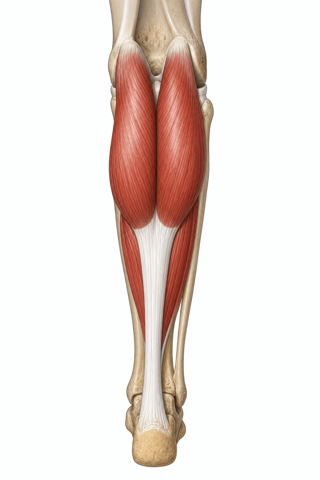
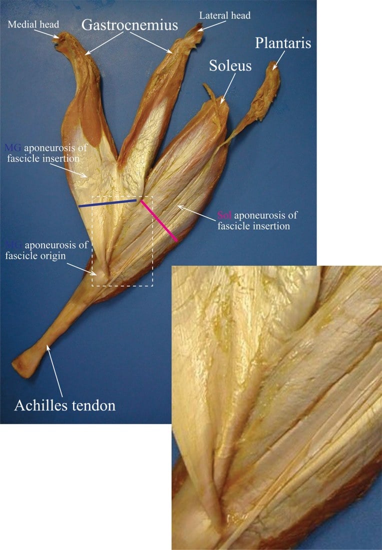
The gastrocnemius arises from the femoral condyles (above the knee) and joins the soleus to form the Achilles tendon - so it spans both the knee and the ankle. The soleus arises below the knee and crosses only the ankle. Because of this, a contracted gastrocnemius restricts ankle dorsiflexion most when the knee is straight and is "let out" when the knee bends - the basis of the Silfverskiöld test.
- Equinus = limited ankle dorsiflexion (commonly defined as less than ~10 degrees with the knee extended and the hindfoot held neutral).
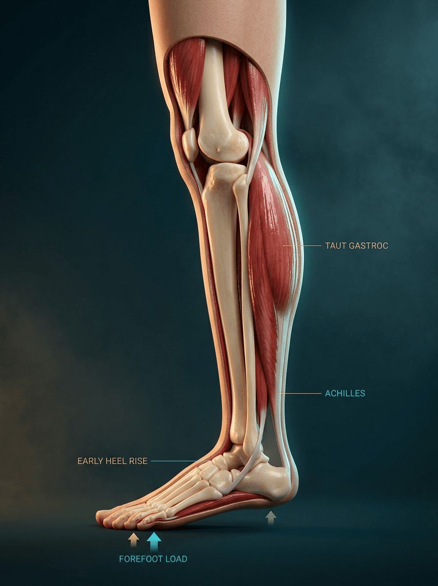
- A tight gastrocnemius forces the foot into relative plantarflexion, transferring load to the forefoot and altering gait (early heel rise, increased toe-extensor recruitment).
- This forefoot overload underlies mechanical metatarsalgia, and contributes to plantar fasciitis, Achilles tendinopathy, posterior tibial tendon dysfunction, midfoot/forefoot deformity, and diabetic forefoot ulceration.
Aetiology of equinus contracture
Equinus is the end-point of several distinct processes, and identifying the cause changes management — a spastic equinus is treated very differently from a constitutional one.
The commonest pattern in adult foot-and-ankle practice — a constitutionally tight gastrocnemius that increases with age, sedentary lifestyle and sustained high-heel wear, presenting as a Silfverskiöld-positive isolated contracture. This is the group that responds to stretching and, if refractory, gastrocnemius recession.
Upper-motor-neuron spasticity — cerebral palsy (the classic paediatric cause of equinus), stroke, traumatic brain injury — produces a dynamic then fixed equinus. Treatment integrates tone management (botulinum toxin, serial casting) with lengthening, and tends to involve the whole gastroc-soleus complex.
Prolonged immobilisation (a cast/splint in plantarflexion), compartment syndrome / calf fibrosis, deep posterior compartment scarring, or healing in equinus after tibial/pilon/calcaneal trauma can fix the ankle in plantarflexion.
Diabetes stiffens the Achilles–gastrocnemius complex through non-enzymatic glycosylation of collagen, raising forefoot pressure and ulcer risk; congenital causes (e.g. residual or relapsed clubfoot) and a congenitally short gastrocnemius also present with fixed equinus.
Clinical Assessment
Examination
- Measure ankle dorsiflexion with the hindfoot held in neutral (inversion) to lock the midfoot and avoid false dorsiflexion through the midfoot.
- Perform the Silfverskiöld test: dorsiflexion with the knee extended then with the knee flexed to ~90 degrees. Improvement with knee flexion = isolated gastrocnemius contracture.
- Examine the forefoot for metatarsalgia/callosities and assess gait (early heel rise).
Investigations
Gastrocnemius equinus is fundamentally a clinical diagnosis — the goniometric dorsiflexion measurement (hindfoot neutral, knee extended then flexed) and the Silfverskiöld test are the core "investigations." Adjuncts have a supporting role:
- Weight-bearing radiographs of the foot/ankle to identify associated or alternative pathology (forefoot deformity, midfoot arthritis, flatfoot) — equinus itself is not seen radiographically.
- Pedobarography / plantar pressure mapping can objectively demonstrate the elevated forefoot pressures that the contracture produces, useful in research and in the diabetic foot.
- Gait analysis may show early heel rise and increased toe-extensor recruitment.
- There is no role for routine MRI/ultrasound for the contracture itself, though they may be used to assess an associated tendinopathy or the plantar fascia.
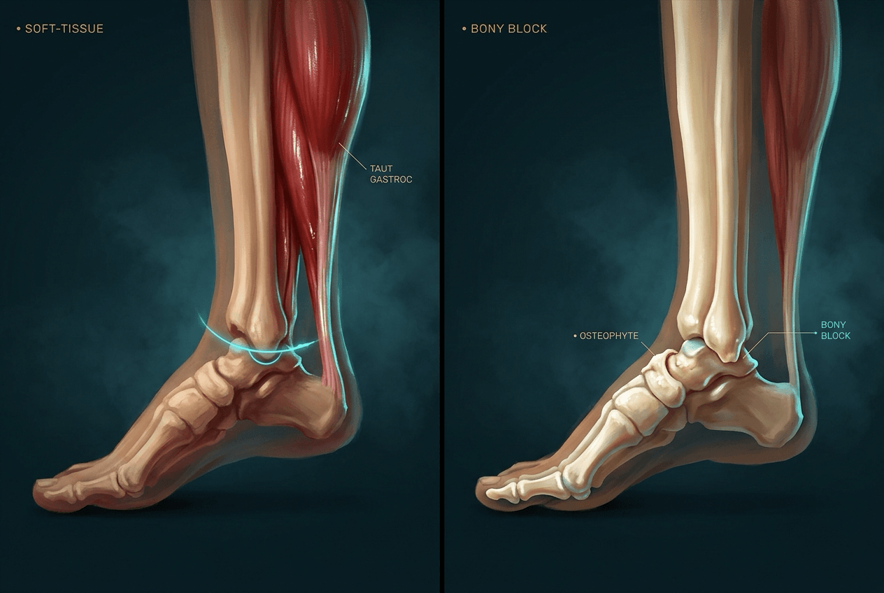
A muscle lengthening only helps soft-tissue equinus. Before attributing limited dorsiflexion to a tight gastroc-soleus, exclude a structural (bony) block and a pseudoequinus:
- Bony anterior-ankle block: anterior tibiotalar osteophytes / anterior ankle impingement or tibiotalar osteoarthritis limit dorsiflexion with a hard, abrupt end-feel that does not change with knee position (so it can masquerade as a Silfverskiöld-negative "combined" contracture) and will not respond to a gastrocnemius recession or TAL — it needs a cheilectomy/osteophyte excision instead. Look for the spur on a weight-bearing lateral radiograph and feel for the abrupt (versus springy/soft) end-point.
- Pseudoequinus: apparent equinus generated through the forefoot/midfoot — a plantarflexed first ray or forefoot equinus in a cavus foot, or simply measuring dorsiflexion through an unlocked midfoot — which is why dorsiflexion is measured with the hindfoot held in neutral/inversion to lock the transverse tarsal joint.
So the sequence is: confirm the limit is real (hindfoot neutral), confirm it is soft-tissue (soft end-feel, no blocking spur), then localise it with the Silfverskiöld test.
Management
Non-operative
- Stretching programmes (knee-extended gastrocnemius stretches), eccentric loading, and night splints/dorsiflexion orthoses.
- Heel lifts/orthoses and footwear modification to off-load the forefoot symptomatically.
- Most patients improve; these are the mainstay before any surgery.
Equinus correction in cerebral palsy carries a specific, examinable hazard absent in the adult constitutional contracture: over-lengthening the triceps surae causes an iatrogenic calcaneus/crouch gait that is very hard to reverse — "the calf muscle is a one-way street." Because the gastroc-soleus provides the plantarflexion–knee-extension couple that holds the knee straight in stance, an excessive isolated tendo-Achilles lengthening — especially in a spastic DIPLEGIC child already prone to crouch — collapses the child into excessive ankle dorsiflexion and knee flexion. The principles:
- Prefer the most muscle-selective / proximal lengthening (gastrocnemius recession — Strayer/Baumann — over a tendo-Achilles lengthening) guided by the Silfverskiöld test and ideally instrumented gait analysis; reserve a true TAL for a genuine fixed combined contracture.
- Address dynamic equinus first with tone management — botulinum toxin and serial casting — before it becomes fixed; integrate with the child's GMFCS level and the Winters-Gage hemiplegic gait pattern.
- Isolated TAL is more dangerous in diplegia than hemiplegia (the diplegic child has bilateral crouch tendency); never lengthen the Achilles aggressively in a child who already over-dorsiflexes.
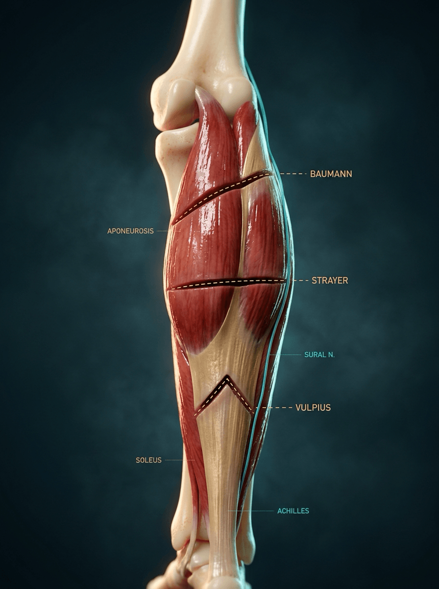
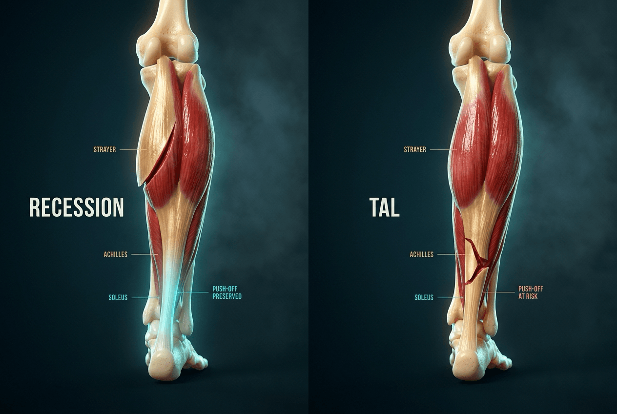
Baumann → Strayer → Vulpius → (Achilles)Lengthening procedures, proximal to distal
Hook:Achilles-sparing recessions (Baumann/Strayer/Vulpius) for an isolated gastrocnemius contracture; reserve true tendo-Achilles lengthening for the combined contracture.
Complications
The contracture's main "complication" is the forefoot/hindfoot pathology it drives (above); the surgical complications are level-specific.
- Note
- The key risk in a Strayer recession — the nerve lies near the musculotendinous junction; numbness or a painful neuroma
- Note
- Especially after tendo-Achilles lengthening (gastrocnemius recession largely preserves power); usually transient but can persist if over-lengthened
- Note
- Excessive Achilles lengthening can produce a weak, calcaneus-gait foot — avoid in the combined contracture by lengthening judiciously
- Note
- Inadequate release or failure to address the correct level (gastroc vs combined)
- Note
- Including at the posteromedial calf incision
BENDSilfverskiöld Interpretation
Hook:BEND the knee: if dorsiflexion eases, it is the (biarticular) gastrocnemius.
Guidelines, Registries & Global Practice
Global Practice Picture
Isolated gastrocnemius contracture is increasingly recognised worldwide as an under-appreciated, correctable driver of forefoot and hindfoot pathology. The internationally consistent approach: measure dorsiflexion in hindfoot neutral, apply the Silfverskiöld test to localise the contracture, treat conservatively first, and use an Achilles-sparing gastrocnemius recession (rather than Achilles lengthening) for refractory isolated gastrocnemius equinus.
Side-by-Side Synthesis
- Isolated gastrocnemius
- Limited
- Combined gastroc-soleus
- Limited
- Isolated gastrocnemius
- Improves
- Combined gastroc-soleus
- Still limited
- Isolated gastrocnemius
- Positive
- Combined gastroc-soleus
- Negative
- Isolated gastrocnemius
- Gastrocnemius recession (Strayer)
- Combined gastroc-soleus
- Tendo-Achilles lengthening
- Isolated gastrocnemius
- Preserved
- Combined gastroc-soleus
- May be weakened
MCQ Practice Points
A Silfverskiöld test is recorded as 15 degrees of dorsiflexion with the knee extended, so equinus is excluded - yet the patient has classic forefoot overload. What is the likeliest technical error?
The hindfoot was not held in neutral. Allowing the heel to evert unlocks the transverse tarsal joints, and the foot then dorsiflexes through the midfoot rather than the ankle. The measured angle is real movement but it is not tibiotalar, so a genuine gastrocnemius contracture reads as normal. Hold the hindfoot inverted to lock the midfoot, and measure the angle between the tibia and the lateral border of the foot, not the plantar arch.
Does a negative Silfverskiöld test mean a gastrocnemius recession will not gain dorsiflexion?
No - and this is the counterintuitive one. DeOrio and Lewis tested 29 ankles under anaesthesia before and after a Strayer. The 23 with a negative preoperative test still gained 11.3 degrees with the knee extended (p less than 0.0001); those with a markedly positive test gained more (17.8 degrees), but the negative group was not a non-responder group. The likely reason is anatomical: the gastrocnemius aponeurosis is confluent with the soleal fascia, so releasing it lengthens more than a test of knee-position dependence predicts.
it tells you where the dominant restraint sits and therefore whether you can expect to correct the deformity without touching the Achilles - which is the point, because it is Achilles lengthening that carries the push-off weakness and calcaneal-gait risk. Read a negative test as "a recession alone may not be enough", not as "a recession will do nothing".
In the Mueller RCT of Achilles lengthening for diabetic neuropathic forefoot ulcers, which outcome reached significance?
Recurrence, not healing. Ulcers healed in 100 percent with lengthening versus 88 percent with total-contact cast alone, and that difference was not significant. Recurrence was: 15 versus 59 percent at 7 months and 38 versus 81 percent at 2 years, both significant. Quote it as a recurrence trial.
the peak forefoot pressure reduction - the presumed mechanism - had returned to baseline by 7 months, while the recurrence benefit persisted to 2 years. Plantarflexor torque dipped and also recovered. The variable that was still improved at 7 months was dorsiflexion range, which is the one to protect with stretching, splinting and footwear after the ulcer heals.
What decides between a Baumann, a Strayer, a Vulpius and an Achilles lengthening?
How much correction is needed, traded against how much push-off you are willing to lose. They form an ascending ladder: Baumann (intramuscular, proximal, most conservative), Strayer (distal gastrocnemius aponeurosis - the workhorse), Vulpius (gastrocsoleus aponeurosis, so it lengthens the soleus too), then tendo-Achilles lengthening, which lengthens everything and carries the greatest risk of overlengthening, plantarflexion weakness and calcaneal gait. The rule is to take the most proximal procedure that achieves the correction, because complications climb as you move distally. Sural nerve injury is the characteristic complication of the Strayer - the nerve lies immediately deep and lateral to the aponeurosis at that level.
Viva Scenarios
Practise clinical reasoning and management decisions out loud
“A patient has persistent forefoot pain and callosities under the metatarsal heads despite orthoses, and you notice limited ankle dorsiflexion. How do you assess and treat this?”
“A patient with diabetic neuropathy has a recurrent neuropathic ulcer under the metatarsal heads that heals in a total-contact cast but breaks down again. Examination shows ankle dorsiflexion limited to about 0 degrees that does not improve with knee flexion. How does the equinus contribute, and what would you do?”
Concept
- Gastrocnemius is biarticular (knee + ankle)
- Equinus = limited ankle dorsiflexion
- Measure DF in hindfoot neutral
- Drives forefoot overload
Silfverskiöld Test
- Positive: DF limited knee-extended, improves knee-flexed
- = isolated gastrocnemius contracture
- Negative: limited regardless = combined
- Determines recession vs TAL
Management
- Stretch / night splint / orthoses first
- Gastroc recession (Strayer) - Achilles-sparing
- TAL for combined contracture
- Protect the sural nerve
Evidence Base
All four citations were checked against PubMed. The strongest is the Mueller RCT (Level 1) showing that Achilles tendon lengthening cuts diabetic forefoot-ulcer recurrence (15 vs 59% at 7 months) — the firmest proof that correcting equinus changes outcomes, and the one to quote. The recession evidence (DeOrio, Maskill) is Level IV case-series: gastrocnemius recession reliably gains dorsiflexion and relieves recalcitrant isolated foot pain while sparing push-off. Cortina is a narrative review tying the mechanism (forefoot overload) to the operation. The honest line: equinus correction is RCT-proven for the diabetic ulcer and well-supported by case series for mechanical foot pain — but the recession-versus-lengthening choice rests on the Silfverskiöld test and expert consensus, not head-to-head trials.
Effect of Achilles tendon lengthening on neuropathic plantar ulcers. A randomized clinical trial
- Randomised trial of 64 diabetic patients with a neuropathic forefoot ulcer: total-contact cast alone versus cast plus percutaneous Achilles tendon lengthening
- Ulcer recurrence was far lower with Achilles lengthening: 15 versus 59 percent at 7 months and 38 versus 81 percent at 2 years
- Achilles lengthening increased ankle dorsiflexion and reduced peak forefoot pressure; plantarflexor torque fell transiently then recovered by 7 months
- THE PRESSURE REDUCTION WAS ALSO TRANSIENT - peak forefoot pressure had returned to baseline by 7 months, yet the recurrence benefit was still present at 2 years, so peak plantar pressure at a single timepoint does not explain the durable effect. The dorsiflexion gain, which was still present at 7 months, is the better candidate and the better thing to protect in follow-up
- Healing itself did not differ significantly: 100 percent versus 88 percent (p greater than 0.05). The trial is evidence about RECURRENCE, not about getting an ulcer closed
Gastrocnemius recession to treat isolated foot pain
- Retrospective series of 29 patients (34 feet) with chronic foot pain and an isolated gastrocnemius contracture but no structural deformity, treated with gastrocnemius recession
- Mean pain fell from 8/10 to 2/10
- 93 percent were satisfied and would recommend the procedure; 92 percent of unilateral cases would have the other side done
- Supports recession for recalcitrant foot pain attributable to isolated gastrocnemius contracture
Silfverskiold's test in total ankle replacement with gastrocnemius recession
- Prospective series of 29 patients undergoing Strayer gastrocnemius recession (during total ankle replacement) with Silfverskiöld testing
- Recession increased dorsiflexion by an average of 12.6 degrees with the knee extended and 10.1 degrees with the knee flexed
- Gain occurred regardless of the Silfverskiöld result, with markedly positive tests gaining most
- Gastrocnemius recession avoids the push-off/plantarflexion weakness of Achilles lengthening
Gastrocnemius Recession for Metatarsalgia
- Gastrocnemius contracture causes mechanical metatarsalgia by overloading the forefoot
- Altered gait increases toe-extensor recruitment and forefoot pressure
- Patients with mechanical metatarsalgia and gastrocnemius contracture show ankle equinus and a positive Silfverskiöld test
- Non-operative therapy is first-line; gastrocnemius lengthening is an option when it fails