Gruen Zones (Femoral) and DeLee-Charnley Zones (Acetabular)
Examiners expect you to draw the Gruen zones on a diagram (numbered 1–7 clockwise from proximal-lateral) and the DeLee-Charnley zones (1–3 from superolateral to inferomedial). You must then interpret a radiograph showing lucency in specific zones: for example, lucency in Gruen zones 1 and 7 with calcar resorption in a cemented Exeter stem suggests taper-slip subsidence — which may be normal. Progressive lucency in all seven zones of a cemented stem indicates gross loosening. Know the difference between stable fibrous fixation (non-progressive, less than 2 mm lucency) and aseptic loosening (progressive, migratory).
The Gruen Zone Classification (Femoral Stem)
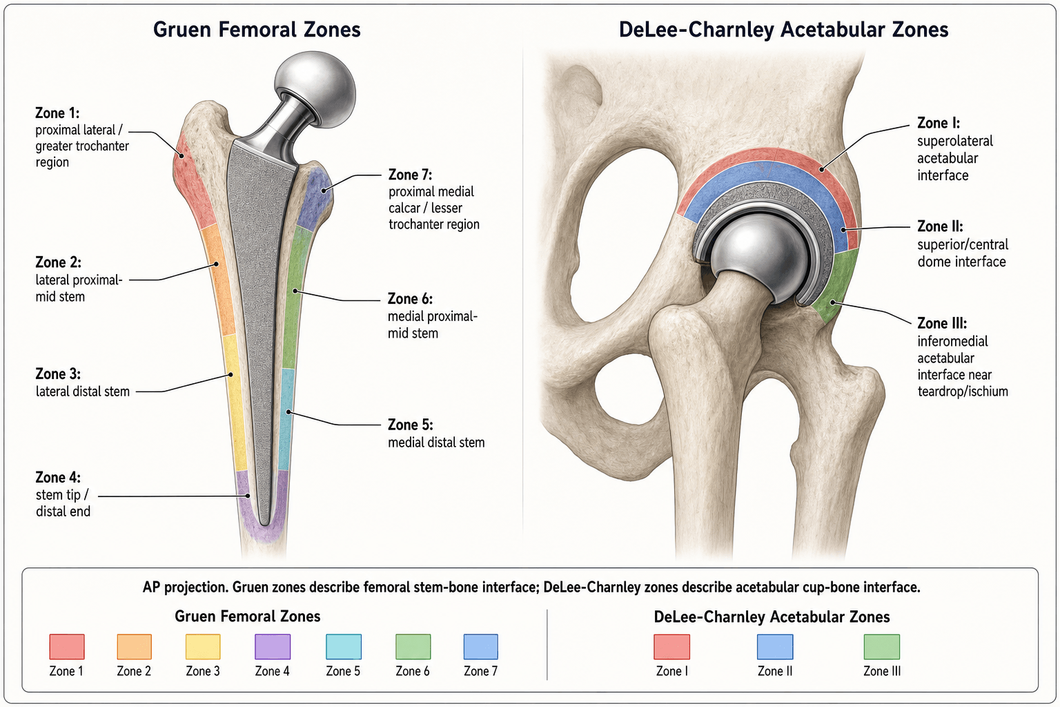
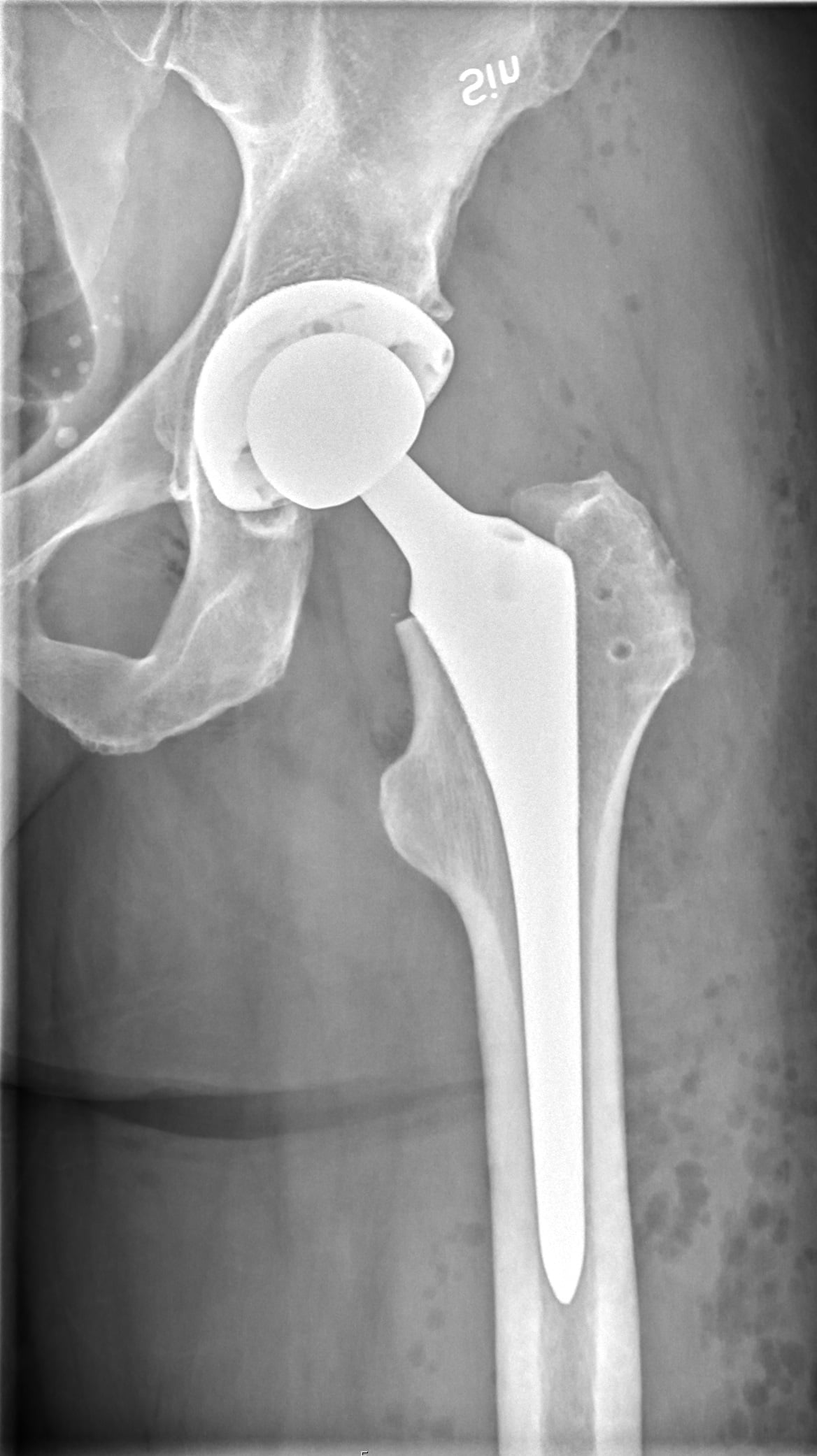
Gruen, McNeice, and Amstutz (1979) defined seven zones around the femoral stem on the anteroposterior radiograph to standardise the description of the bone–cement or bone–stem interface. The zones are numbered clockwise starting from the proximal-lateral cortex.
- Location
- Proximal-lateral
- Anatomical Landmark
- Greater trochanter region
- Key Pathology if Lucency Present
- Abductor osteolysis, polyethylene debris, trochanteric bursitis
- Location
- Mid-lateral
- Anatomical Landmark
- Mid-femoral shaft, lateral cortex
- Key Pathology if Lucency Present
- Mid-stem debonding (cemented), diaphyseal stress shielding
- Location
- Distal-lateral
- Anatomical Landmark
- Supra-distal lateral cortex
- Key Pathology if Lucency Present
- Distal debonding or cantilever loading
- Location
- Distal (tip)
- Anatomical Landmark
- Stem tip
- Key Pathology if Lucency Present
- Pedestal formation, tip pain, distal osteolysis or hypertrophy
- Location
- Distal-medial
- Anatomical Landmark
- Supra-distal medial cortex
- Key Pathology if Lucency Present
- Distal medial stress shielding or point loading
- Location
- Mid-medial
- Anatomical Landmark
- Mid-femoral shaft, medial cortex
- Key Pathology if Lucency Present
- Mid-stem debonding (cemented), loosening
- Location
- Proximal-medial
- Anatomical Landmark
- Calcar and lesser trochanter region
- Key Pathology if Lucency Present
- Calcar resorption, stress shielding, subsidence
Down lateral, round the tip, up medialGruen zones — clockwise from proximal-lateral
Hook:Walk down the lateral side (1-2-3), round the tip (4), back up the medial side (5-6-7) — like tracing the letter U clockwise
The lateral radiograph adds zones 8 through 14 (proximal-anterior through to proximal-posterior), but examiners almost always ask about the AP zones 1–7 only. If you mention zones 8–14, you earn extra credit — but do not confuse the two views.
The DeLee-Charnley Zone Classification (Acetabular Cup)
DeLee and Charnley (1976) divided the acetabular bone–cement (or bone–implant) interface into three zones on the AP pelvis or hip radiograph. The cup is divided by two lines drawn from the centre of the cup to its margins.
- Location
- Superolateral
- Anatomical Landmark
- Superior dome, lateral sourcil
- Key Pathology if Lucency Present
- Polyethylene wear debris, superior migration, most common loosening zone
- Location
- Superior-central (middle)
- Anatomical Landmark
- Central dome of the cup
- Key Pathology if Lucency Present
- Cement mantle deficiency, central osteolysis, medial wall breach
- Location
- Inferomedial
- Anatomical Landmark
- Inferior medial aspect, teardrop region
- Key Pathology if Lucency Present
- Medial migration, protrusion, column deficiency
Superolateral · Central · InferomedialDeLee-Charnley zones — superolateral to inferomedial
Hook:Think 10-12-3 on a clock face: zone 1 at 10 o'clock (superolateral), zone 2 at 12 (central dome), zone 3 at 3 o'clock (inferomedial)
Zone 1 acetabular lucency is the most commonly reported finding in asymptomatic total hip arthroplasties followed long-term. Not all zone 1 lucency means the cup is loose — less than 2 mm non-progressive lucency may be stable. The key is change on serial radiographs.
Clinical Interpretation: Zone-Specific Patterns of Loosening
The pattern of zones involved tells you the mode of failure. Interpreting the radiograph is not just about spotting lucency — it is about understanding why it is there.
- Gruen Zones
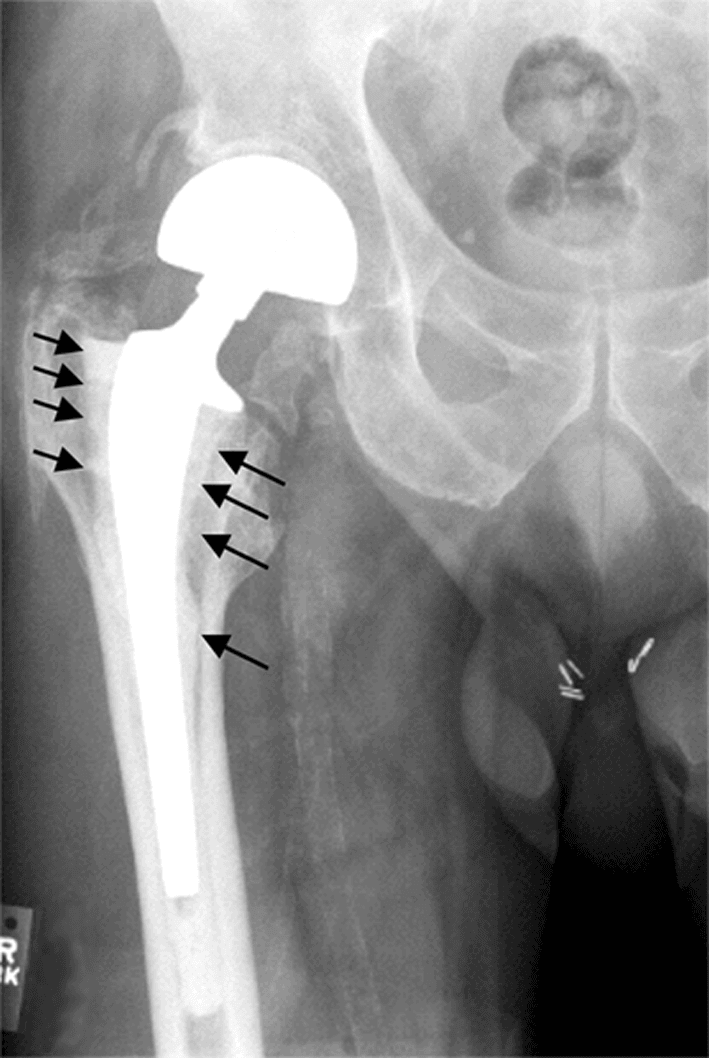
- 1 and 7 widening, stem migrates distally
- DeLee-Charnley Zones
- Usually none
- Interpretation
- Expected to a degree in Exeter-type stems; progressive subsidence over 5 mm is abnormal
- Gruen Zones
- All zones 1–7, or 2-3-5-6 circumferential
- DeLee-Charnley Zones
- May coexist with acetabular loosening
- Interpretation
- Aseptic loosening; revision indicated if symptomatic or progressive
- Gruen Zones
- Non-progressive, less than 2 mm, parallel to stem
- DeLee-Charnley Zones
- None
- Interpretation
- Stable; no revision unless symptoms change
- Gruen Zones
- Progressive, migratory, pedestal plus divergent lucency
- DeLee-Charnley Zones
- May coexist
- Interpretation
- Aseptic loosening; revision when symptomatic
- Gruen Zones
- Zone 1 (granuloma, trochanteric), zone 7 (calcar)
- DeLee-Charnley Zones
- Zone 1 (superolateral)
- Interpretation
- Debris-mediated osteolysis; liner exchange or revision if bone loss threatens fixation
- Gruen Zones
- N/A
- DeLee-Charnley Zones
- Zone 1 or all zones 1–2–3
- Interpretation
- Migration, screw breakage, or component tilt confirms loosening
Do not dismiss new-onset thigh pain in a patient with a well-fixed uncemented stem and distal Gruen zone 4 lucency. A distal pedestal with surrounding lucency may indicate a loose stem with distal point-loading. Always compare with prior films — progressive change is the key to diagnosing aseptic loosening.
222 — two mm, two films, two yearsLoosening vs stable — the 2 mm rule
Hook:222: 2 mm threshold, 2 films compared, 2 years stable — if any '2' is exceeded, suspect loosening
The zones describe where the lucency is; examiners then want the grading and the mechanism:
- Radiographic loosening grade for a cemented stem (the Hip Society / Harris criteria):
- Definite — stem migration/subsidence, a cement-mantle fracture, or a fracture of the stem itself (or a new radiolucent line at the metal–cement interface).
- Probable — a continuous (complete) radiolucent line around the entire cement–bone interface.
- Possible — a radiolucent line covering 50 to 99 per cent of the cement–bone interface.
- Gruen's four modes of cemented-stem failure (the patterns named in his 1979 paper):
- Mode I — pistoning. Ia: the stem subsides within the cement mantle (distal stem–cement lucency with proximal zone 1 changes). Ib: the stem and cement subside together within the femur (cement–bone lucency in all zones).
- Mode II — medial midstem pivot. The stem pivots about its midpoint — the proximal stem moves medially (zones 6–7) while the tip moves laterally (zones 3–4).
- Mode III — calcar pivot ("windshield-wiper"). The proximal stem is held at the calcar but the tip toggles medial–lateral, giving distal-zone lucency (often with a pedestal).
- Mode IV — cantilever/bending fatigue. Loss of proximal (calcar) support with a still-fixed tip leaves the unsupported proximal stem to bend — progressive proximal-medial bone loss and a real risk of stem fracture at mid-stem.
The zones map the lucency; the marks come from explaining the biology:
- Particle disease (the osteolysis cascade): wear particles — predominantly submicron polyethylene (also metal and PMMA cement) — are phagocytosed by macrophages, which release pro-inflammatory mediators (TNF-α, IL-1, IL-6, prostaglandin E2) and upregulate RANKL, driving osteoclast differentiation and periprosthetic bone resorption. This is the mechanistic reason that lowering wear below a threshold (cross-linked polyethylene; roughly 0.05 to 0.1 mm per year, per Dumbleton) dramatically reduces osteolysis.
- The "effective joint space" (Schmalzried): pressurised, particle-laden joint fluid tracks along any interface the implant communicates with — down the stem, through screw holes, into distal zones — so osteolysis can appear in non-contiguous or distal zones far from the bearing surface, not just zones 1 and 7.
- Stress shielding vs osteolysis — the distinction examiners test:
- Stress shielding is diffuse proximal bone loss, cortical thinning and calcar rounding (typically zone 7) from load bypassing the proximal femur into a stiff, often extensively-coated or large-diameter distal stem (Wolff's law). It is benign, stabilises after remodelling, and is graded by Engh.
- Osteolysis is focal, scalloped, expansile and well-demarcated lytic lesion(s) from particle disease — potentially progressive, threatening fixation and predisposing to periprosthetic fracture; it needs surveillance and may need liner exchange, grafting or revision.
- Do not label aggressive scalloped lysis as "stress shielding" — the management is completely different.
Limitations and Modern Context
- Two-dimensional limitation. Gruen and DeLee-Charnley zones are defined on plain AP radiographs and do not capture the three-dimensional extent of osteolysis. CT (with metal-artefact reduction) and MRI detect substantially more osteolysis than plain films, particularly in the posterior column and greater trochanter.
- Inter-observer variability is moderate, especially for narrow lucent lines at the cement–bone interface. Standardised positioning (internally rotated AP hip, centred on the mid-femur) improves consistency.
- Modern implants move differently. Highly polished tapered stems (e.g., Exeter, CPT) are designed to subside within the cement mantle — a degree of proximal-lateral and proximal-medial (zones 1 and 7) lucency is an expected loading pattern, not loosening. The examiner will distinguish this from a roughened cemented stem where the same lucency pattern is pathological.
- Cross-linked polyethylene and ceramic bearings have dramatically reduced the volume of wear debris and therefore the prevalence of periprosthetic osteolysis in zones 1 and 7 (femoral) and zone 1 (acetabular). The zones remain relevant because millions of older bearings are still in situ.
- Uncemented fixation uses different criteria. Engh's classification (bone ingrowth, fibrous stable, unstable) is applied alongside Gruen zones for porous-coated stems. Look for spot welds, cortical hypertrophy, and pedestal formation rather than just radiolucent lines.
- Registry reporting. The NJR (UK), AOANJRR (Australia), and AJRR (US) all use Gruen and DeLee-Charnley zones as the standard nomenclature for radiographic follow-up in revision risk models and survivorship analyses.
- EBRA (Einzel-Bild-Roentgen-Analyse) and RSA (Roentgen Stereophotogrammetric Analysis) are more sensitive than manual zone measurement for detecting early migration, but are used in research and specialist centres, not routine follow-up.
Guidelines, Registries and Global Practice
- Global radiographic follow-up after total hip arthroplasty is standard practice, though the frequency and duration vary. Most national guidelines recommend radiographs at 6 weeks, 1 year, and then at intervals of 1 to 5 years, with Gruen and DeLee-Charnley zone assessment at each visit.
- AAOS (US) and NICE/BOA-BOAST (UK) both recommend standardised AP and lateral hip radiographs with zone-based reporting for surveillance. NICE NG226 (2023) advises that asymptomatic patients with well-fixed components may extend follow-up intervals where registry data supports it.
- Registry evidence — the AOANJRR (Australia), NJR (UK), and AJRR (US) all record revision procedures using Gruen and DeLee-Charnley zone descriptors in their radiographic follow-up modules. Registry survivorship data consistently show that early detection of progressive zone lucency correlates with lower revision complexity and better outcomes.
- EBRA and RSA are used in European centres (particularly Scandinavian registries and the Swiss Hip Registry) for research-grade early migration detection, providing sensitivity an order of magnitude greater than manual zone measurement. These tools have established migration thresholds (for example, greater than 1.2 mm subsidence at two years) that predict late revision.
- Resource variation — in high-volume arthroplasty centres, CT with metal-artefact reduction sequences is increasingly used to quantify three-dimensional osteolysis that plain film Gruen zones underestimate. In resource-limited settings, serial AP radiographs with zone-based assessment remain the standard.
Exam Viva
Practise clinical reasoning and management decisions out loud
“A 72-year-old woman presents with gradually worsening right thigh pain eight years after a cemented total hip arthroplasty. An AP radiograph shows a well-positioned femoral stem with a 3 mm radiolucent line at Gruen zones 1 and 7, and calcar resorption. The acetabular component shows 2 mm lucency in DeLee-Charnley zone 1 only. How do you interpret these findings and what is your management plan?”
“A 58-year-old man is referred with an asymptomatic radiolucent line around the distal half of his uncemented femoral stem on a five-year follow-up AP radiograph. Gruen zones 3, 4, and 5 show a 1.5 mm lucent line that is non-progressive compared with his two-year film. The proximal zones show spot welds and no lucency. What is your interpretation and how do you counsel the patient?”
Gruen femoral zones (AP, 1–7, clockwise)
- Zone 1: proximal-lateral (greater trochanter) — osteolysis, poly debris, abductor compromise
- Zone 2: mid-lateral — mid-stem debonding
- Zone 3: distal-lateral — distal debonding
- Zone 4: distal tip — pedestal, tip loading, pain
- Zone 5: distal-medial — distal stress shielding
- Zone 6: mid-medial — mid-stem debonding, loosening
- Zone 7: proximal-medial (calcar) — calcar resorption, stress shielding, subsidence
DeLee-Charnley acetabular zones (AP, 1–3)
- Zone 1: superolateral — most common site of poly wear and loosening
- Zone 2: central dome — cement mantle failure, medial wall
- Zone 3: inferomedial — protrusion, column deficiency
- Grading: 0 (none), 1 (incomplete), 2 (complete, less than 2 mm), 3 (complete, greater than 2 mm or progressive)
Loosening criteria — the 2 mm rule
- Lucency less than 2 mm, non-progressive on serial films: stable (no revision)
- Lucency greater than 2 mm or progressive on serial films: aseptic loosening (plan revision)
- Always compare with prior films — never diagnose loosening on one radiograph
- Cemented polished taper stems: proximal zones 1 and 7 subsidence is expected, not pathological
Uncemented stem radiographic signs (Engh)
- Bone ingrowth: spot welds, no progressive lucency, stable position
- Fibrous stable: non-progressive lucent lines less than 2 mm, no migration
- Unstable: progressive lucent lines, pedestal with divergent lines, subsidence or tilt
High-yield patterns for the exam
- Gruen 1 and 7 lucency in cemented stem: likely debonding or subsidence — compare with prior films
- Gruen zone 1 granuloma with DeLee-Charnley zone 1 lucency: polyethylene wear and debris-mediated osteolysis
- Gruen zone 4 pedestal with circumferential lucency: loose uncemented stem
- DeLee-Charnley all three zones: gross acetabular loosening — plan cup revision
Evidence Base
Every citation below has been checked against its source record in PubMed. The two foundational papers (Gruen 1979, DeLee-Charnley 1976) created the zone nomenclature; Johnston 1990 (the Hip Society consensus) locked it in as the universal reporting standard; Engh 1990 extended radiographic assessment to uncemented stems; and Dumbleton 2002 established the wear-rate threshold below which osteolysis is rare.
'Modes of failure' of cemented stem-type femoral components: a radiographic analysis of loosening
- Retrospective zonal analysis of 389 total hip replacements: 19.5 percent (76 hips) showed radiographic evidence of mechanical loosening, with progressive loosening in 56
- Defined seven radiographic zones around the femoral stem on the AP radiograph to standardise reporting of the stem–cement and cement–bone interface
- Identified the characteristic modes of stem failure — pistoning (stem within cement, or stem-plus-cement within the femur), medial midstem pivot, calcar pivot, and bending (fatigue) cantilever
Radiological demarcation of cemented sockets in total hip replacement
- Divided the acetabular bone–cement interface into three zones on the AP radiograph for standardised demarcation/loosening assessment
- In 141 Charnley arthroplasties followed for a mean of 10.1 years, 69 percent showed demarcation of some degree and 9.2 percent showed progressive socket migration — the vast majority of demarcation was asymptomatic
- Nearly 30 percent showed no demarcation even at 10 years, supporting cemented acetabular fixation as sound when technique is good
Clinical and radiographic evaluation of total hip replacement: a standard system of terminology for reporting results
- Consensus statement from the Hip Society standardising clinical and radiographic outcome reporting after THA
- Adopted the Gruen femoral zones and the DeLee-Charnley acetabular zones as the standard nomenclature for radiographic assessment
- Provided a uniform terminology for reporting loosening based on zone involvement, lucency extent, and progression