Parathyroid Hormone Deficiency | Hypocalcaemia | Tetany | Basal Ganglia Calcification
- Biochemistry: Low calcium AND low PTH (distinguishes from vitamin D deficiency)
- Tetany triad: Carpopedal spasm, Chvostek sign, Trousseau sign (neuromuscular irritability)
- Emergency: Severe hypocalcaemia (Ca less than 1.8) causes seizures, laryngospasm, cardiac arrhythmias
- Treatment: IV calcium gluconate for acute, oral calcium carbonate plus calcitriol for chronic
- Orthopaedic relevance: Basal ganglia calcification, increased bone density, perioperative complications
- “Low calcium WITH low PTH distinguishes hypoparathyroidism from vitamin D deficiency or malabsorption
- “Chvostek sign is facial twitching when tapping facial nerve; Trousseau is carpopedal spasm with BP cuff inflation
- “Post-thyroidectomy hypocalcaemia may be transient (temporary ischaemia) or permanent (gland removal)
- “Basal ganglia calcification on CT is characteristic finding in chronic hypoparathyroidism
Overview and Epidemiology
Hypoparathyroidism is a disorder of insufficient parathyroid hormone (PTH) secretion, leading to hypocalcaemia, hyperphosphataemia and disrupted calcium homeostasis. Its signature is a PTH that is inappropriately low or absent in the face of hypocalcaemia. That separates it from the more common secondary hyperparathyroidism, in which chronic hypocalcaemia drives PTH up.
How common. It is rare: 70,000-90,000 cases in the United States.
- Post-surgical - 75%. Iatrogenic, after thyroid or parathyroid surgery. Permanent hypoparathyroidism follows 1-2% of total thyroidectomies, and more after reoperative surgery or central neck dissection, up to 10%.
- Autoimmune. A substantial minority of cases, often as part of autoimmune polyglandular syndrome type 1.
- Genetic or congenital - 5-10%. Includes DiGeorge syndrome.
Why it reaches an orthopaedic list. Calcium that has not been optimised causes perioperative complications, hypocalcaemic seizures and arrhythmias among them. Chronic disease brings basal ganglia calcification, with its extrapyramidal movement disorders, and bone that is paradoxically dense because remodelling is reduced. Recognising the condition before surgery is what allows preoperative optimisation and avoids a metabolic crisis in theatre.
Physiology and Pathophysiology
What PTH Normally Does
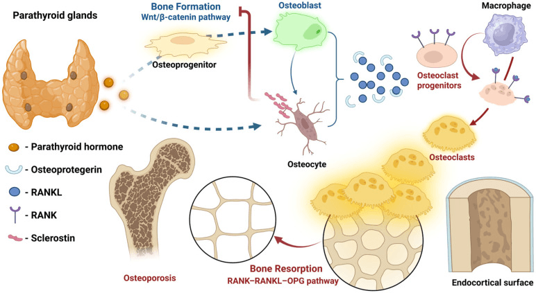
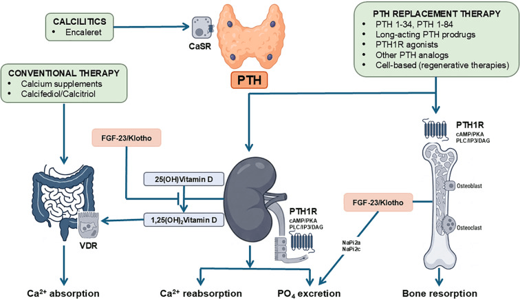
PTH acts on bone and kidney directly, and on the gut through vitamin D activation. Hypoparathyroidism is what happens when all of those actions are lost at once.
- Normal PTH Action
- Activates osteoclasts (calcium release)
- Effect of PTH Deficiency
- Reduced bone resorption
- Result
- Paradoxically increased bone density, low turnover
- Normal PTH Action
- Promotes phosphate excretion
- Effect of PTH Deficiency
- Phosphate retention
- Result
- Hyperphosphataemia (elevated serum phosphate)
- Normal PTH Action
- Increases calcium reabsorption
- Effect of PTH Deficiency
- Reduced calcium reabsorption
- Result
- Hypocalcaemia, hypocalciuria
- Normal PTH Action
- Activates vitamin D to calcitriol
- Effect of PTH Deficiency
- Reduced active vitamin D
- Result
- Impaired intestinal calcium absorption
The net result is low serum calcium, high serum phosphate and low urine calcium.
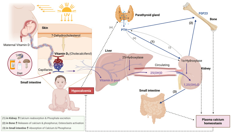
Why Calcium Falls and Phosphate Rises
Calcium. The losses all pull the same way. With no PTH-mediated osteoclast activation, calcium stays sequestered in bone; the distal tubule lets calcium go that it would have reclaimed; and with little calcitriol the gut absorbs poorly despite the low serum calcium. The result is a net calcium loss.
Phosphate. PTH normally promotes renal phosphate excretion by inhibiting the sodium-phosphate cotransporter in the proximal tubule, so without it phosphate is retained. The high serum phosphate then suppresses 1-alpha hydroxylase further, which worsens vitamin D activation.
Nerve and muscle. Ionised calcium is critical for normal neuronal and muscle function. Low calcium reduces the action potential threshold and increases neuronal excitability, and the result is tetany, seizures and cardiac conduction abnormalities.
Causes of Hypoparathyroidism
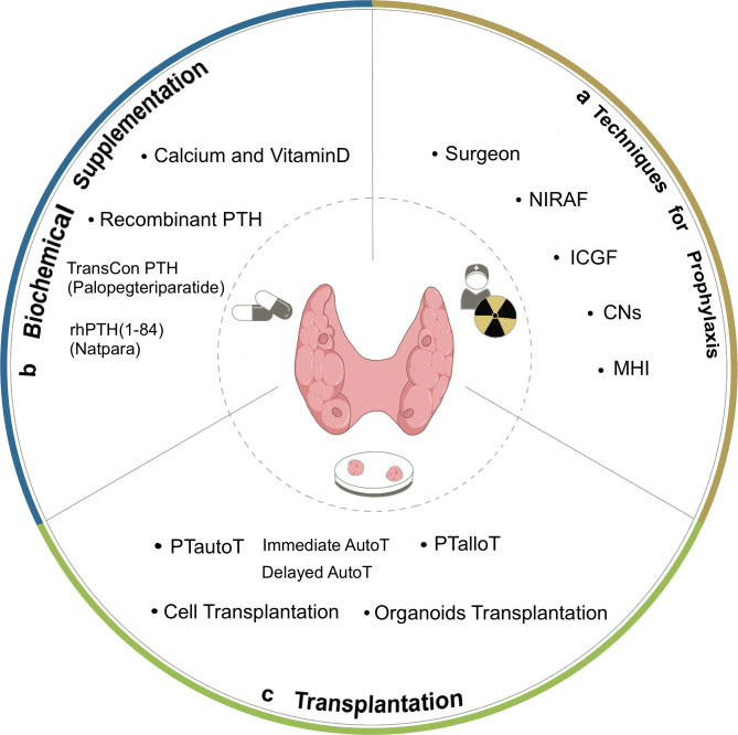
How the glands are lost. By one of three routes:
- Gland removal - inadvertent excision during thyroidectomy, parathyroidectomy for hyperparathyroidism or radical neck dissection
- Vascular injury - devascularisation of the glands during surgery, temporary or permanent
- Autotransplantation failure - glands deliberately autotransplanted may not engraft
Who is left with permanent disease. The risk rises with:
- Total thyroidectomy (higher than hemithyroidectomy)
- Central lymph node dissection for thyroid cancer
- Reoperative surgery, through scar tissue and a difficult dissection
- Parathyroids not identified or preserved at operation
- Concurrent parathyroid pathology
Transient or permanent. Transient hypocalcaemia comes from temporary ischaemia or stunning of the glands: symptoms develop within 24-72 hours of surgery and resolve within 6 months. Permanent hypoparathyroidism follows gland removal, persists beyond that and needs lifelong replacement. Where the line falls has varied between 6 and 12 months from study to study, which inflates the heterogeneity in reported incidence; the 2022 international guideline sets it at more than 12 months.
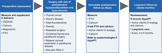
Surveillance. After thyroid or parathyroid surgery, calcium is checked daily for 48-72 hours and supplemented as needed during recovery.
Clinical Assessment
Acute Hypocalcaemia
Severe hypocalcaemia (Ca less than 1.8 mmol/L) is a medical emergency. Laryngospasm causes stridor and potential airway obstruction. Seizures (tonic-clonic) can occur without warning. Cardiac arrhythmias (prolonged QT, torsades de pointes, ventricular fibrillation) may be fatal. Immediate IV calcium gluconate is required.
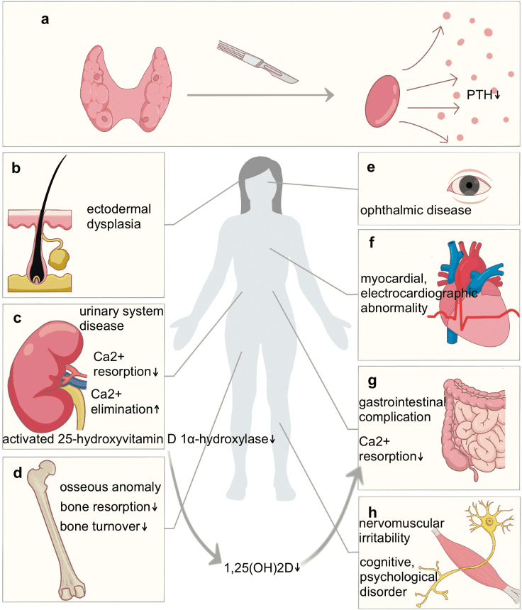
Tetany. This is the neuromuscular irritability of hypocalcaemia. Tingling or numbness around the mouth and lips is often the earliest symptom, and carpopedal spasm the most characteristic finding.
- Perioral paraesthesia - often first
- Carpopedal spasm - wrist flexion, finger extension and thumb adduction, the obstetrician's hand
- Muscle cramps - painful, in the hands, feet, back and legs
- Laryngospasm - throat tightness and stridor, a high-pitched inspiratory sound; bronchospasm causes wheeze
- Seizures - generalised tonic-clonic, without focal features
Muscle and gut. Muscle weakness with a proximal myopathy and fatigue; abdominal cramps and diarrhoea (rare).
The heart. Arrhythmias are the lethal end of acute hypocalcaemia, and the ECG is covered under Investigations. Rarely, severe chronic hypocalcaemia causes a dilated cardiomyopathy and heart failure.
Mood and cognition. Anxiety, irritability, depression, cognitive impairment and confusion; psychosis is rare.
Examination
- Technique
- Tap facial nerve 2 cm anterior to ear lobe
- Positive Finding
- Ipsilateral facial muscle twitching (orbicularis oculi, nasalis)
- Sensitivity
- 70% in hypocalcaemia (10% false positive in normals)
- Technique
- Inflate BP cuff 20 mmHg above systolic for 3 minutes
- Positive Finding
- Carpopedal spasm (wrist flexion, finger extension, thumb adduction)
- Sensitivity
- 94% in hypocalcaemia (more specific than Chvostek)
- Technique
- Tap peroneal nerve at fibular head
- Positive Finding
- Foot dorsiflexion and toe extension
- Sensitivity
- Less commonly tested but present in severe cases
Chvostek's sign is tapping over the facial nerve (VII) in front of the ear. Beyond the three classic signs, the deep tendon reflexes are brisk, and tapping the ulnar nerve can elicit twitching of the hand muscles.
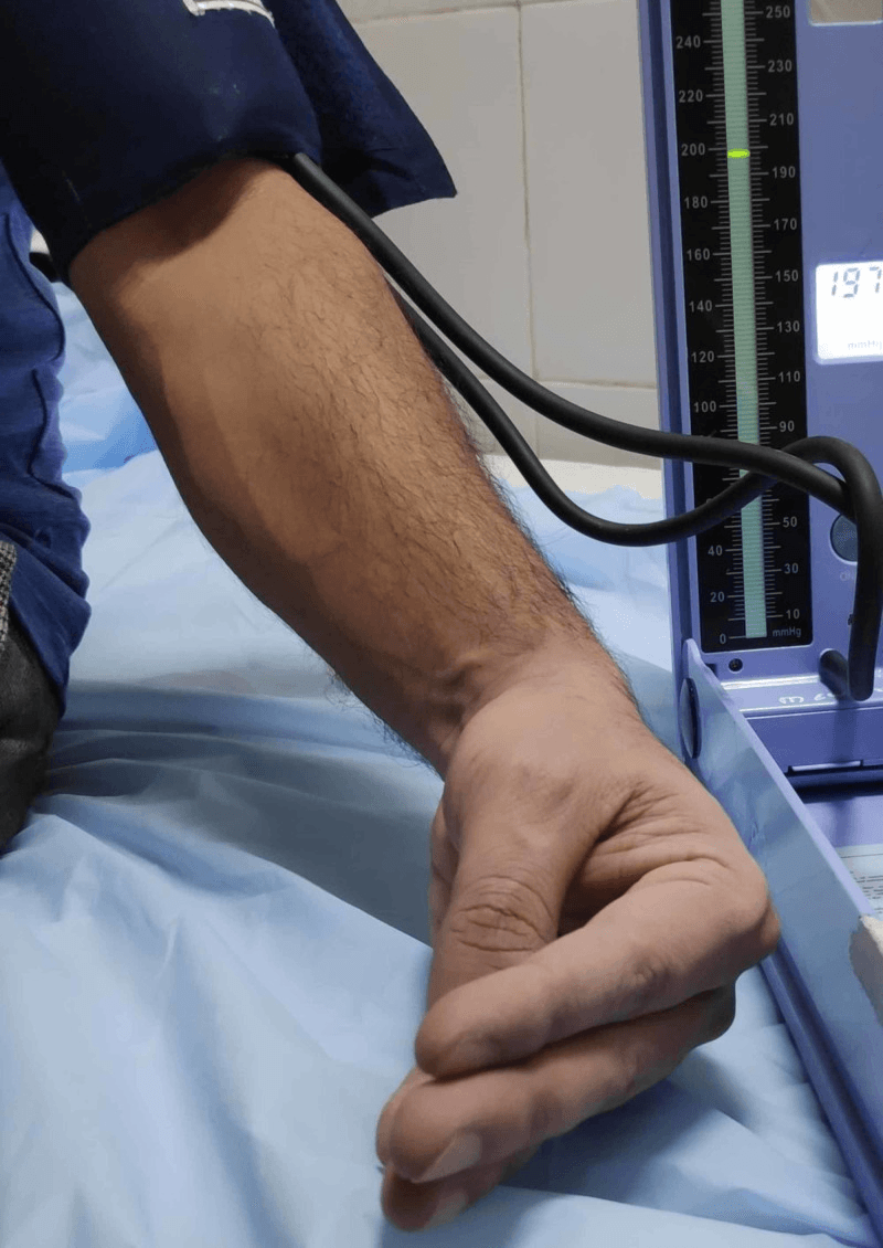
When testing for hypocalcaemia, Trousseau's sign is the one to prefer: it is the more specific, with a low false-positive rate, whereas Chvostek's is positive in some normocalcaemic people. Both may be negative in very severe hypocalcaemia, where muscle weakness prevents spasm, or in chronic cases with adaptation.
Chronic Hypoparathyroidism
Skeleton. Bone turnover is low, with low alkaline phosphatase and low bone formation markers. Despite the high density, bone quality is abnormal and strength may be impaired, a static bone disease.
Brain. Basal ganglia calcification (see Imaging) can produce extrapyramidal disease: parkinsonism with tremor, rigidity and bradykinesia, chorea and dystonia. Seizures recur if calcium is not well controlled, and papilloedema from raised intracranial pressure (pseudotumour cerebri) is rare.
Eyes. Chronic hypocalcaemia causes posterior subcapsular cataracts, the lens calcium deposition visible on slit-lamp examination.
Teeth. Enamel hypoplasia follows if the disease is present while the teeth develop in childhood. Tooth eruption is delayed in congenital cases, and the risk of caries is increased.
Skin. Dry, coarse skin, brittle nails and hair loss (alopecia).
Biochemical and Imaging Investigations
Biochemistry
The diagnosis rests on two numbers together: a low calcium AND a low PTH. In vitamin D deficiency or malabsorption the PTH is elevated, as it should be; in hypoparathyroidism it is not.
- Calcium
- Low (less than 2.0)
- PTH
- Low or undetectable
- Phosphate
- High (over 1.5)
- Vitamin D
- Low calcitriol, normal 25-OH
- Calcium
- Low
- PTH
- High (appropriate)
- Phosphate
- Low/normal
- Vitamin D
- Low 25-OH vitamin D
- Calcium
- Low
- PTH
- High (secondary HPT)
- Phosphate
- High
- Vitamin D
- Low calcitriol (impaired 1-alpha hydroxylase)
- Calcium
- Low
- PTH
- High (PTH resistance)
- Phosphate
- High
- Vitamin D
- Normal/low
- Calcium
- Low
- PTH
- Low (functional hypoparathyroidism)
- Phosphate
- Variable
- Vitamin D
- Normal
Calcium. A total calcium below 2.0 mmol/L indicates hypocalcaemia (normal 2.2-2.5). Ionised calcium is more accurate because albumin does not affect it, and below 1.1 mmol/L is abnormal. Correct a total calcium for albumin: corrected Ca = measured Ca + 0.02 x (40 - albumin in g/L).
PTH. Measured with the standard intact PTH assay, it is inappropriately low or undetectable. A normal PTH in the face of hypocalcaemia is itself inappropriate, because it should be elevated.
Phosphate. Elevated, above 1.5 mmol/L (normal 0.8-1.5), and characteristic of the disease.
Vitamin D. 25-OH vitamin D is usually normal, which distinguishes hypoparathyroidism from vitamin D deficiency; if it is low, there is a concurrent deficiency that needs treating separately. Calcitriol is low despite the hypocalcaemia. It is not routinely measured, but it explains why calcitriol has to be supplemented.
Magnesium. It must be checked. Hypomagnesaemia below 0.7 mmol/L impairs both PTH secretion and PTH action and so causes a functional hypoparathyroidism; correct the magnesium before diagnosing true hypoparathyroidism.
Alkaline phosphatase. Normal or low, reflecting reduced bone turnover, the opposite of hyperparathyroidism.
24-hour urine calcium. Low, because the filtered load falls, despite the reduced renal reabsorption. This hypocalciuria, the opposite of hyperparathyroidism, distinguishes hypoparathyroidism from other causes of hypocalcaemia.
Electrocardiography
A prolonged QT interval is the most characteristic finding, a QTc over 450 ms in men or 470 ms in women. A prolonged ST segment increases the QT duration, and T waves may be flattened or inverted. The arrhythmias that follow are torsades de pointes (polymorphic VT with a prolonged QT), ventricular tachycardia and ventricular fibrillation.
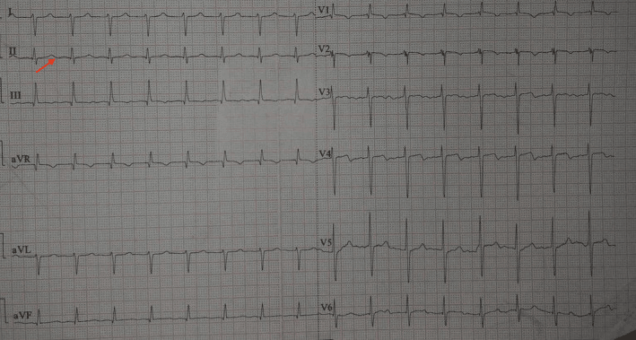
A QTc over 500 ms significantly increases the risk of torsades de pointes and sudden cardiac death. Avoid QT-prolonging drugs (antiarrhythmics, antipsychotics, antibiotics), monitor the ECG while calcium is corrected, and remember that severe cases may need temporary pacing.
Imaging
CT brain (non-contrast). Basal ganglia calcification is bilateral and symmetrical, in the globus pallidus, putamen and caudate, with calcification of the cerebellar dentate nuclei as well. It is seen in 50% of chronic cases. Goswami's series of long-standing idiopathic hypoparathyroidism found 73.8% (95% CI 66.6-81.0), symptomatic in only 3 of 145 patients; prevalence in post-surgical disease, which is recognised and treated earlier, is likely lower. Calcification correlates with the duration and severity of hypocalcaemia and is not always symptomatic.
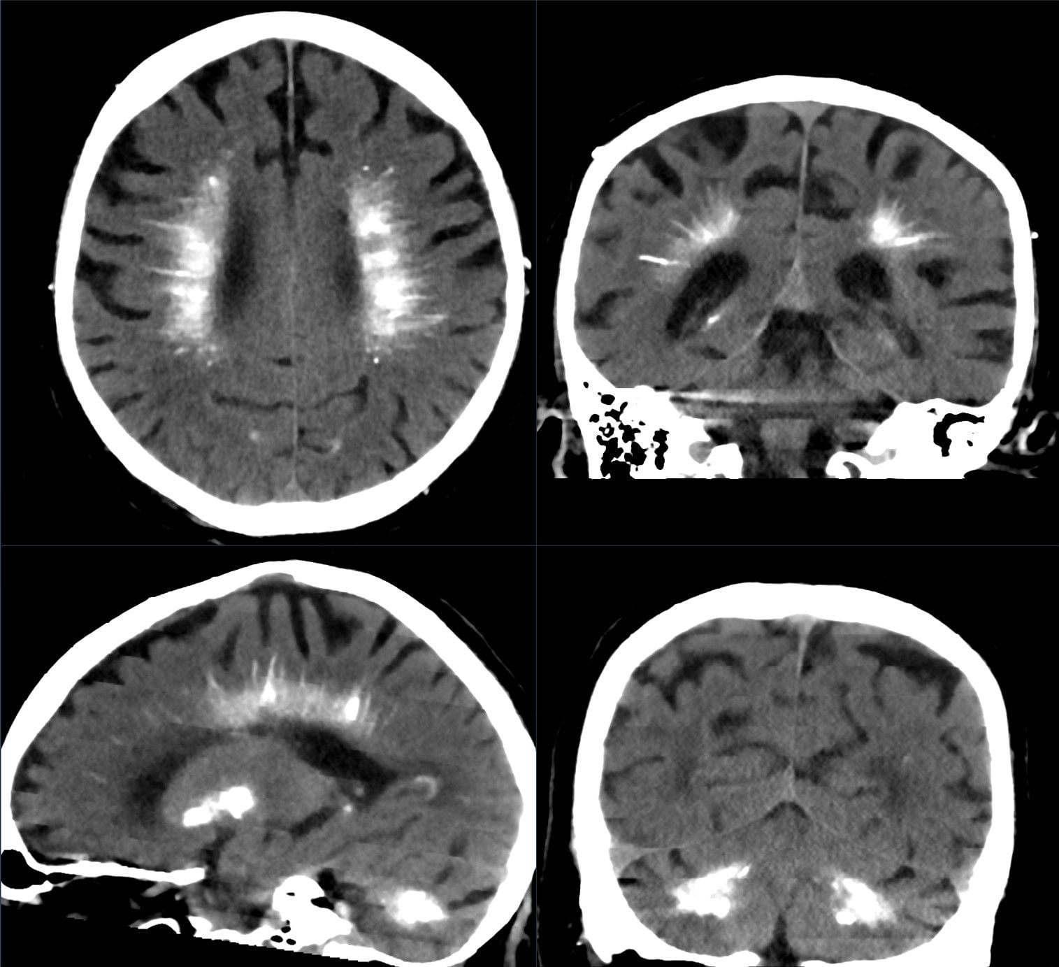
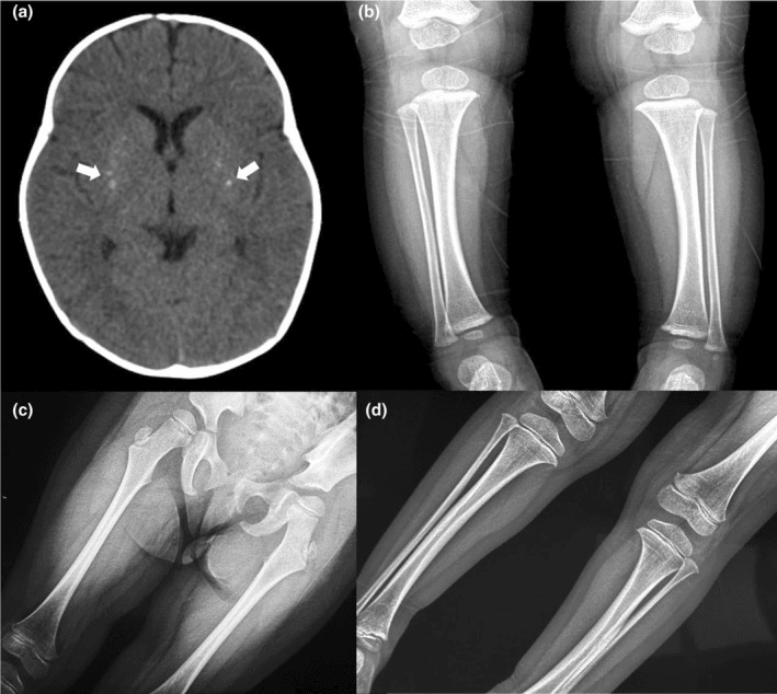
DEXA. Bone mineral density is paradoxically increased, with T-scores often +2 to +4 SD above normal. Z-scores, which compare with age-matched controls, are the more relevant figure, and a high density does not mean normal bone quality in this low-turnover state.
Parathyroid imaging. If the aetiology is unclear. Neck ultrasound looks for parathyroid tissue and may show atrophic or absent glands; a sestamibi scan is not useful, because it needs functioning parathyroid tissue.
Acute and Chronic Management
Acute Hypocalcaemia
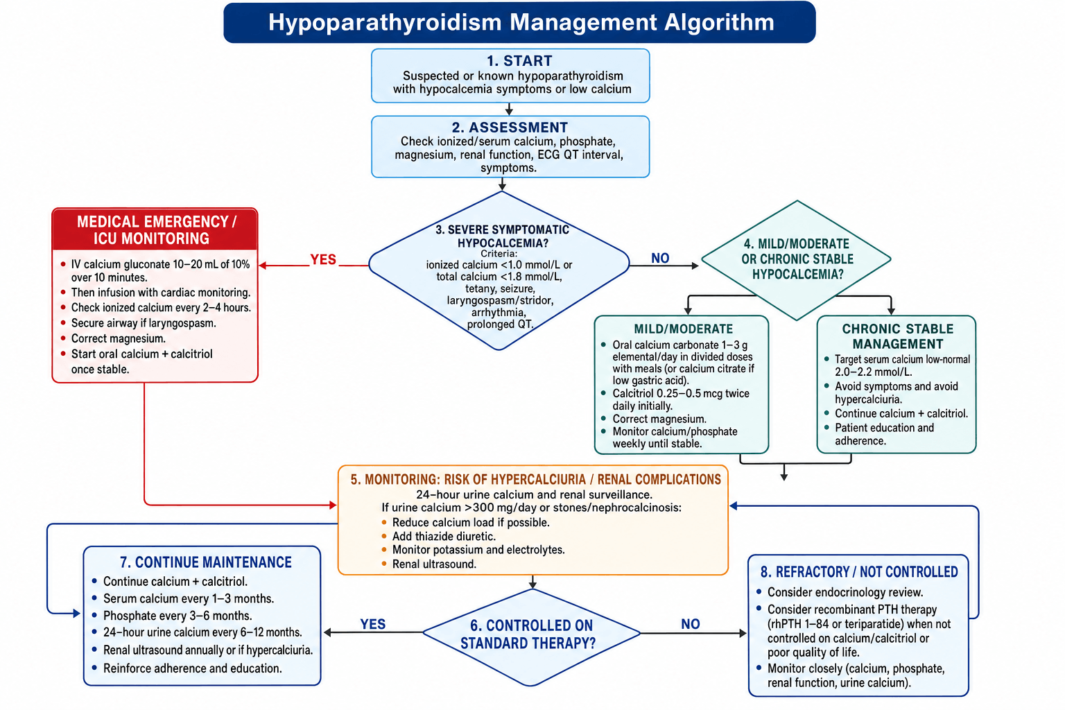
When IV calcium is needed. Severe symptomatic hypocalcaemia is a medical emergency, and the treatment is life-saving and needs ICU monitoring. The indications:
- Ionised calcium below 1.0 mmol/L (total below 1.8)
- Tetany or carpopedal spasm
- Seizures
- Laryngospasm or stridor
- Cardiac arrhythmias or a prolonged QT
Calcium gluconate. Load with 10-20 mL of 10% calcium gluconate (90-180 mg elemental calcium) IV over 10 minutes. Follow with an infusion of 50-100 mL calcium gluconate in 500 mL D5W or normal saline at 50 mL/hour, adjusted to maintain the calcium. Keep the patient on continuous cardiac monitoring and check the ionised calcium every 2-4 hours at first.
Hazards. IV calcium has three:
- Bradycardia if it is infused too quickly
- Tissue necrosis if it extravasates, so secure good IV access
- Precipitation with bicarbonate, with which it is incompatible
Treat the cause. Check the magnesium and correct it if low, with magnesium sulfate 2 g IV over 15 minutes, then 1-2 g/hour by infusion. Start oral calcium and calcitriol once the patient is stable.
The airway. If laryngospasm develops:
- High-flow oxygen
- Heliox (helium-oxygen) may relieve the stridor
- Emergent intubation if the airway is completely obstructed
Chronic Hypoparathyroidism
The goals. Hold the serum calcium in the low-normal range, 2.0-2.2 mmol/L, and avoid hypercalciuria by keeping urine calcium below 300 mg/24 hours. Beyond the numbers, the aim is to prevent the long-term complications (nephrolithiasis, nephrocalcinosis, basal ganglia calcification) and to maintain quality of life.
Calcium. Calcium carbonate gives 1-3 g elemental calcium daily in divided doses with meals, because it needs acid to be absorbed; 1250 mg of calcium carbonate is 500 mg elemental calcium. Do not take it with levothyroxine, whose absorption it decreases. Calcium citrate does not need acid and is the alternative when gastric acid is poor. Titrate the dose to the serum and urine calcium.
Activated vitamin D - essential. Calcitriol (1,25-dihydroxy vitamin D), 0.25-1 mcg twice daily, is the active form and bypasses the PTH-mediated activation the patient no longer has. It increases intestinal calcium absorption and is more potent than cholecalciferol or ergocalciferol, but it carries a risk of hypercalciuria, so the 24-hour urine calcium is monitored. Alfacalcidol (1-alpha calcidiol) is the alternative.
Thiazides. Hydrochlorothiazide 25-50 mg daily increases renal calcium reabsorption and reduces urine calcium. It is useful if hypercalciuria develops despite dose optimisation, but may cause hypokalaemia, so the electrolytes are monitored.
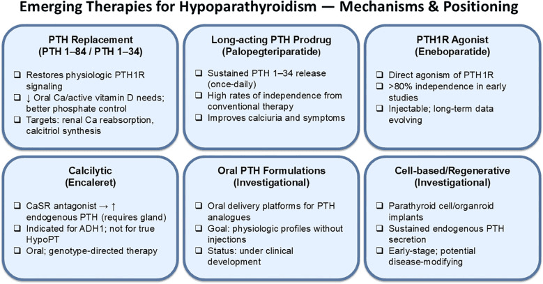
PTH replacement. Teriparatide or rhPTH 1-84, by subcutaneous injection, improves calcium control and may reduce calcium and calcitriol requirements. It is expensive and not widely available, and is reserved for refractory cases not controlled on standard therapy.
Magnesium. Replace if deficient, with oral magnesium oxide 400-800 mg daily.
Monitoring. The schedule once the patient is stable:
- Frequency
- Every 1-3 months when stable
- Target
- 2.0-2.2 mmol/L (low-normal)
- Action if Abnormal
- Adjust calcium or calcitriol dose
- Frequency
- Every 3-6 months
- Target
- Within normal range (less than 1.5)
- Action if Abnormal
- High phosphate suggests overtreatment
- Frequency
- Every 6-12 months
- Target
- Less than 300 mg/day
- Action if Abnormal
- Add thiazide if over 300, reduce calcium dose
- Frequency
- Annually or if hypercalciuria
- Target
- No stones or nephrocalcinosis
- Action if Abnormal
- Reduce calcium/calcitriol if stones develop
- Frequency
- Every 6-12 months
- Target
- Normal eGFR (over 60)
- Action if Abnormal
- Reduce calcium load if renal impairment
- Frequency
- Every 1-2 years
- Target
- No cataracts
- Action if Abnormal
- Cataract surgery if vision impaired
Perioperative Considerations for Orthopaedic Surgery
Before Surgery
Target serum calcium 2.0-2.2 mmol/L before elective orthopaedic surgery. Verify compliance with calcium and calcitriol. Check ionised calcium (more accurate than total calcium). Postpone elective surgery if calcium is less than 1.9 mmol/L (risk of perioperative complications). Have IV calcium available in theatre.
Assessment. Confirm the diagnosis and the control:
- Confirm the diagnosis from the history, and that the patient is on calcium and calcitriol replacement
- Serum total and ionised calcium, phosphate and magnesium
- ECG for the baseline QT interval, since a prolonged QT increases anaesthetic risk
- Medication review for compliance
- Endocrinology consultation for complex or poorly controlled cases
On the day. Continue calcium and calcitriol on the day of surgery if oral intake is permitted. If the patient will be nil by mouth for more than 24 hours, IV calcium supplementation may be needed.
In Theatre
Monitoring. Continuous ECG for QT prolongation and arrhythmias. Consider ionised calcium monitoring in a prolonged case or with significant fluid shifts; large-volume transfusion of citrated blood products can chelate calcium (see Citrate-Related Hypocalcaemia below).
The anaesthetic. Muscle relaxants may act for longer in hypocalcaemia, so neuromuscular blockade is monitored. Rapid alkalosis is avoided (see the pearl below).
Rescue. Keep 10% calcium gluconate immediately available. If the calcium drops or tetany or an arrhythmia develops, give 10 mL IV over 10 minutes.
Hyperventilation during spine surgery (to reduce epidural venous bleeding) can precipitate hypocalcaemic crisis in patients with hypoparathyroidism. Respiratory alkalosis increases calcium binding to albumin, lowering ionised calcium. Monitor ionised calcium intraoperatively if hyperventilating. Have IV calcium available. Avoid excessive hyperventilation.
After Surgery
Monitoring. Check the ionised calcium on arrival in recovery, especially after a long period nil by mouth or a large fluid resuscitation. Continue cardiac monitoring if the QT is prolonged, and resume oral calcium and calcitriol as soon as oral intake is tolerated.
What to watch for. The acute complications of hypocalcaemia:
- Tetany - carpopedal spasm and perioral numbness; test Trousseau's and Chvostek's signs
- Seizures - especially if the calcium drops acutely
- Arrhythmias - torsades de pointes if the QT is prolonged
IV to oral. A patient nil by mouth for a prolonged period after surgery may need a temporary IV calcium infusion. Move to oral once medication is taken reliably, and check the calcium 24-48 hours after stopping IV calcium to confirm the oral therapy is adequate.
Complications of Hypoparathyroidism
- Mechanism
- Neuronal hyperexcitability from low calcium
- Presentation
- Generalised tonic-clonic seizures
- Management
- IV calcium gluconate, anticonvulsants if recurrent
- Mechanism
- Vocal cord spasm from hypocalcaemia
- Presentation
- Stridor, respiratory distress, airway obstruction
- Management
- IV calcium, heliox, emergent intubation if severe
- Mechanism
- Prolonged QT interval, altered repolarisation
- Presentation
- Torsades de pointes, ventricular fibrillation, sudden death
- Management
- IV calcium, magnesium, avoid QT-prolonging drugs, pacing
- Mechanism
- Chronic hypocalcaemia causes calcium deposition
- Presentation
- Parkinsonism, chorea, dystonia, cognitive impairment
- Management
- Optimise calcium control, symptomatic treatment
- Mechanism
- Lens calcium deposition (subcapsular)
- Presentation
- Progressive vision impairment
- Management
- Cataract surgery if vision affected
- Mechanism
- Hypercalciuria from high calcium doses
- Presentation
- Renal stones, renal impairment
- Management
- Reduce calcium dose, add thiazide, increase hydration
- Mechanism
- Severe chronic hypocalcaemia (rare)
- Presentation
- Dilated cardiomyopathy, reduced ejection fraction
- Management
- Optimise calcium, standard heart failure management
- Mechanism
- Increased intracranial pressure (rare)
- Presentation
- Headache, papilloedema, vision changes
- Management
- Lumbar puncture, acetazolamide, optimise calcium
Hungry Bone Syndrome versus Surgical Hypoparathyroidism
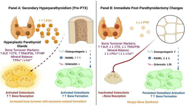
Not every hypocalcaemia after neck surgery is PTH deficiency. After parathyroidectomy the other major mechanism is hungry bone syndrome (HBS), and biochemically it is the mirror image.
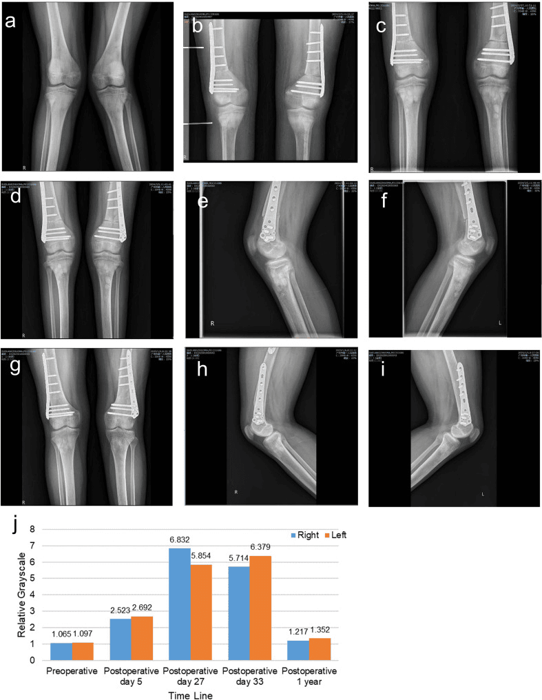
Mechanism. After parathyroidectomy for severe or long-standing hyperparathyroidism, or occasionally after thyroidectomy for thyrotoxicosis with high-turnover bone, the abrupt fall in PTH removes the drive to bone resorption while osteoblastic bone formation continues. The demineralised, high-turnover "hungry" skeleton avidly re-mineralises, taking calcium, phosphate and magnesium out of the circulation. The result is a profound, prolonged hypocalcaemia, often lasting days to weeks.
Q: A patient is profoundly hypocalcaemic after parathyroidectomy for severe hyperparathyroidism - how do you tell hungry bone syndrome from surgical hypoparathyroidism? A: Check phosphate and PTH. Hungry bone syndrome = low calcium + LOW phosphate + low magnesium + HIGH alkaline phosphatase (ongoing bone formation) + PTH normal or appropriately elevated/recovering. Surgical hypoparathyroidism = low calcium + HIGH phosphate + LOW or undetectable PTH + low/normal alkaline phosphatase (low-turnover state). The phosphate direction and the PTH level separate them at the bedside.
Who gets it. The risk factors:
- Severe pre-operative hyperparathyroidism, with very high PTH and calcium
- Radiological or biochemical bone disease - osteitis fibrosa cystica, high alkaline phosphatase
- A large adenoma or parathyroid carcinoma
- Older age
- Vitamin D deficiency
Management. Aggressive and prolonged calcium, large intravenous doses then oral, with activated vitamin D and repletion of magnesium and phosphate, continued until the skeleton has re-mineralised. HBS is self-limiting as the bone refills; surgical hypoparathyroidism may be permanent.
Citrate-Related Hypocalcaemia in Massive Transfusion
Citrate from transfused blood is the orthopaedic surgeon's commonest encounter with acute hypocalcaemia: revision arthroplasty, pelvic and spine surgery, polytrauma resuscitation.
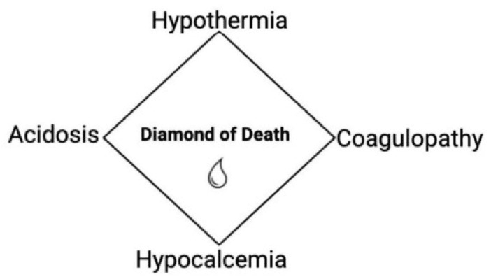
Mechanism. Stored blood products contain citrate anticoagulant, which chelates ionised calcium. The liver normally metabolises citrate rapidly, to bicarbonate, so transfusion at an ordinary rate causes no problem. With rapid, massive transfusion (roughly one blood volume, or faster than the liver can clear the citrate), made worse by hypothermia, hepatic impairment or the low hepatic flow of shock, citrate accumulates and ionised calcium falls. The risks are those of any hypocalcaemia: tetany, QT prolongation and myocardial depression.
In hypoparathyroidism. There is no PTH reserve to defend the calcium, so the citrate load of major surgery hits harder and faster. Anticipate it and pre-empt it.
Management. During massive transfusion, monitor the ionised calcium and give intravenous calcium, chloride or gluconate, to keep it normal; this is the calcium limb of the lethal-triad and massive-transfusion protocols. Keep the patient warm to preserve citrate clearance. Detailed transfusion protocols belong to the blood-loss-management topic.
Total calcium can read normal or even high during massive transfusion, because the citrate-bound calcium is still counted. Only an ionised calcium detects the fall - a classic pitfall.
Guidelines, Registries & Global Practice
Global Epidemiology
- Prevalence: roughly 25-40 per 100,000 population in Western registry data; defined as an orphan/rare disease in Europe and the US
- Dominant cause worldwide: post-surgical, accounting for around 75% of chronic cases; permanent hypoparathyroidism follows about 1-3% of total thyroidectomies and more after central neck dissection or reoperation
- Non-surgical causes (autoimmune, genetic, infiltrative, hypomagnesaemia-related) predominate in regions with lower thyroid-surgery volumes and in paediatric cohorts
- Idiopathic/non-surgical disease is relatively more represented in South Asian and Middle Eastern series, where it presents with prominent basal ganglia calcification and seizures
Major Guidelines Side by Side
- Calcium target
- Low-normal serum calcium, symptom-guided
- First-line therapy
- Calcium plus activated vitamin D (calcitriol/alfacalcidol)
- Distinctive emphasis
- GRADE-based; monitor 24h urine calcium and renal function
- Calcium target
- Lower half of reference range
- First-line therapy
- Calcium plus active vitamin D; PTH if poorly controlled
- Distinctive emphasis
- Unified diagnostic criteria and complication surveillance
- Calcium target
- Asymptomatic low-normal calcium
- First-line therapy
- Conventional therapy; PTH analogues reserved for refractory cases
- Distinctive emphasis
- Quality of life and long-term renal/CNS endpoints
- Calcium target
- Avoid symptomatic hypocalcaemia perioperatively
- First-line therapy
- Routine post-thyroidectomy calcium plus or minus PTH protocols
- Distinctive emphasis
- Parathyroid identification, preservation and autotransplantation
Registry and Database Signals
National registries are emerging rather than mature for hypoparathyroidism, but population datasets are informative. Danish national-registry cohorts (Underbjerg and colleagues) link postsurgical disease to excess infection and neuropsychiatric morbidity and to increased renal stones/insufficiency in idiopathic disease, while showing no overall fracture excess despite high bone mineral density. The international PARADIGHM observational registry tracks long-term outcomes and PTH-replacement safety across multiple countries.
High- vs Limited-Resource Practice Variation
- High-resource settings: intraoperative or early postoperative PTH-guided protocols, ionised calcium assays, calcitriol availability, and access to PTH analogues (rhPTH 1-84 / palopegteriparatide) for refractory disease
- Limited-resource settings: reliance on total serum calcium with albumin correction, plain (parent) vitamin D or alfacalcidol where calcitriol is scarce, and greater burden of late-presenting idiopathic disease with established basal ganglia calcification, cataract and seizures
- Universal priorities: surgeon experience and meticulous parathyroid preservation remain the single most effective preventive measure regardless of resource level
Controversies and Areas of Uncertainty
- Is PTH replacement disease-modifying? rhPTH(1-84) and the long-acting prodrug palopegteriparatide reduce calcium/calcitriol requirements and improve biochemistry, but whether they prevent hard endpoints (renal impairment, basal ganglia calcification, fractures) is unproven. PTH analogues also carry a historical osteosarcoma class warning from rodent data.
- Optimal calcium target: guidelines recommend low-normal calcium, yet no trial links any specific target to clinical outcomes. The balance between symptom relief and hypercalciuria/nephrocalcinosis is individualised.
- Phosphate versus calcium control: calcium-phosphate product, not calcium alone, drives basal ganglia and ectopic calcification, but routine phosphate-lowering strategies are not standardised.
- Bone paradox: bone mineral density is high and fracture risk is not increased in cohort data, but bone is low-turnover with abnormal microarchitecture; the true fracture and quality implications remain debated.
- Routine post-thyroidectomy PTH protocols: early PTH measurement can triage supplementation and discharge, but cost-effectiveness and the best cut-offs differ between surgical units.
MCQ Practice Points
Q: A patient has serum calcium 1.8 mmol/L and PTH 5 pg/mL (low). What is the most likely diagnosis? A: Hypoparathyroidism. Low calcium with low PTH indicates insufficient PTH secretion. In hypocalcemia, PTH should be elevated (secondary hyperparathyroidism); if it's low, that's inappropriate and diagnostic of hypoparathyroidism. Check phosphate (expect high) and magnesium (low Mg causes functional hypoparathyroidism).
Q: Which sign is more specific for hypocalcemia: Trousseau or Chvostek? A: Trousseau sign (94% sensitivity, very low false positive rate). Chvostek sign has 70% sensitivity but 10% false positive rate in normocalcemic individuals. Trousseau is induced carpopedal spasm with BP cuff inflation above systolic for 3 minutes. Chvostek is facial twitching when tapping facial nerve.
Q: How do you differentiate transient from permanent post-thyroidectomy hypoparathyroidism? A: Time course: Transient hypocalcemia recovers within 6 months (parathyroid gland ischemia or stunning with eventual recovery). Permanent hypoparathyroidism persists beyond 6 months (gland removal or permanent damage) and requires lifelong calcium and calcitriol replacement. Check PTH at 6 months - if still low, permanent.
Q: Why does hypomagnesemia cause hypocalcemia? A: Hypomagnesemia (Mg less than 0.7 mmol/L) causes functional hypoparathyroidism through two mechanisms: (1) Impaired PTH secretion from parathyroid glands (magnesium required for hormone release), and (2) End-organ PTH resistance (skeletal and renal PTH resistance). Result is low calcium despite low PTH. Correct magnesium first before diagnosing true hypoparathyroidism.
Q: A patient with hypoparathyroidism is undergoing spine surgery. The anesthesiologist hyperventilates the patient to reduce epidural bleeding. What effect does this have on calcium? A: Hyperventilation causes respiratory alkalosis (increased pH) which increases calcium binding to albumin, lowering ionized calcium. This can precipitate hypocalcemic crisis (tetany, seizures, laryngospasm) in patients with hypoparathyroidism who have limited calcium reserve. Monitor ionized calcium intraoperatively and avoid excessive hyperventilation. Have IV calcium available.
Exam Viva Scenarios
Practise clinical reasoning and management decisions out loud
“A 45-year-old woman underwent total thyroidectomy for multinodular goiter 48 hours ago. She now complains of tingling around her mouth and muscle cramps in her hands. On examination, Trousseau sign is positive. Blood tests show calcium 1.7 mmol/L (normal 2.2-2.5), phosphate 1.8 mmol/L, and PTH 8 pg/mL (normal 10-65). How do you assess and manage this patient?”
“A 28-year-old man with known hypoparathyroidism (post-parathyroidectomy for severe hyperparathyroidism 2 years ago) presents to the emergency department with a generalized tonic-clonic seizure. He admits he ran out of his medications 2 weeks ago and has not been taking calcium or calcitriol. Blood tests show calcium 1.5 mmol/L, ionized calcium 0.9 mmol/L, phosphate 2.0 mmol/L, PTH undetectable, magnesium 0.6 mmol/L (low). ECG shows QTc 520 ms. How do you manage this patient?”
“A 55-year-old woman with chronic hypoparathyroidism (autoimmune, diagnosed 10 years ago) is scheduled for elective lumbar fusion for degenerative spondylolisthesis. She takes calcium carbonate 1500 mg three times daily and calcitriol 0.5 mcg twice daily. Preoperative labs show calcium 1.9 mmol/L, ionized calcium 1.05 mmol/L, phosphate 1.6 mmol/L. ECG shows QTc 480 ms. Her surgery is scheduled in 3 days. How do you proceed?”
Key Biochemistry
- Hypoparathyroidism: Low Ca (less than 2.0), Low PTH, High phosphate (over 1.5)
- Vitamin D deficiency: Low Ca, High PTH (appropriate), Low 25-OH vit D
- Pseudohypoparathyroidism: Low Ca, High PTH (PTH resistance), High phosphate
- Always check magnesium - low Mg causes functional hypoparathyroidism
Clinical Features
- Convulsions (seizures from neuronal irritability)
- Arrhythmias (prolonged QT, torsades de pointes)
- Tetany (carpopedal spasm, Chvostek/Trousseau signs)
- Spasm (laryngospasm with stridor, bronchospasm)
- Basal ganglia calcification (Parkinsonism, chorea)
Causes
- Post-surgical (75%): Thyroidectomy, parathyroidectomy, neck surgery
- Autoimmune: Isolated or polyglandular syndrome type 1
- Genetic (5-10%): DiGeorge syndrome, X-linked, CASR mutations
- Hypomagnesemia: Functional hypoparathyroidism (reversible with Mg)
Examination Signs
- Chvostek sign: Facial twitch when tapping facial nerve (70% sensitive, 10% false positive)
- Trousseau sign: Carpopedal spasm with BP cuff inflation for 3 min (94% sensitive, more specific)
- Prolonged QT interval on ECG (QTc over 450-470 ms)
- Carpopedal spasm: Wrist flexion, finger extension, thumb adduction (obstetrician hand)
Acute Management
- IV calcium gluconate: 10-20 mL of 10% over 10 min, then infusion at 50 mL/hr
- Correct magnesium if low (Mg sulfate 2 g IV, then infusion)
- Continuous cardiac monitoring (watch for bradycardia with rapid infusion)
- Transition to oral: Calcium carbonate 1-3 g/day + calcitriol 0.25-0.5 mcg bid
Chronic Management
- Target serum calcium 2.0-2.2 mmol/L (low-normal, avoid hypercalciuria)
- Calcium carbonate 1-3 g elemental calcium/day (with meals)
- Calcitriol 0.25-2 mcg/day (activated vitamin D, essential)
- Thiazide diuretics if hypercalciuria (increases renal Ca reabsorption)
- Monitor: Calcium every 1-3 months, urine calcium every 6-12 months
Perioperative Management
- Target preop calcium 2.0-2.2 mmol/L, postpone elective surgery if less than 1.9
- Check ECG (prolonged QT increases anesthetic risk)
- Continue oral Ca/calcitriol on day of surgery if permitted
- Have IV calcium available in OR, monitor ionized Ca if prolonged case
- Avoid hyperventilation (alkalosis lowers ionized calcium)
Complications
- Seizures (hypocalcemic, generalized tonic-clonic)
- Laryngospasm (airway obstruction, stridor)
- Cardiac arrhythmias (torsades de pointes, VF, sudden death)
- Basal ganglia calcification (Parkinsonism, dystonia)
- Cataracts (subcapsular from chronic hypocalcemia)
- Nephrolithiasis (from hypercalciuria with treatment)
Evidence Base and Key Studies
Predictors of Post-Thyroidectomy Hypocalcaemia (Systematic Review)
- Systematic review and meta-analysis of 115 observational studies of post-thyroidectomy hypocalcaemia
- Median incidence: transient hypocalcaemia 27% (IQR 19-38), permanent 1% (IQR 0-3)
- Independent predictors of permanent hypocalcaemia: 24h calcium below 1.88 mmol/L, fewer than two parathyroid glands identified, reoperation for bleeding, Graves disease, heavier specimens
- Meta-analysis: inadvertent parathyroid excision (OR 1.90), gland autotransplantation (OR 2.03), Graves disease (OR 1.75) and female sex (OR 2.28) raise transient hypocalcaemia risk
ESE Clinical Guideline: Treatment of Chronic Hypoparathyroidism in Adults (2015 - superseded by the Second International Workshop, 2022)
- European Society of Endocrinology GRADE-based guideline on chronic hypoparathyroidism
- Activated vitamin D analogues (calcitriol/alfacalcidol) plus calcium are standard therapy, not hormone replacement
- Target serum calcium in the low-normal range to relieve symptoms while avoiding hypercalciuria
- Monitor 24-hour urinary calcium and renal function to limit nephrolithiasis and nephrocalcinosis
- Few high-quality trials exist; no studies link a specific calcium target to clinical endpoints
Second International Workshop: Evaluation and Management of Hypoparathyroidism (current guideline)
- Four task forces, 50 international experts; seven recommendations built on GRADE methodology with formal systematic review, and 20 non-GRADEd recommendations from narrative review
- PREDICTION RULE: measure serum PTH 12 to 24 hours after total thyroidectomy - a PTH above 10 pg/mL (1.05 pmol/L) virtually excludes long-term hypoparathyroidism (strong recommendation, moderate-quality evidence)
- Postsurgical hypoparathyroidism is considered permanent when it persists beyond 12 MONTHS, settling the 6-versus-12-month inconsistency in the older literature
- Conventional calcium plus active vitamin D remains first-line (weak recommendation, LOW-quality evidence); PTH is considered when conventional therapy is unsatisfactory
- Genetic testing is worth considering in non-surgical disease with a positive family history, syndromic features, or onset under 40 years
Basal Ganglia Calcification in Idiopathic Hypoparathyroidism
- Study of 145 patients with idiopathic hypoparathyroidism; subset of 49 followed for a mean of 6.9 years
- Basal ganglia calcification present in 73.8% (95% CI 66.6-81.0), most often globus pallidus (68.8%), putamen (55.9%) and caudate (54.8%)
- Calcification associated with choroid plexus calcification, cataract and increased seizure risk
- Progression of calcification related to the calcium/phosphorus ratio, highlighting the value of phosphate control
- Overt parkinsonism/dystonia was uncommon (3 cases) despite the high imaging prevalence
REPLACE: Recombinant Human PTH(1-84) for Hypoparathyroidism
- Phase 3 double-blind RCT (REPLACE): 134 adults randomised 2:1 to rhPTH(1-84) 50-100 mcg/day vs placebo for 24 weeks
- Primary endpoint met by 48/90 (53%) on rhPTH(1-84) vs 1/44 (2%) on placebo (difference 51.1%, 95% CI 39.9-62.3; p<0.0001)
- Endpoint = at least 50% reduction in oral calcium and active vitamin D while maintaining serum calcium
- Most common adverse events: hypocalcaemia, muscle spasm, paraesthesia, headache, nausea; serious AE rates similar between groups
- Daily subcutaneous injection; high cost and limited availability
Co-morbidity in Postsurgical Hypoparathyroidism
- Danish registry cohort of postsurgical hypoparathyroidism matched 1:3 to general-population controls
- Increased risk of hospitalisation for infection (HR 1.42, 95% CI 1.20-1.67) and depression/bipolar disorder (HR 1.99, 95% CI 1.14-3.46)
- No increase in overall fracture risk; upper-extremity fracture risk was lower (HR 0.69, 95% CI 0.49-0.97)
- No excess of cataract, spinal stenosis or overall malignancy
- Reflects the low-turnover, dense-bone state of chronic PTH deficiency