Innate, Adaptive, HLA and Transplant Immunology
- The immune system has two arms. INNATE immunity is the IMMEDIATE, NON-SPECIFIC first line - physical/chemical barriers, PHAGOCYTES (neutrophils and macrophages), NATURAL KILLER (NK) cells and the COMPLEMENT system - and it has NO immunological MEMORY; ADAPTIVE immunity is SLOWER but SPECIFIC and has MEMORY - T LYMPHOCYTES (cell-mediated) and B LYMPHOCYTES that produce ANTIBODIES (humoral immunity).
- The COMPLEMENT system is a cascade of plasma proteins (classical, alternative and lectin pathways converging on C3) that OPSONISES microbes, recruits inflammatory cells (chemotaxis, e.g. C5a), and forms the MEMBRANE ATTACK COMPLEX (C5b-9) to lyse targets - it is part of innate immunity and also contributes to ANTIBODY-MEDIATED (humoral) tissue injury and transplant rejection.
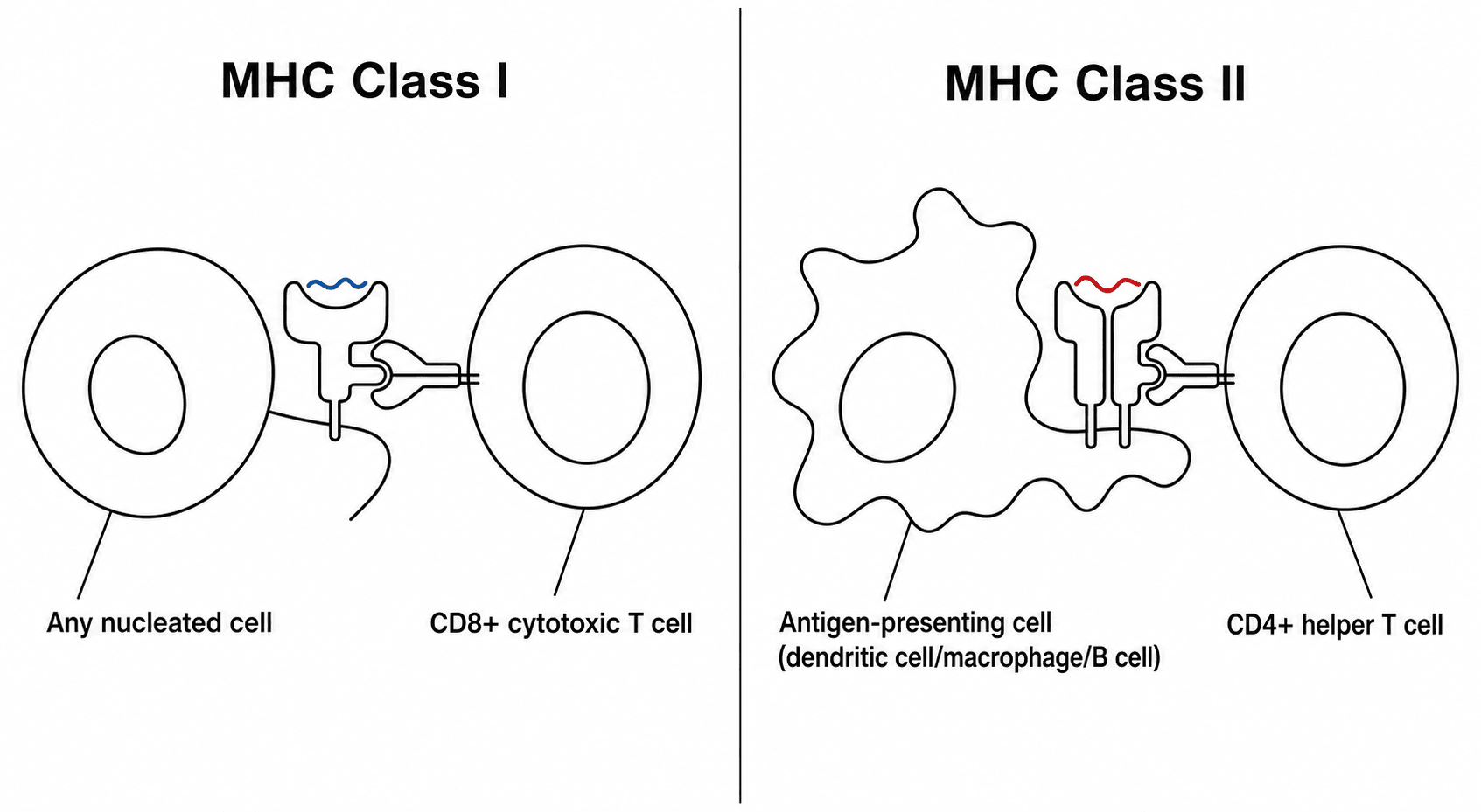
- ADAPTIVE immunity centres on ANTIGEN PRESENTATION via the MAJOR HISTOCOMPATIBILITY COMPLEX (MHC), called HLA in humans: MHC class I (HLA-A/B/C) on all nucleated cells presents to CD8 cytotoxic T cells, and MHC class II (HLA-DR/DQ/DP) on antigen-presenting cells presents to CD4 helper T cells; B cells then make antibodies, and MEMORY cells give a faster, stronger secondary response.
- ALLOGRAFT REJECTION results from recognition of FOREIGN HLA: HYPERACUTE rejection (minutes-hours) is from PRE-FORMED antibodies (e.g. ABO/HLA) activating complement; ACUTE rejection (days-weeks) is T-cell-mediated (cellular) and/or antibody-mediated (humoral, with donor-specific anti-HLA antibodies and complement); CHRONIC rejection (months-years) is a slow fibro-proliferative immune injury - HLA mismatch and donor-specific antibodies drive these processes.
- ORTHOPAEDIC APPLICATION - bone allografts: standard PROCESSED bone allograft (fresh-frozen, freeze-dried/lyophilised, or irradiated) is only WEAKLY IMMUNOGENIC because processing removes/kills the immunogenic cellular and marrow elements, so bone allografts are NOT routinely ABO/HLA matched and act largely as an osteoconductive (and variably osteoinductive) scaffold incorporated by creeping substitution.
- By contrast, FRESH OSTEOCHONDRAL ALLOGRAFTS (transplanted with viable chondrocytes) are MORE IMMUNOGENIC and can provoke a host immune response; the cartilage matrix is relatively immunoprivileged (chondrocytes shielded from immune cells), but exposed subchondral bone/marrow elements and HLA antigens can elicit a response affecting incorporation - a reason fresh osteochondral allografts are used judiciously and are not HLA-matched but are size-matched and used promptly.
- “Innate = immediate, non-specific, NO memory (barriers, phagocytes, NK cells, complement); Adaptive = specific, MEMORY (T cells, B cells/antibodies, MHC/HLA).
- “Complement: opsonisation + chemotaxis (C5a) + membrane attack complex (C5b-9); MHC I → CD8, MHC II → CD4.
- “Allograft rejection: hyperacute (pre-formed antibody/complement), acute (cellular T-cell +/- humoral DSA), chronic (fibroproliferative). Processed bone allograft weakly immunogenic (not HLA-matched); fresh osteochondral more immunogenic.
Barriers, phagocytes (neutrophils/macrophages), NK cells, complement. Fast, non-specific, no memory.
T cells (cell-mediated), B cells/antibodies (humoral), MHC/HLA antigen presentation, immunological memory. Slower, specific.
Innate & Adaptive Immunity
Innate immunity is the body's immediate defence: physical and chemical barriers (skin, mucosa), phagocytes (neutrophils and macrophages) that engulf pathogens, natural killer (NK) cells that kill infected/stressed cells, and the complement system; it is rapid and non-specific and has no memory. Adaptive immunity develops over days but is specific and remembers: T lymphocytes mediate cell-mediated immunity (CD4 helper T cells coordinate the response; CD8 cytotoxic T cells kill target cells), while B lymphocytes differentiate into plasma cells that secrete antibodies (humoral immunity), and memory T and B cells provide a faster, stronger response on re-exposure. The two arms are linked - innate cells (especially antigen-presenting cells) instruct the adaptive response.
Complement is a cascade of plasma proteins activated by three pathways - classical (antibody- triggered), alternative and lectin - that converge on C3. Its key effects are OPSONISATION (C3b coats microbes for phagocytosis), CHEMOTAXIS/inflammation (anaphylatoxins such as C5a recruit neutrophils), and the MEMBRANE ATTACK COMPLEX (C5b-9) that lyses target cells. As an innate mechanism it defends against infection, but it is also a major effector of antibody-mediated (humoral) tissue injury, including in transplant rejection, where donor-specific antibodies fix complement on graft endothelium.
MHC/HLA & Antigen Presentation
The major histocompatibility complex (MHC) - the human leukocyte antigen (HLA) system - presents peptide antigens to T cells and is the basis of allorecognition. MHC class I (HLA-A, -B, -C) is on all nucleated cells and presents intracellular peptides to CD8 cytotoxic T cells; MHC class II (HLA-DR, -DQ, -DP) is on antigen-presenting cells (dendritic cells, macrophages, B cells) and presents extracellular peptides to CD4 helper T cells, which then orchestrate the B-cell antibody response and cytotoxic responses. Because HLA is highly polymorphic, an individual's HLA is recognised as foreign by another's immune system - the central problem in transplantation. (HLA associations are also high-yield in rheumatology, e.g. HLA-B27 with the seronegative spondyloarthropathies.)
Allograft Rejection & Orthopaedic Application
- Hyperacute (minutes-hours): PRE-FORMED recipient antibodies (e.g. ABO or anti-HLA) bind donor endothelium and activate complement → rapid thrombosis/graft loss.
- Acute (days-weeks): cellular (T-cell-mediated) and/or humoral/antibody-mediated rejection (donor-specific anti-HLA antibodies, DSA, with complement) - the main form prevented by immunosuppression in solid-organ transplants.
- Chronic (months-years): a slow fibro-proliferative immune-mediated injury causing graft fibrosis and failure. HLA mismatch and donor-specific antibodies drive these processes; NK cells (via KIR-HLA interactions) also contribute.
- Processed bone allograft (fresh-frozen, freeze-dried/lyophilised, irradiated) is only WEAKLY IMMUNOGENIC: processing removes or kills the immunogenic cells and marrow, leaving a largely acellular matrix that acts as an osteoconductive (and variably osteoinductive) scaffold incorporated by creeping substitution. For this reason bone allografts are NOT routinely ABO/HLA matched - unlike solid organs - and immunosuppression is not used.
- Fresh OSTEOCHONDRAL allograft (transplanted with viable chondrocytes) is MORE immunogenic: the hyaline cartilage matrix is relatively immunoprivileged (chondrocytes are shielded within matrix from immune cells), but exposed subchondral bone/marrow and HLA antigens can provoke a host response that may affect incorporation; these grafts are size-matched and used promptly but are not HLA-matched.
- Implant materials and wear debris also engage the immune system (e.g. macrophage-driven osteolysis, metal hypersensitivity) - see our Wear/Osteolysis and Metal Hypersensitivity topics.
Hypersensitivity: Gell & Coombs Types I-IV
Innate and adaptive immunity, complement, rejection and metal hypersensitivity are tied together by one framework - the Gell & Coombs hypersensitivity classification, core applied-basic-science viva material:
- Type I (immediate, IgE-mediated): allergen cross-links IgE on mast cells → degranulation (histamine) → anaphylaxis/urticaria within minutes. Orthopaedic examples: latex allergy, drug/antibiotic anaphylaxis.
- Type II (antibody-mediated, cytotoxic): IgG/IgM against cell-surface antigens, activating complement and ADCC. Examples: transfusion (ABO) reactions, hyperacute graft rejection, autoimmune cytopenias.
- Type III (immune-complex): circulating antigen-antibody complexes deposit in tissues and fix complement → vasculitis/synovitis. Examples: rheumatoid arthritis, SLE, serum sickness.
- Type IV (delayed, cell-mediated): T-cell/macrophage response peaking at 48 to 72 hours, with no antibody. Examples: metal hypersensitivity (nickel/cobalt-chromium implant reactions, including ALVAL - an aseptic lymphocyte-dominated vasculitis-associated lesion - around metal-on-metal implants), contact dermatitis, the tuberculin (Mantoux) skin test, and acute cellular transplant rejection.
The orthopaedic punchline: metal hypersensitivity / ALVAL is Type IV, hyperacute rejection is Type II, and the inflammatory arthropathies (RA, SLE) involve Type III. (Implant metal hypersensitivity and ALVAL in depth belong to our Metal Hypersensitivity / Wear-Osteolysis topics; here is the framework.)
Gell & Coombs: I immediate IgE (latex/drug anaphylaxis); II antibody-cytotoxic + complement (transfusion, hyperacute rejection); III immune-complex (RA, SLE); IV delayed T-cell, 48 to 72 hours, no antibody (metal hypersensitivity/ALVAL, contact dermatitis, Mantoux, acute cellular rejection).
The Immune Status of the Orthopaedic Patient
An "immunology in orthopaedics" topic must also cover the patient's own immune competence, which changes peri-operative decisions:
- The immunosuppressed / on-biologics patient: disease-modifying biologics (anti-TNF, anti-IL-6, rituximab) and corticosteroids raise the infection risk of surgery. The usual approach is to hold biologic agents around surgery (timed to the drug's dosing cycle) to reduce infection while balancing a disease flare, often continue methotrexate, and give peri-operative steroid cover for those on long-term steroids; restart biologics once the wound is healing and there is no infection.
- The asplenic / hyposplenic patient (e.g. after post-traumatic splenectomy): loss of the spleen's filtering and antibody function leaves a lifelong risk of overwhelming post-splenectomy infection (OPSI) from encapsulated organisms (pneumococcus, meningococcus, Haemophilus). Manage with vaccination against these organisms (ideally before, or after, splenectomy), antibiotic prophylaxis/standby, and patient education - relevant whenever the orthopaedic/trauma team is responsible for such a patient.
- General principle: the immunocompromised surgical patient (also diabetes, chemotherapy, transplant immunosuppression) has a higher infection risk and a blunted response, demanding meticulous antisepsis, optimisation, and a low threshold for investigating infection.
(The cytokines and their inhibitors are covered in our Inflammation/Cytokines topic; the point here is the peri-operative immune status.)
Mind the patient's immunity: hold biologics around surgery (continue methotrexate, give steroid cover) to cut infection risk; the asplenic patient (post-traumatic splenectomy) needs vaccination against encapsulated organisms and antibiotic prophylaxis against OPSI; the immunocompromised patient has a higher, blunted-response infection risk.
The Two Immune Responses That Decide Whether an Implant Survives
Most immunology taught to surgeons is transplant immunology borrowed from other specialties. The two immune responses that actually determine orthopaedic outcomes are different, and neither involves a donor: both are reactions to a piece of inert material the body cannot remove.
Polyethylene wear particles are the wrong size and the wrong chemistry for the immune system. They are small enough to be ingested by macrophages and impossible to degrade, so the macrophage mounts a response it can never complete - the classic frustrated phagocytosis - and keeps signalling.
The sequence is worth knowing in order because each step is a place the process could be interrupted:
- Particles are phagocytosed by periprosthetic macrophages, which cannot digest them.
- Inflammatory cytokines are released - TNF-alpha, IL-1, IL-6 and PGE2 - producing a chronic, self-sustaining inflammatory membrane at the bone-implant interface.
- RANKL rises relative to osteoprotegerin (OPG). OPG is the decoy receptor that normally mops RANKL up, so what matters is the RANKL/OPG ratio, not RANKL alone.
- RANKL binds RANK on osteoclast precursors, driving their differentiation and activation.
- Osteoclasts resorb bone around a prosthesis that is still perfectly well fixed - producing the expanding, non-infective lucency of periprosthetic osteolysis, and eventually aseptic loosening.
Why this matters for a decision you actually make: every intervention that has reduced osteolysis works at step 1, by reducing the particle burden - crosslinking the polyethylene above all. Nothing works at steps 2 to 5, because there is no licensed way to interrupt the immune response itself, and attempts to repurpose antiresorptives have not changed practice. Bearing choice is the immunological intervention.
Implant infection behaves unlike infection in native tissue, and the reason is immunological rather than microbiological. An implant surface has no blood supply, so the cellular and humoral defences that clear bacteria elsewhere reach it poorly. Neutrophils that do arrive are functionally impaired at a foreign surface, and organisms within a mature biofilm are metabolically quiescent, physically shielded by their matrix, and therefore invisible both to those defences and to antibiotics that require dividing cells.
Three consequences follow directly, and they explain the whole management algorithm:
- Host defence will not clear it, and nor will antibiotics alone, however long they are given. The question is surgical from the outset.
- Biofilm maturity, not symptom duration alone, drives the decision between debridement with implant retention and exchange of the implant.
- Biofilm-active agents matter - rifampicin combinations for staphylococci, and never rifampicin as monotherapy because resistance emerges rapidly.
The unifying idea across both halves of this section: in orthopaedics the immune system is usually failing not because it is deficient but because it has been asked to deal with something it cannot remove, in a place it cannot reach.
Mnemonics & Memory Aids
INNATE
Hook:INNATE = immediate, non-specific, no memory.
MHC 1-8 / 2-4
Hook:Rule of 8: MHC I x CD8 = 8; MHC II x CD4 = 8.
Clinical Decision Scenarios
Practise clinical reasoning and management decisions out loud
“Outline the difference between innate and adaptive immunity, and explain the role of complement and MHC/HLA.”
“Why are bone allografts not HLA-matched, and how does this differ for fresh osteochondral allografts?”
Innate immunity
- Immediate, non-specific, NO memory
- Barriers, phagocytes (neutrophils/macrophages), NK cells, complement
- Complement: opsonisation (C3b), chemotaxis (C5a), membrane attack complex (C5b-9)
Adaptive immunity
- Specific, has MEMORY
- T cells (CD4 helper, CD8 cytotoxic) - cell-mediated; B cells → antibodies (humoral)
- MHC I (HLA-A/B/C) → CD8; MHC II (HLA-DR/DQ/DP) → CD4
Allograft rejection
- Hyperacute: pre-formed antibody + complement (minutes-hours)
- Acute: cellular (T-cell) +/- humoral (DSA + complement) (days-weeks)
- Chronic: fibro-proliferative immune injury (months-years)
Orthopaedic allografts
- Processed bone allograft: weakly immunogenic - NOT HLA-matched; osteoconductive scaffold (creeping substitution)
- Fresh osteochondral allograft: viable chondrocytes - more immunogenic; cartilage relatively immunoprivileged
- Osteochondral grafts size-matched + used promptly (not HLA-matched)
Evidence & Key Studies
KIR and their HLA class I ligands in chronic rejection and graft loss in transplantation
- Incompatibility between donor and recipient for HLA (MHC) class I generates complex cellular and humoral immune responses largely responsible for rejection and graft loss.
- Alloreactive natural killer (NK) cells, regulated by killer immunoglobulin-like receptors (KIR) interacting with HLA class I, contribute to rejection - linking innate (NK) and adaptive (HLA) immunity.
- Demonstrates that HLA matching and KIR-HLA combinations influence rejection risk - the immunological basis of allorecognition.
Consensus recommendations on donor-specific anti-HLA antibodies (DSA) in transplantation
- Donor-specific anti-HLA antibodies (DSA) are an important cause of graft (engraftment) failure, reflecting antibody-mediated (humoral) rejection.
- DSA are detected by multiplex bead arrays and complement-binding (C1q) assays, linking antibody, HLA and the complement cascade.
- Higher DSA levels confer higher rejection/failure risk, and desensitization removes/neutralises antibodies and inhibits complement activation - illustrating humoral allorejection mechanisms.
Wear-particle-induced periprosthetic osteolysis and the RANKL/RANK/OPG axis
- Periprosthetic osteolysis induced by WEAR PARTICLES is an important cause of aseptic loosening after joint replacement, and its central mechanism is an imbalance between osteogenesis and osteoclastic resorption.
- The cells involved are osteoclasts and the macrophages they derive from, alongside osteoblasts, osteocytes and fibroblasts - so the process is an immune response to a non-degradable foreign particle.
- The RANKL/RANK/OPG axis is the typical pathway to osteolysis: particle-activated macrophages release inflammatory cytokines and drive RANKL expression, tipping the balance towards osteoclastogenesis.
- Autophagy is activated in periprosthetic cells and modulates cytokine release, osteoclast activation and osteoblast differentiation, but whether its net effect is protective or harmful remains unresolved.
Periprosthetic joint infection: biofilm and the limits of the local host immune response
- Periprosthetic joint infection complicates 1% to 2% of primary arthroplasties and requires interdisciplinary treatment.
- The defining problem is BIOFILM - organisms persisting on the implant surface in a form that resists both antimicrobials and, critically, the LIMITATIONS OF THE LOCAL HOST IMMUNE RESPONSE.
- Understanding the phases of biofilm formation and those immune limitations is what allows a rational choice between debridement with implant retention and exchange.
- Sonication improves diagnostic sensitivity, particularly in chronic low-virulence infection; biofilm-active antibiotics permit eradication in the presence of a foreign body, and total antibiotic duration after revision should not exceed 12 weeks.
Read the four cards in two pairs, because they answer different questions and only one pair is orthopaedic. Littera and Kongtim are transplant immunology from other specialties - a KIDNEY transplant immunogenetics study and a HAEMATOPOIETIC STEM CELL TRANSPLANT consensus. Neither contains any musculoskeletal content, and they are cited only for the general mechanisms of cellular and humoral allorecognition. They do not support any statement about bone or cartilage grafting, where the graft is usually avascular, often acellular after processing, and implanted without immunosuppression.
The two orthopaedic cards carry the immunology that actually decides outcomes in this specialty. Yin supplies the wear-particle route to aseptic loosening - macrophage activation, cytokine release, RANKL/RANK/OPG, osteoclastogenesis, periprosthetic osteolysis. Izakovicova supplies the other half, biofilm and the limits of local host defence, which is why an infected implant is not cleared by immunity and antibiotics alone. If you take two things from this page into a viva, take those.
The innate/adaptive framework, complement cascade, MHC/HLA antigen presentation and the immunogenicity of processed bone versus fresh osteochondral allograft are standard, well-established teaching. (See also our Inflammation/Cytokines, Bone Grafts and Osteochondral Allograft topics.)