Nucleus Pulposus | Annulus Fibrosus | Endplates | Nutrition | Degeneration
PFIRRMANN CLASSIFICATION OF DISC DEGENERATION
Critical Must-Knows
- Nucleus pulposus: 80% water, Type II collagen, gelatinous, resists compression
- Annulus fibrosus: 15-20 lamellae, Type I collagen at ±30°, resists tension and torsion
- Avascular after age 20 - nutrients diffuse through endplates (major limitation to healing)
- Disc height loss with degeneration → facet joint overload → osteoarthritis cascade
- Endplate injury disrupts nutrition → accelerates degeneration (Modic changes)
Clinical Pearls
- "Viva opener: 'Draw the intervertebral disc showing annular fiber orientation'
- "Key biomechanical function: Compression resistance (nucleus) + tensile/torsional resistance (annulus)
- "Disc degeneration is biochemical before structural - proteoglycan loss → water loss → height loss
- "Pfirrmann grading (MRI T2): Grade I (white) → Grade V (black, collapsed)
Critical Disc Biology Exam Points
Nucleus Pulposus Structure
80% water, Type II collagen, aggrecan proteoglycans. Gelatinous consistency provides hydrostatic compression resistance. Water content decreases with age (80% at birth → 70% by age 60).
Annulus Fibrosus Architecture
15-20 concentric lamellae of Type I collagen oriented at ±30° alternating layers. This crossed-fiber arrangement resists tensile and torsional loads.
Nutrition Pathway
Avascular after age 20 - largest avascular structure in body. Nutrients (oxygen, glucose) diffuse through vertebral endplates. Diffusion pathway is 8mm → cells furthest from blood supply.
Degeneration Cascade
Biochemical → Structural → Biomechanical failure. Proteoglycan loss → water loss → height loss → facet overload → OA. Endplate injury accelerates process.
NAEDisc Components and Functions
| N | Nucleus pulposus Central gelatinous core - resists compression (80% water, Type II collagen) |
| A | Annulus fibrosus Outer fibrous ring - resists tension and torsion (Type I collagen, ±30° orientation) |
| E | Endplates (cartilaginous) Superior and inferior boundaries - nutrition pathway (diffusion only route) |
Hook:NAE the disc! Nucleus for compression, Annulus for tension, Endplates for nutrition.
PANDADisc Degeneration Cascade
| P | Proteoglycan loss Aggrecan degradation by MMPs |
| A | Apoptosis of cells Nutrient deprivation → cell death |
| N | Nucleus dehydration Water content drops from 80% to under 60% |
| D | Disc height loss Structural collapse, decreased T2 signal |
| A | Adjacent facet arthritis Load transfer to facets → OA |
| P | Proteoglycan loss Aggrecan degradation by MMPs | D | Disc height loss Structural collapse, decreased T2 signal |
| A | Apoptosis of cells Nutrient deprivation → cell death | A | Adjacent facet arthritis Load transfer to facets → OA |
| N | Nucleus dehydration Water content drops from 80% to under 60% |
Hook:Disc degeneration follows the PANDA pathway from biochemical to structural failure.
Overview and Introduction
Gross Anatomy
The intervertebral disc comprises three distinct components:
1. Nucleus Pulposus (Central):
- Gelatinous, hydrated core
- 80% water at birth, decreasing to 70% by age 60
- Type II collagen (random orientation) and aggrecan
- Resists compressive loads through hydrostatic pressure
- Located slightly posterior to anatomical center (important for herniation pathomechanics)
2. Annulus Fibrosus (Peripheral):
- 15-20 concentric lamellae of Type I collagen
- Fibers oriented at ±30° from horizontal in alternating layers
- Outer annulus contains pain fibers (recurrent meningeal nerve of Luschka)
- Inner annulus blends with nucleus (no clear boundary)
- Anterior annulus thicker than posterior (posterior more prone to tear)
3. Cartilaginous Endplates (Superior/Inferior):
- Thin layer of hyaline cartilage (0.6-1.0 mm thick)
- Covers vertebral body endplate
- Primary route for disc nutrition (diffusion pathway)
- Weakest structural component (vulnerable to trauma)
Microstructure and Composition
Nucleus Pulposus Composition
- Water: 70-80% (age-dependent)
- Proteoglycans: 50-65% dry weight (mainly aggrecan)
- Collagen: 15-20% dry weight (Type II, random orientation)
- Cells: Notochordal cells (young), chondrocyte-like cells (adult)
Annulus Fibrosus Composition
- Water: 60-70%
- Collagen: 50-60% dry weight (Type I)
- Proteoglycans: 10-20% dry weight
- Cells: Fibrochondrocytes
The crossed-fiber arrangement of the annulus (alternating ±30° layers) creates a structure that:
- Resists tensile loads in any direction
- Resists torsional (rotational) loads
- Confines the nucleus centrally under compression
- Prevents nuclear herniation (when intact)
Concepts and Molecular Biology
Core Biochemical Concepts
Extracellular Matrix Composition:
The disc matrix determines function and undergoes significant changes with age and degeneration:
- Nucleus pulposus: Aggrecan proteoglycan (water retention), Type II collagen (scaffold)
- Annulus fibrosus: Type I collagen (tensile strength), elastin (elasticity)
- Endplate: Hyaline cartilage, collagen, proteoglycans (nutrient passage)
Cellular Biology:
- Notochordal cells (embryonic) → replaced by chondrocyte-like cells by adulthood
- Cell density very low (sparse population)
- Cells rely entirely on diffusion for oxygen and glucose (8mm diffusion distance)
- Hypoxic environment with anaerobic metabolism
Molecular Degeneration Pathway:
- Matrix metalloproteinases (MMPs) and ADAMTS degrade proteoglycans
- Aggrecan loss → decreased water retention (80% → under 60%)
- Type II collagen replaced by Type I collagen
- Disc height decreases → altered load distribution
- Pro-inflammatory cytokines (IL-1beta, TNF-alpha) perpetuate cycle
Biomechanical Function
Load Distribution
The disc functions as a load-bearing hydraulic system:
Under Compression:
- Nucleus pulposus subjected to hydrostatic pressure
- Nucleus attempts to expand radially
- Annulus fibrosus resists radial expansion (hoop stress in fibers)
- Load distributed evenly across vertebral endplate
Under Flexion/Extension:
- Anterior annulus compressed, posterior annulus tensioned (flexion)
- Posterior annulus compressed, anterior annulus tensioned (extension)
- Nucleus shifts posteriorly (flexion) or anteriorly (extension)
Under Lateral Bending and Rotation:
- Asymmetric loading of annular lamellae
- Crossed-fiber orientation resists torsion
- Rotation is the most damaging motion (shear forces on annulus)
Disc Response to Loading
| Load Type | Nucleus Response | Annulus Response | Clinical Significance |
|---|---|---|---|
| Compression | Hydrostatic pressure increases | Hoop stress in fibers | Normal weight-bearing |
| Flexion | Shifts posteriorly | Anterior compressed, posterior tensioned | Posterior annular stress |
| Rotation | Minimal response | Shear stress on lamellae | Most damaging motion |
Viscoelastic Properties
The disc demonstrates time-dependent behavior:
- Creep: Under constant load, disc height decreases over time (fluid exudation)
- Stress relaxation: Under constant deformation, internal stress decreases over time
- Diurnal variation: Fluid is expressed during the day and reabsorbed overnight, producing a diurnal stature change of roughly 15-20mm across all discs combined (each disc losing on the order of 1mm), with intradiscal pressure highest after a night of rest
- Age-related changes: Decreased water content → decreased viscoelasticity
This viscoelastic behavior explains why back pain is often worse in the morning (disc fully hydrated, maximum intradiscal pressure) and why prolonged standing/sitting causes symptoms (creep deformation).
Nutrition and Metabolism
Avascular Nature
The adult intervertebral disc is the largest avascular structure in the human body:
- Vascularized in childhood: Blood vessels penetrate endplates
- Avascular by age 20: Vessels regress, diffusion becomes only nutrition route
- Longest diffusion pathway: Up to 8mm from endplate to central nucleus
- Metabolic challenge: Cells in center of disc furthest from blood supply in body
Nutritional Limitation to Healing
The avascular nature of the disc is the primary reason discs do not heal once injured:
- No blood supply → no inflammatory cells → no repair cascade
- Cells rely on diffusion for oxygen and glucose
- Cells furthest from blood supply are most vulnerable to nutrient deprivation
- Endplate injury or sclerosis → disrupts already tenuous nutrition → accelerated degeneration
This is fundamentally different from other musculoskeletal tissues (bone, tendon, ligament) which have robust healing capacity due to vascularity.
Nutrient Transport
Diffusion Pathway for Disc Nutrition
Nutrients (oxygen, glucose) in vertebral body capillaries adjacent to endplate.
Nutrients diffuse through porous cartilaginous endplate (0.6-1.0mm thick).
Nutrients diffuse through disc matrix (up to 8mm to reach central nucleus cells).
Nucleus pulposus cells metabolize nutrients anaerobically (low oxygen environment).
Factors Affecting Disc Nutrition:
- Endplate permeability: Sclerosis or calcification → decreased diffusion → cell death
- Disc height: Thicker discs → longer diffusion pathway → poorer nutrition
- Loading: Cyclic loading enhances diffusion (pumping action), static loading impairs it
- Smoking: Decreases endplate blood supply → accelerates degeneration
Disc Degeneration
Degeneration Cascade
Disc degeneration follows a predictable biochemical → structural → biomechanical pathway:
Early Degeneration (Pfirrmann I-II)
Proteoglycan Loss:
- Matrix metalloproteinases (MMPs) degrade aggrecan
- Loss of aggrecan → decreased osmotic pressure
- Decreased water-binding capacity
Cellular Changes:
- Nutrient deprivation → increased apoptosis
- Decreased cell density
- Shift from anabolic to catabolic metabolism
- Inflammatory cytokines (IL-1, TNF-α) upregulated
Biochemical markers:
- Decreased proteoglycan content (MRI: decreased T2 signal)
- Increased collagen cross-linking
- pH decreases (lactate accumulation from anaerobic metabolism)
Early changes are reversible with improved nutrition (theoretical).
Pfirrmann Classification (MRI Grading)
Pfirrmann MRI Classification of Disc Degeneration
| Grade | Structure | Signal (T2) | Disc Height | Interpretation |
|---|---|---|---|---|
| I | Homogeneous bright white | Hyperintense | Normal | Normal disc |
| II | Inhomogeneous with horizontal band | Hyperintense | Normal | Early degeneration |
| III | Inhomogeneous gray | Intermediate | Normal/decreased | Moderate degeneration |
| IV | Inhomogeneous dark gray | Hypointense | Decreased | Advanced degeneration |
| V | Inhomogeneous black | Hypointense | Collapsed | Severe degeneration |


Modic Endplate Changes
Modic changes represent endplate and adjacent bone marrow pathology associated with disc degeneration:
- Type I (T1 low, T2 high): Edema and inflammation → acute/active degeneration
- Type II (T1 high, T2 high): Fatty replacement → chronic degeneration
- Type III (T1 low, T2 low): Sclerosis → end-stage degeneration
Modic Type I changes are associated with discogenic back pain and may represent an inflammatory phenotype amenable to treatment.
Clinical Relevance and Applications
Differential Diagnosis of a Low-Signal ("Dark") Disc or Suspected Discogenic Pain
A degenerate disc on MRI is common and frequently asymptomatic. The exam-relevant skill is distinguishing benign age-related change from pathology that mimics it.
Differentiating Disc Degeneration from Mimics
| Entity | Key MRI / Clinical Feature | Distinguishing Point | Action |
|---|---|---|---|
| Age-related disc degeneration | Low T2 signal, mild height loss, no Modic or only Type II | Very common, often asymptomatic; prevalence rises with age | Reassure; treat symptoms, not the image |
| Symptomatic discogenic pain | Modic Type I, high-intensity zone in posterior annulus, single-level concordant pain | Inflammatory/active phenotype; pain reproduced by that level | Targeted rehab; selected injection/fusion in refractory cases |
| Infective discitis / spondylodiscitis | T2-high disc and endplate, endplate destruction, paravertebral/epidural collection, contrast enhancement | Pain at rest and night, raised CRP/ESR, fever; crosses the disc space | Urgent - blood cultures, biopsy, antibiotics |
| Vertebral metastasis / myeloma | Marrow replacement on T1 but disc space PRESERVED | Tumour spares the avascular disc; infection destroys it | Staging, biopsy, oncology referral |
| Modic Type I (non-infective) | T1 low, T2 high endplate, NO disc destruction or collection | Mimics early discitis; normal inflammatory markers | Conservative; correlate with markers before treating as infection |
Disc Space: The Key Discriminator
Pyogenic infection crosses and destroys the disc space (the organism reaches the avascular disc via the endplate); metastatic and myeloma deposits spare the disc and replace vertebral marrow. "Marrow disease that respects the disc is tumour until proven otherwise; marrow disease that eats the disc is infection until proven otherwise."
Understanding Disc Biology Informs Clinical Practice
Why Discs Don't Heal:
- Avascular after age 20 → no inflammatory healing response
- Low cell density → insufficient regenerative capacity
- Hostile biochemical environment with catabolic cytokines
- This explains why conservative treatment focuses on symptom management
Surgical Implications:
- Discectomy removes herniating nucleus but accelerates degeneration
- Fusion eliminates motion segment but increases adjacent level stress
- Disc replacement aims to preserve motion but long-term outcomes variable
- Biologic therapies (stem cells, growth factors) target regeneration
Prevention and Modification:
- Smoking cessation (nicotine impairs already limited nutrition)
- Weight management (reduces compressive load)
- Avoid repetitive flexion-rotation (most damaging motion)
- Core strengthening (dynamic stabilization)
Controversies and Areas of Uncertainty
The biology of the disc is mature, but its clinical translation remains contested. High-yield viva controversies:
Does degeneration cause pain?
Disc degeneration on MRI is highly prevalent in asymptomatic people and rises with age. The correlation between Pfirrmann grade and back pain is weak. Modic Type I and a posterior annular high-intensity zone are the features most linked to pain, but neither is specific. Treating images rather than patients is a recognised pitfall.
Degeneration vs ageing
Whether disc degeneration is a distinct disease or simply accelerated normal ageing is unresolved. Boos et al showed an early, nutrition-driven histological continuum from the second decade, blurring the line between "normal ageing" and "pathology".
Biological regeneration
MSC and growth-factor injection (e.g. allogeneic MSC RCT, Noriega 2017) show proof-of-concept disc-quality and pain improvement, but benefit is confined to a responder subgroup and no large definitive trial yet supports routine use. Re-injuring an avascular, hostile, low-pH niche to deliver cells remains a fundamental hurdle.
Fusion vs motion preservation
Total disc replacement aims to avoid adjacent-segment degeneration seen after fusion, but registry and trial evidence show comparable medium-term outcomes and its own failure modes. Whether adjacent-segment change is caused by fusion or reflects the patient's underlying degenerative biology is debated.
Guidelines, Registries & Global Practice
Global Epidemiology
- Low back pain is the leading cause of years lived with disability worldwide (Global Burden of Disease), and disc degeneration is its commonest structural correlate.
- Degenerative MRI findings are near-universal with age: disc signal loss and bulging are found in the majority of asymptomatic adults over 50-60 years.
- Heritability of disc degeneration is substantial (29-54% in twin data), with smoking, obesity, heavy repetitive loading, and vibration as modifiable contributors.
Side-by-Side Guidance on Degenerative Disc Disease / Low Back Pain
Major Society Positions Relevant to Disc Degeneration
| Body (region) | Imaging stance | Management emphasis |
|---|---|---|
| NICE / BOA (UK) | Do NOT routinely image non-specific low back pain; image only if result changes management or red flags present | Self-management, exercise, manual therapy; surgery reserved for radiculopathy/stenosis failing conservative care |
| ACP / NASS (US) | Avoid early imaging without red flags or progressive deficit | First-line non-pharmacological care; injections and fusion in selected refractory discogenic cases |
| AO Spine (global) | Standardised MRI grading (Pfirrmann) and endplate (Modic) reporting for research and surgical planning | Classification-led, evidence-graded surgical decision frameworks |
| EFORT / European consensus | Reserve advanced imaging for surgical candidates | Motion-preserving options (disc arthroplasty) vs fusion individualised to pathology |
Registry and Resource-Setting Notes
- Spine device registries (e.g. national arthroplasty/spine registries, the international Spine Tango registry) track disc-replacement and fusion survival and adjacent-segment reoperation; medium-term data show broadly comparable outcomes for well-selected fusion versus disc arthroplasty.
- High-resource settings debate motion-preservation, biologics, and intradiscal therapies; limited-resource settings prioritise affordable conservative care and prioritising surgery for neurological compromise rather than axial discogenic pain.
- Across all settings, the consistent message is restraint with imaging and surgery for non-specific axial pain, reflecting the weak image-symptom correlation.
Evidence Base
Degeneration of the Intervertebral Disc (Urban & Roberts review)
- Adult disc is the largest avascular structure - nutrition is by diffusion through the endplate only
- Cells in the disc centre lie up to 8mm from the nearest blood supply, with the lowest oxygen and glucose concentrations of any tissue
- Disc shows degenerative and ageing change earlier than any other connective tissue in the body
- Falling nutrient supply and rising lactate (low pH) drive cell death and a shift to a catabolic matrix-degrading phenotype
- Disc degeneration is associated with low back pain but the relationship is inconsistent
Pfirrmann MRI Classification of Lumbar Disc Degeneration
- Five-grade T2-weighted MRI system (I-V) based on nucleus structure, signal, and disc height; tested on 300 discs in 60 patients
- Grade I: bright homogeneous nucleus, normal height (normal disc)
- Grade V: black heterogeneous signal with collapsed disc space (severe degeneration)
- Intra-observer agreement kappa 0.84-0.90; inter-observer agreement kappa 0.69-0.81 (substantial to excellent)
- Complete agreement in 83.8% of discs; a two-grade discrepancy in only 1.3%
Modic Vertebral Endplate Marrow Changes
- Reviewed 474 consecutive lumbar MRI studies and defined endplate marrow change types
- Type I (T1 low, T2 high) in 4%: fissured endplates with vascularised fibrous tissue (active)
- Type II (T1 high, T2 high) in 16%: yellow marrow (fatty) replacement (chronic stable)
- Type I converted to Type II over 14 months to 3 years in 5 of 6 followed patients; Type II remained stable
- All cases had degenerative disc disease at the involved level
Histologic Classification of Age-Related Disc Change (2002 Volvo Award)
- Histologic study of 180 sagittal lumbar motion-segment slices from 44 individuals (fetal to 88 years)
- Tissue breakdown begins in the nucleus pulposus in the second decade of life
- Diminished blood supply to the endplate in the first half of the second decade appears to initiate disc breakdown
- Ageing and degeneration form a histological continuum rather than discrete processes
- Macroscopic grading scattered widely against histology, favouring a histology-based reference standard
Genetic vs Environmental Influences on Disc Degeneration (Twin Study)
- Classic twin study of 152 monozygotic and 148 dizygotic male twin pairs (Finnish Twin Cohort)
- Heritability of disc degeneration phenotypes ranged 29-54% depending on phenotype and level
- Signal loss, height narrowing, and bulging shared a common genetic pathway
- Genetic and environmental influences differed substantially between upper and lower lumbar levels
- Genetics and environment were of broadly similar overall importance
Allogeneic MSC Intradiscal Injection for Degenerative Disc Disease (RCT)
- Phase II randomised controlled trial: 24 patients, intradiscal allogeneic bone-marrow MSC versus sham paravertebral anaesthetic
- Feasibility and safety confirmed at 1 year
- Pain and disability improved rapidly in the MSC group, but benefit was restricted to a ~40% responder subgroup
- Pfirrmann grade improved in MSC-treated discs and worsened in controls
- Allogeneic cells offer a logistically simpler off-the-shelf alternative to autologous MSC
Exam Viva Scenarios
Use these scenarios to practise clinical reasoning and management decisions
Scenario 1: Basic Disc Structure and Function
"The examiner shows you a sagittal MRI of the lumbar spine and asks: 'Describe the structure and biomechanical function of the intervertebral disc.'"
Scenario 2: Disc Degeneration Cascade
"The examiner shows you sequential MRI scans showing progressive disc degeneration and asks: 'Describe the pathophysiology of disc degeneration and the Pfirrmann classification.'"
Scenario 3: Dark Disc, Pain Correlation and the Future of Disc Repair
"A 45-year-old presents with chronic axial low back pain. The MRI shows a single dark (Pfirrmann IV) disc at L5/S1 with Modic Type I endplate change. The examiner asks: 'How confident are you that this disc is the source of pain, and what does disc biology tell us about treatment options?'"
MCQ Practice Points
Collagen Type Question
Q: What is the predominant collagen type in the nucleus pulposus versus the annulus fibrosus?
A: Nucleus pulposus: Type II collagen (random orientation). Annulus fibrosus: Type I collagen (organized in ±30° alternating lamellae). This reflects their different biomechanical roles: nucleus resists compression, annulus resists tension and torsion.
Nutrition Question
Q: How does the adult intervertebral disc receive nutrition, and what is the significance?
A: Diffusion through cartilaginous endplates - the disc is avascular after age 20. This is the longest diffusion pathway in the body (up to 8mm). The avascular nature explains why discs cannot heal once injured and why endplate injury accelerates degeneration by disrupting the already tenuous nutrition supply.
Pfirrmann Classification Question
Q: Describe the Pfirrmann Grade I and Grade V disc on MRI.
A: Grade I: Bright white (hyperintense) homogeneous nucleus on T2, normal disc height - represents a normal disc. Grade V: Black (hypointense) heterogeneous signal on T2, collapsed disc height - represents severe degeneration. The classification progresses from I to V based on decreasing T2 signal (water content) and disc height.
Modic Changes Question
Q: What is a Modic Type I change and its clinical significance?
A: Modic Type I: T1 low signal, T2 high signal - represents edema and inflammation in the vertebral endplate and adjacent bone marrow. This indicates active/acute degeneration and correlates with discogenic back pain. It may represent an inflammatory phenotype amenable to targeted treatment.
Biomechanics Question
Q: Why is the annular fiber orientation at ±30° biomechanically important?
A: The alternating ±30° crossed-fiber arrangement allows the annulus to resist loads in multiple directions: tensile forces in any direction, torsional (rotational) forces, and radial expansion of the nucleus under compression. This creates hoop stress that confines the nucleus and distributes load evenly.
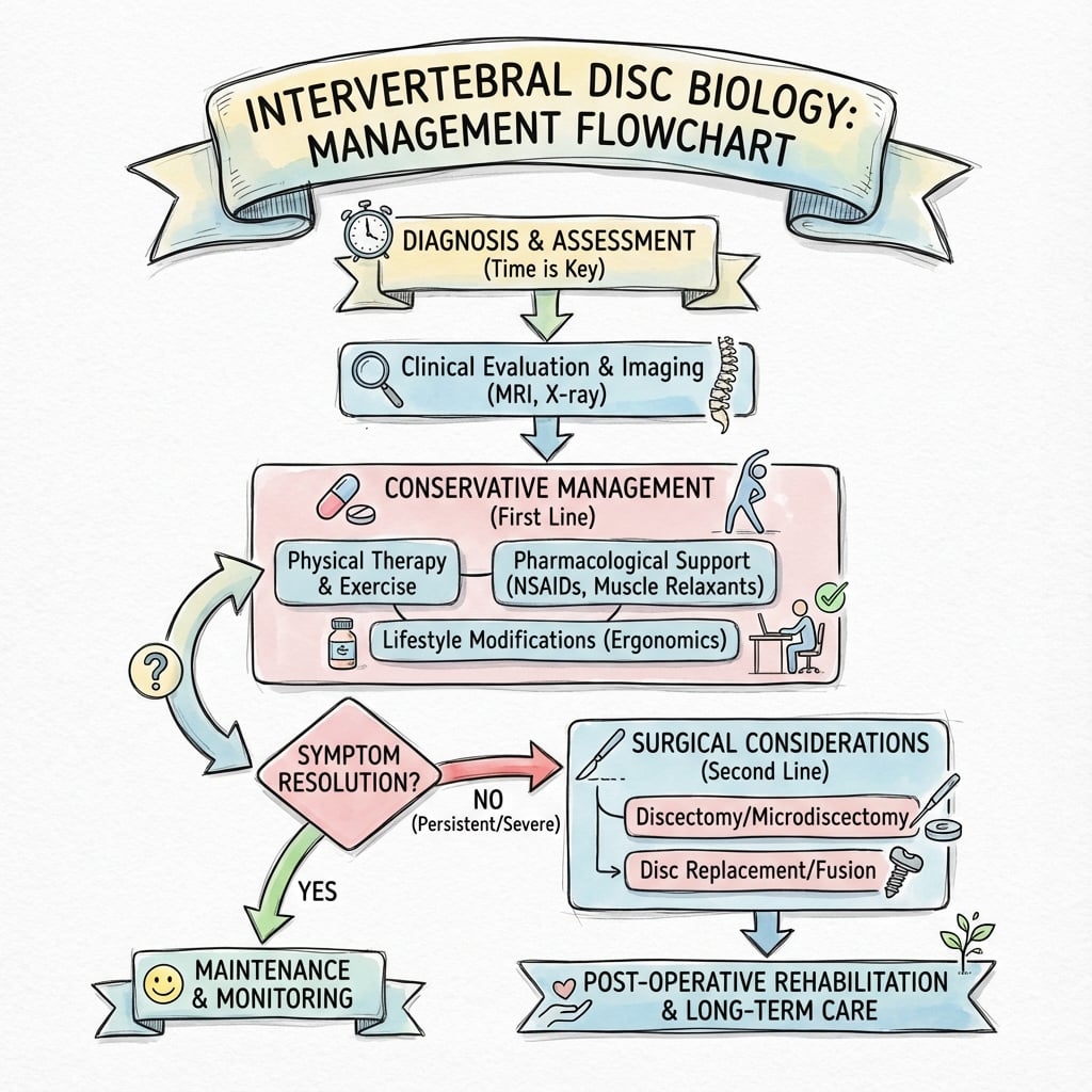
Management Algorithm
INTERVERTEBRAL DISC BIOLOGY
Clinical summary
Key Structures
- •Nucleus pulposus: 80% water, Type II collagen, aggrecan, resists compression
- •Annulus fibrosus: 15-20 lamellae, Type I collagen at ±30°, resists tension/torsion
- •Cartilaginous endplates: 0.6-1.0mm thick, nutrition pathway
- •Avascular after age 20 - largest avascular structure in body
Biomechanical Functions
- •Nucleus: Hydrostatic compression resistance
- •Annulus: Tensile and torsional resistance via hoop stress
- •Crossed fibers (±30°): Resist multi-directional loads
- •Viscoelastic: Creep, stress relaxation, 20mm diurnal variation
Nutrition Pathway
- •Diffusion only route (no blood supply)
- •Pathway: Vertebral capillaries → Endplate → Disc matrix (up to 8mm)
- •Endplate injury/sclerosis → disrupts nutrition → accelerates degeneration
- •Smoking decreases endplate blood supply
Degeneration Cascade
- •Biochemical: Proteoglycan loss → water loss
- •Structural: Nucleus fibrosis, height loss, annular tears
- •Biomechanical: Instability → facet overload → OA
- •Irreversible once structural changes occur
Pfirrmann Classification
- •Grade I: Bright white homogeneous (normal)
- •Grade II: Inhomogeneous with band (early degeneration)
- •Grade III: Gray inhomogeneous (moderate)
- •Grade IV: Dark gray, decreased height (advanced)
- •Grade V: Black, collapsed (severe)
Modic Changes
- •Type I: T1 low, T2 high = edema/inflammation (active, painful)
- •Type II: T1 high, T2 high = fatty replacement (chronic stable)
- •Type III: T1 low, T2 low = sclerosis (end-stage)