Glenohumeral Stability | Rotator Cuff Force Couples | Scapulothoracic Rhythm
- Scapulothoracic rhythm: 2:1 ratio (for every 2° GH abduction, 1° ST upward rotation)
- Rotator cuff force couple: Subscapularis/infraspinatus-teres minor balance humeral head centering
- Deltoid-rotator cuff force couple: Deltoid elevates, RC depresses and centers humeral head
- Glenoid covers only 25% of humeral head (inherent instability requires dynamic stabilizers)
- Concavity-compression: RC compresses head into glenoid concavity for stability
- “Critical arc of abduction 60-120°: supraspinatus most active, impingement risk highest
- “Capsular volume 2x humeral head volume allows large ROM but sacrifices stability
- “Labrum deepens glenoid socket 50% (increases stability)
- “Inferior glenohumeral ligament (IGHL) is primary anterior stabilizer in abduction-external rotation
Overview and Introduction
Shoulder biomechanics is the interplay of several joints, muscles and ligaments that together produce the greatest range of motion of any joint in the body. Understanding those principles is critical to managing instability, rotator cuff pathology, arthroplasty and rehabilitation.
The shoulder complex. Four articulations work in concert to achieve 180° of abduction:
- Glenohumeral (GH)
- Scapulothoracic (ST)
- Acromioclavicular (AC)
- Sternoclavicular (SC)
Glenohumeral Joint Geometry and Stability
The trade-off. The glenohumeral joint is the most mobile and the least stable joint in the body, because the bone offers so little constraint. The glenoid covers only 25% of the surface of the humeral head, the head's radius is greater than that of the glenoid concavity, and the glenoid sits in an average of 5° of retroversion. The result is minimal intrinsic bony stability.
The capsule. Its volume is about twice that of the humeral head. That redundancy, taken up in capsular folds, is what allows 180° of abduction and 90° of external and internal rotation without the capsule coming under tension; the price of a loose capsule is less static restraint.
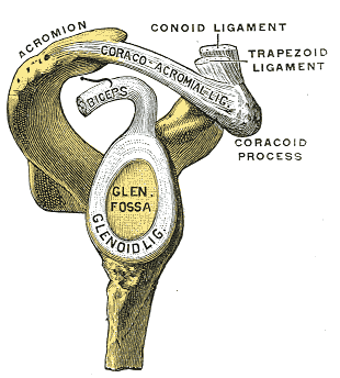
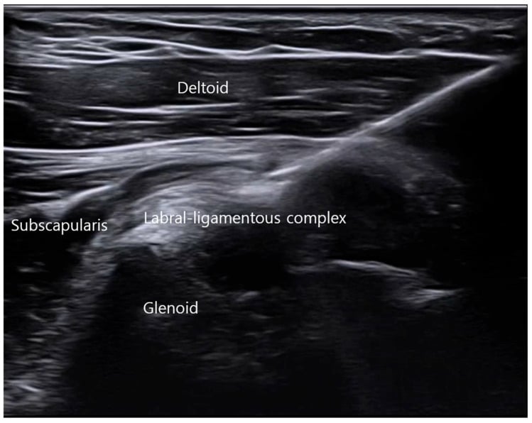
The labrum. The labrum supplies about 50% of the socket's depth. This increases contact area and creates a suction seal. Get the two measurements the right way round; they are orthogonal planes, not a before and after. The combined glenoid-labral socket is roughly 9 mm deep superoinferiorly and 5 mm deep anteroposteriorly (Howell and Galinat). The labrum's share is the difference between about 2.5 mm of bony depth and the 5 mm socket. Detaching the labrum anteriorly, as in a Bankart lesion, drops the anteroposterior depth from 5.0 mm to 2.4 mm, which is the quantitative reason a Bankart lesion destabilises the joint. Superiorly the labrum is the biceps anchor, so SLAP tears compromise the biceps anchor and superior stability.
Static and dynamic stabilisers. Stability is conventionally split 50% static, 50% dynamic; the table sets out which structures do what, and how each group fails.
- Structures
- Capsule, ligaments (IGHL, MGHL, SGHL), labrum, glenoid concavity
- Contribution
- Passive restraint at end ROM
- Failure Mechanism
- Capsular laxity, labral tear, Bankart lesion
- Structures
- Rotator cuff (SITS), deltoid, scapular stabilizers (serratus, trapezius)
- Contribution
- Active concavity-compression, force couples
- Failure Mechanism
- RC tear, scapular dyskinesis, nerve injury
Concavity-compression is the primary dynamic mechanism. The rotator cuff compresses the humeral head into the glenoid concavity, creating a suction seal and depth, and the increased friction and depth resist translating loads; it accounts for approximately 50% of glenohumeral stability. In Lippitt and Matsen's analysis the effect rises with the compressive muscle load and with the depth of the concavity, and an intact labrum is essential to it, especially in the lax mid-range.
The remaining stabilisers complete the picture:
- Adhesion-cohesion: synovial fluid creates a suction effect
- Proprioception: neuromuscular control and reflex stabilisation
- Scapular stability: serratus anterior and trapezius hold the glenoid as a stable platform
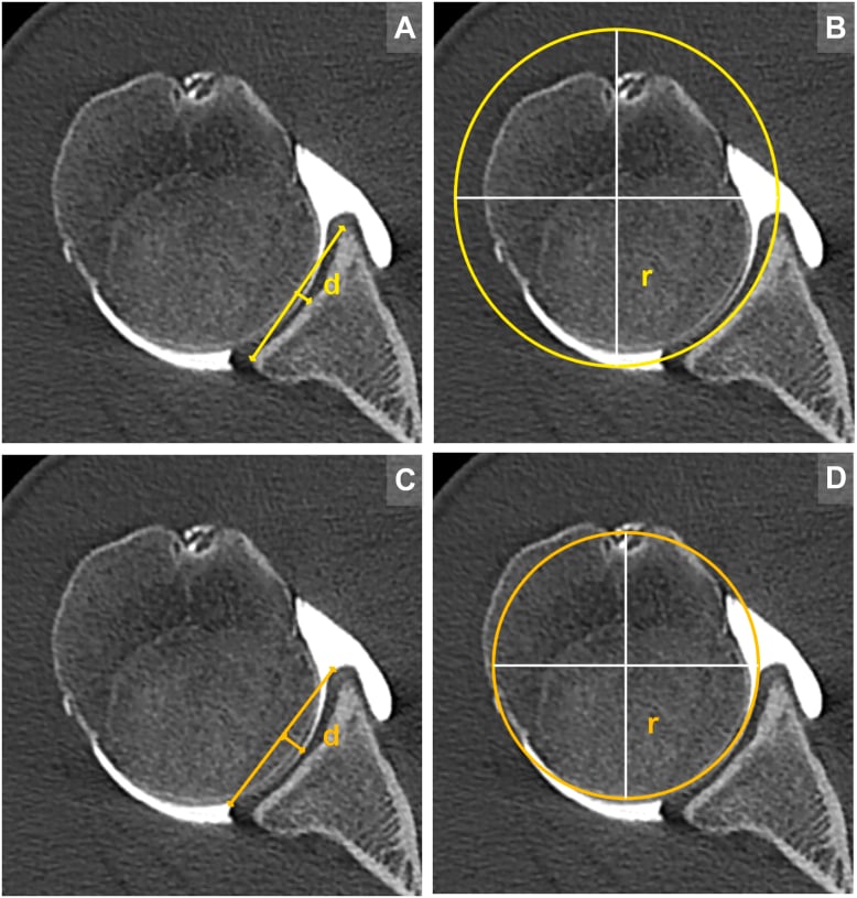
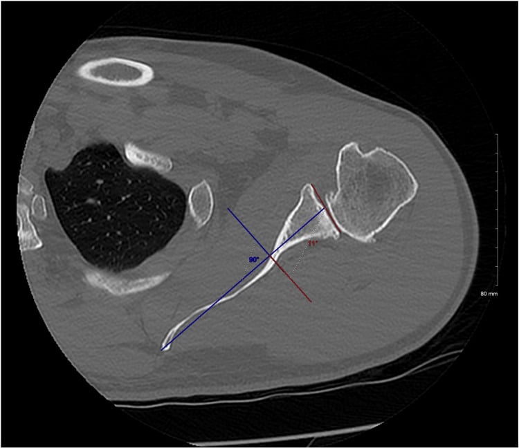
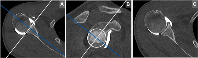
Glenoid version. Version is the angle between the glenoid face and the perpendicular to the scapular axis, normally a few degrees of retroversion. Increasing retroversion shifts the humeral head posteriorly and reduces the effective arc of containment, which is why it is measured before any arthroplasty or instability procedure. Every glenoid measurement depends on the slice being taken in the true scapular plane.
Scapulothoracic Rhythm
The 2:1 ratio. Normal elevation requires coordinated motion of the glenohumeral and scapulothoracic articulations, and the classic ratio is 2:1: for every 2° of glenohumeral abduction there is 1° of scapular upward rotation. Over 180° of elevation that is 120° glenohumeral and 60° scapulothoracic, the scapula rotating upward so that the glenoid tracks under the humeral head.
Read the ratio correctly. It does not hold throughout the arc. The first 0-30° is a setting phase in which motion is predominantly glenohumeral, and the 2:1 relationship describes 30-180°. The ratio is also only fixed at low speed: measured with the arm moving quickly it is not constant, glenohumeral motion dominating early and then falling away (Sugamoto). Quote 2:1 as a low-speed average across the whole arc, not an instantaneous rule.
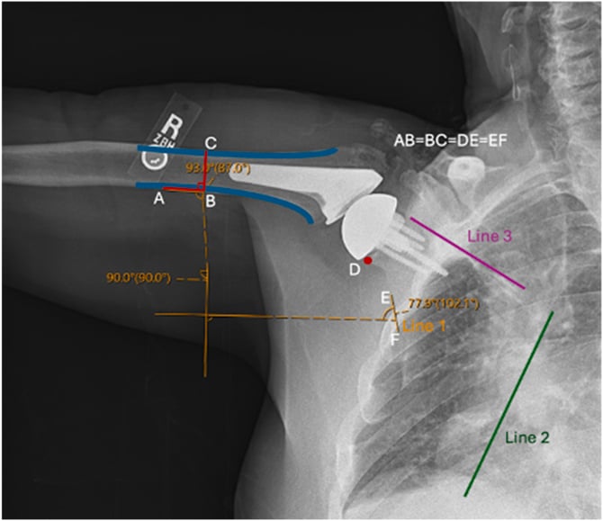
Scapular motion. The scapula does more than rotate upward:
- Upward rotation 60°: the inferior angle moves laterally
- Posterior tilt 20-30°: the acromion tilts posteriorly
- External rotation 15-25°: the medial border moves away from the spine
Serratus anterior produces the upward rotation and trapezius the elevation.
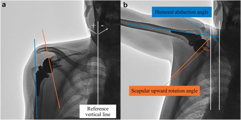
Loss of normal ST rhythm causes impingement and instability. Serratus anterior palsy (long thoracic nerve injury) causes medial scapular winging and loss of upward rotation. This reduces subacromial space and increases impingement risk. Rehabilitation must restore scapular stability before addressing GH pathology.
The acromioclavicular and sternoclavicular joints. The AC joint allows scapular protraction, retraction and rotation, and clavicular rotation at the sternoclavicular joint contributes 40-50° to overhead motion. Disruption of the AC joint (separation) or the SC joint affects scapular position and can cause secondary impingement.
Planes of Elevation and the Scapular Plane (Scaption)
The plane in which the arm is elevated changes the demand on the capsule, the cuff and bony clearance, and is a favourite basic-science point.
The scapular plane ("scaption") lies roughly 30 to 45 degrees anterior to the coronal plane, matching the resting orientation of the scapula on the curved thorax. Elevation in this plane is the most efficient and least provocative because:
- The inferior capsule is not twisted, so the capsuloligamentous structures stay relatively relaxed and range of motion is maximal
- Supraspinatus and deltoid are optimally aligned with the plane of the scapula, improving their mechanical advantage
- The greater tuberosity clears the acromion without the obligatory external rotation that pure coronal abduction requires, so impingement is least
The other planes. True coronal-plane abduction requires external rotation of the humerus to move the greater tuberosity out from under the acromion; without it the tuberosity impinges through the mid-arc. Forward flexion in the sagittal plane progressively tightens the posterior capsule. This is why rehabilitation and isolated strength testing are usually performed in the scapular plane, and why the "empty can" (Jobe) test positions the arm in scaption to load the supraspinatus.
Rotator Cuff Force Couples
The rotator cuff works as two force couples to keep the humeral head centred during motion: one in the coronal plane, against the deltoid, and one in the transverse plane, between the anterior and posterior cuff.
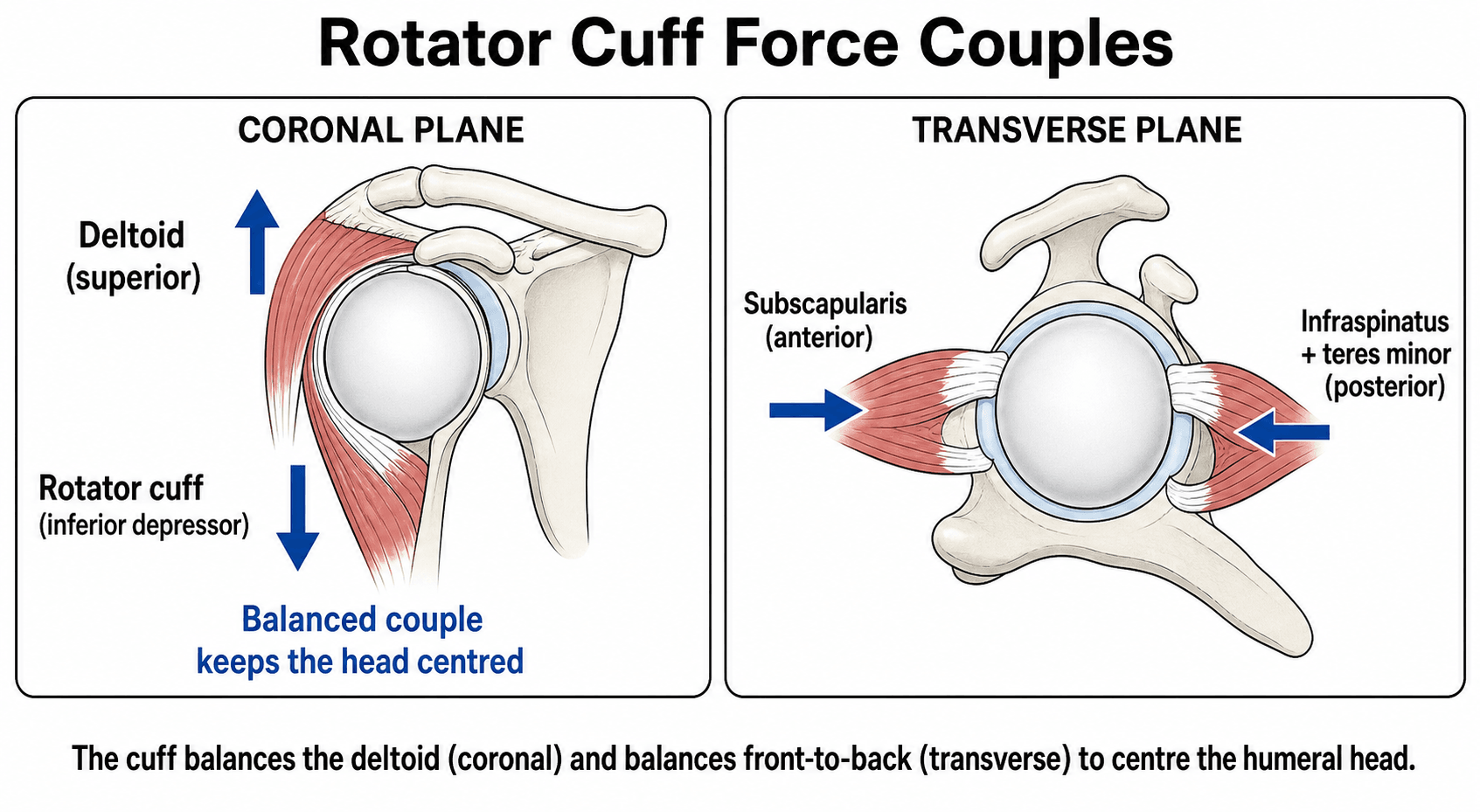
Coronal plane: deltoid against the inferior cuff. The deltoid is the primary abductor, and the upward force it generates translates the humeral head superiorly. Acting alone it would cause superior migration and impingement. Subscapularis and infraspinatus-teres minor pull inferiorly, a depressor force that counteracts the deltoid's superior pull so the head stays centred in the glenoid.
Supraspinatus. Its role is debated. The traditional view makes it the primary initiator of abduction; the modern view makes it primarily a depressor and compressor of the humeral head. The clinical observation behind the modern view is that a supraspinatus tear does not abolish abduction, because the deltoid compensates.
Transverse plane: subscapularis against infraspinatus-teres minor. The anterior and posterior cuff compress the head from either side.
- Action
- Internal rotation, anterior compression
- Force Vector
- Prevents posterior subluxation
- Tear Effect
- Anterior instability, loss of IR strength
- Action
- External rotation, posterior compression
- Force Vector
- Prevents anterior subluxation
- Tear Effect
- Posterior superior migration, loss of ER
SITSRotator Cuff Force Couples
Hook:SITS muscles work as force couples: Supraspinatus-deltoid (coronal), Subscapularis vs Infraspinatus-Teres (transverse)!
Glenohumeral Joint Reaction Force and Force Vectors
Magnitude. Beyond keeping the head centred, the muscles around the shoulder generate a substantial joint reaction force that must be directed into the glenoid for stability. In the classic biomechanical analysis (Poppen and Walker), the resultant glenohumeral joint reaction force rises through abduction to peak at about 0.9 times body weight near 90 degrees of elevation, then falls as the arm rises further, so the mid-range is both the most heavily loaded and the most demanding of the stabilisers.
Direction. The direction of that resultant matters as much as its magnitude. For stability the resultant force vector must fall within the glenoid arc, and the force-couple balance keeps it there (the "scapulohumeral balance" of Lippitt and Matsen). If it falls outside the glenoid rim, as with cuff deficiency or glenoid bone loss, the head subluxes, which is the mechanical basis of the glenoid concavity-arc and "on-track / off-track" concepts.
The deltoid line of pull illustrates this. Early in abduction the deltoid acts almost vertically, producing a large superior shear on the humeral head; the rotator cuff converts this into a compressive, centring force by adding an inferiorly and medially directed vector. As the arm elevates, the deltoid's line of action becomes progressively more compressive and less shearing, which is why superior migration in a cuff-deficient shoulder is worst in the mid-range, and why reverse arthroplasty, by medialising and distalising the centre of rotation, restores a favourable deltoid line of action and lowers the joint reaction force.
Capsular Ligaments and Stability
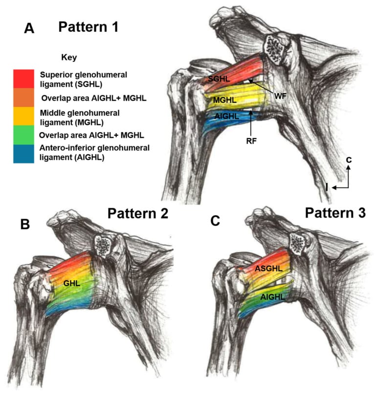
The inferior glenohumeral ligament complex. The IGHLC is the primary anterior stabiliser in the abducted, externally rotated position, the late cocking phase of throwing. It has three parts, and which one is working depends on where the arm is:
- Anterior band: resists anterior translation in abduction-external rotation, maximally loaded at 90° abduction with external rotation
- Posterior band: resists posterior translation in abduction-internal rotation, maximally loaded at 90° abduction with internal rotation
- Axillary pouch: the inferior capsule, resists inferior subluxation
In adduction the complex is redundant and offers minimal restraint. It inserts into the labrum, so a Bankart lesion disrupts the IGHL anchor, and a Bankart repair restores the IGHL to the labrum.
Middle and superior ligaments. The MGHL is the primary restraint to anterior translation at 45-90° of abduction; the SGHL and the coracohumeral ligament restrain inferior subluxation in adduction. Turkel's sequential cutting study puts the hand-over in order: at 0° abduction subscapularis is the main anterior restraint, at 45° subscapularis, the MGHL and the anterosuperior IGHL share it, and at 90° the IGHL is the primary check to anterior dislocation in external rotation.
Clinical Applications
Supraspinatus tear. Loss of compression reduces concavity-compression stability, and with the depressor gone the deltoid is unopposed and the head migrates superiorly. The acromiohumeral interval, normally 7-10 mm, narrows and impingement follows. The deltoid and the remaining cuff can maintain elevation.
Subscapularis tear. Loss of the anterior limb of the transverse force couple produces anterior instability, and the head subluxes anterosuperiorly. Internal rotation strength is lost: the patient cannot internally rotate against resistance, the lift-off test is positive and the belly-press weak.
Massive tear (greater than two tendons or greater than 5 cm) loses both force couples, coronal and transverse, and its consequences follow from that:
- Anterosuperior or posterosuperior escape
- Rotator cuff arthropathy, with acetabularisation of the acromion
Arthroplasty design. Each component decision has a biomechanical reason behind it.
- Design Goal
- Restore native concavity and version
- Biomechanical Basis
- Concavity-compression requires proper depth and orientation
- Design Goal
- Match native radius and offset
- Biomechanical Basis
- Maintains normal center of rotation and deltoid moment arm
- Design Goal
- Medialize center of rotation, distalize humerus
- Biomechanical Basis
- Deltoid becomes primary elevator when RC absent (biomechanical compensation)
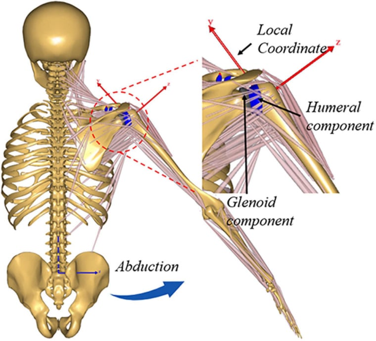
Reverse shoulder arthroplasty reverses the normal geometry. The glenosphere becomes the ball and medialises the centre of rotation; the humeral socket becomes the cup and distalises the humerus. The deltoid becomes the primary elevator, so the cuff is not needed, and its moment arm increases.
Differentiating Biomechanical Failure Patterns
A common exam task is to translate a clinical or imaging finding into the underlying biomechanical lesion. Use the destabilising direction and the failed force couple to reason to the diagnosis.
- Key Mechanism Lost
- Concavity-compression and coronal depressor force
- Direction of Instability/Escape
- Superior migration of the humeral head
- Hallmark Clinical Clue
- Reduced acromiohumeral interval, painful arc, weak elevation
- Key Mechanism Lost
- Anterior limb of transverse force couple
- Direction of Instability/Escape
- Anterosuperior escape
- Hallmark Clinical Clue
- Positive lift-off / belly-press, lost internal-rotation strength
- Key Mechanism Lost
- Anterior static restraint in abduction-external rotation
- Direction of Instability/Escape
- Anterior translation in the apprehension position
- Hallmark Clinical Clue
- Recurrent anterior dislocation, positive apprehension-relocation
- Key Mechanism Lost
- Stable scapular platform and upward rotation
- Direction of Instability/Escape
- Functional impingement, loss of overhead arc
- Hallmark Clinical Clue
- Medial scapular winging, abnormal scapulohumeral rhythm
- Key Mechanism Lost
- Both force couples plus articular cartilage
- Direction of Instability/Escape
- Fixed superior escape with acromial acetabularisation
- Hallmark Clinical Clue
- Pseudoparalysis, superior glenoid/acromial wear, candidate for RSA
Guidelines, Registries and Global Practice
Shoulder biomechanics is a basic-science topic, so formal disease guidelines are limited; the relevant guidance sits within instability, rotator cuff and arthroplasty pathways, and within national joint registries that track the implants these biomechanical principles inform.
Global epidemiology (context)
- Rotator cuff disease prevalence rises steeply with age and is frequently asymptomatic; full-thickness tears become common beyond the sixth decade across populations studied worldwide.
- Anterior glenohumeral instability disproportionately affects young males in contact and overhead sport, the group in whom IGHL/labral biomechanics are most exam-relevant.
- Shoulder arthroplasty volumes, especially reverse arthroplasty for cuff-deficient shoulders, have grown markedly across high-income registries over the past two decades.
Side-by-side society guidance
- Focus
- Rotator cuff and glenohumeral OA management
- Biomechanically Relevant Position
- Evidence-based pathways; recognises irreparable cuff tears with superior escape as a reverse-arthroplasty indication
- Focus
- Instability and cuff care pathways
- Biomechanically Relevant Position
- Structured instability assessment incorporating bone loss and direction-specific restraints before stabilisation
- Focus
- Trauma and reconstruction principles
- Biomechanically Relevant Position
- Restoration of joint line, version and the rotator-cuff force couple as goals of fixation and reconstruction
- Focus
- Arthroplasty and instability consensus
- Biomechanically Relevant Position
- Emphasis on glenoid version/inclination and centre-of-rotation restoration to protect implant survival
Registry evidence
National arthroplasty registries (NJR for England and Wales, AOANJRR in Australia, the Swedish and Norwegian registries, AJRR in the US, NZJR) track shoulder arthroplasty survival and revision. Recurring biomechanically driven signals include glenoid component loosening in anatomic replacement and scapular notching and instability after reverse arthroplasty, which inform component selection, version and centre-of-rotation choices.
High- versus limited-resource practice variation
- In well-resourced settings, advanced imaging, 3D planning and patient-specific instrumentation are increasingly used to restore version, inclination and the centre of rotation when biomechanics are disturbed.
- In limited-resource settings, structured physiotherapy targeting scapular control and the rotator-cuff force couple is the mainstay, with arthroplasty reserved and standard instrumentation used; the underlying biomechanical goals, a stable scapular platform and a centred, compressed humeral head, are identical regardless of resources.
Controversies and Areas of Uncertainty
The headline figures are constructs. The 2:1 rhythm varies with velocity, plane of elevation, load and individual anatomy, so a rigid single ratio overstates the certainty of the data. The "50% static, 50% dynamic" and "concavity-compression provides 50% of stability" figures are likewise teaching constructs derived from specific in vitro models, not precise physiological constants; their value is conceptual rather than literal.
The critical glenoid bone-loss threshold. The amount of glenoid bone loss that mandates a bony procedure rather than soft-tissue repair, historically quoted around 20-25%, with growing attention to "on-track / off-track" and subcritical loss, continues to evolve and is genuinely unsettled.
Optimal reverse arthroplasty configuration. Lateralisation versus medialisation of the centre of rotation, and glenosphere size and tilt, trade off impingement, stability, deltoid efficiency and notching. There is no single agreed optimum and design philosophy continues to change.
MCQ Practice Points
Q: What is the normal scapulothoracic rhythm ratio during shoulder abduction from 30-180 degrees? A: 2:1 (glenohumeral:scapulothoracic) - For every 2 degrees of GH abduction, there is 1 degree of ST upward rotation. Total 180° = 120° GH + 60° ST.
Q: What percentage of glenohumeral stability is provided by concavity-compression from the rotator cuff? A: 50% - Concavity-compression (RC compressing head into glenoid) provides 50% of stability. The other 50% comes from static restraints (capsule, ligaments, labrum).
Q: By what percentage does the labrum deepen the glenoid socket? A: 50% - The labrum increases glenoid depth from approximately 2.5mm to 5mm, a 50% increase. This enhances stability by increasing contact area and creating a suction seal.
Exam Viva Scenarios
Practise clinical reasoning and management decisions out loud
“Examiner asks: Describe the scapulothoracic rhythm during shoulder abduction. What happens when this rhythm is disrupted?”
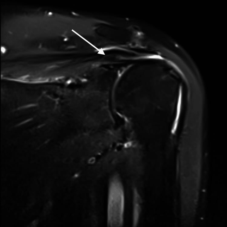
“Examiner shows MRI of massive rotator cuff tear and asks: Explain the biomechanical consequences of this tear and why the patient has superior migration of the humeral head.”
“Examiner asks: A 22-year-old rugby player has recurrent anterior shoulder dislocations. Explain the biomechanical contributions to glenohumeral stability and which structure is the primary restraint in the position of apprehension.”
Stability Mechanisms
- 50% dynamic (RC concavity-compression) + 50% static (capsule/labrum)
- Glenoid covers only 25% of humeral head (inherent instability)
- Labrum deepens socket 50% (2.5mm to 5mm)
- Capsular volume = 2x head volume (allows ROM, sacrifices stability)
Scapulothoracic Rhythm
- 2:1 ratio (GH:ST) from 30-180° abduction
- 0-30° = setting phase (primarily GH)
- Total 180° = 120° GH + 60° ST upward rotation
- Scapular muscles: serratus anterior (upward rotation), trapezius (elevation)
Rotator Cuff Force Couples
- Coronal: Deltoid (superior) vs RC (inferior depressor)
- Transverse: Subscapularis (anterior) vs infraspinatus-teres (posterior)
- SITS muscles: Supraspinatus, Infraspinatus, Teres minor, Subscapularis
- RC tear causes superior migration (deltoid unopposed)
Capsular Ligaments
- IGHL = primary anterior stabilizer in ABD-ER (90°+)
- MGHL = primary restraint at 45-90° abduction
- SGHL + coracohumeral = inferior restraint in adduction
- Bankart lesion = IGHL detachment from labrum
Clinical Applications
- Acromiohumeral interval normal = 7-10mm (reduces with RC tear)
- Reverse shoulder: medializes center, distalizes humerus, deltoid primary elevator
- Scapular dyskinesis causes impingement (loss of upward rotation)
- Massive RC tear (greater than 2 tendons or greater than 5cm) may need RSA
Key Numbers
- Glenoid retroversion: 5° average
- Clavicular rotation (SC joint): 40-50°
- AC joint rotation: 20°
- Supraspinatus critical arc: 60-120° abduction (highest stress)
Evidence Base
The biomechanical understanding of the shoulder rests on a small group of landmark cadaveric, radiographic and modelling studies. Named-society guidance (AAOS, BOA, AO) is summarised in the global practice section.
Concavity-Compression: Mechanisms of Glenohumeral Stability
- Defined two key dynamic mechanisms: concavity-compression and scapulohumeral balance
- Compressing the humeral head into the concave glenoid resists translating loads
- Stability rises with greater compressive (muscle) load and with deeper glenoid concavity
- An intact labrum is essential to both mechanisms, especially in the lax mid-range
The Glenoid-Labral Socket: A Constrained Articular Surface
- Glenoid plus labrum form a socket about 9mm deep superoinferiorly and 5mm anteroposteriorly (n=25)
- The fibrous labrum contributes roughly 50% of total socket depth
- Simulated Bankart lesion reduced anteroposterior depth from about 5.0mm to 2.4mm
- Socket depth is a meaningful structural contributor to glenohumeral stability
Sequential Cutting: Stabilisers Preventing Anterior Dislocation (IGHL)
- Cadaveric sequential-cutting study of 36 embalmed and 10 fresh shoulders
- At 0° abduction subscapularis is the main anterior restraint
- At 45° subscapularis, MGHL and anterosuperior IGHL share the restraint
- At 90° abduction the inferior glenohumeral ligament is the primary check to anterior dislocation in external rotation
Scapulohumeral Rhythm: Motion Velocity and Ratio
- 19 shoulders of 10 healthy men analysed with image intensifier and high-resolution video
- At low speed the glenohumeral-to-scapulothoracic ratio was constant, agreeing with prior work
- At high speed the ratio was not fixed and differed significantly from low speed
- Glenohumeral motion dominated early abduction beyond the setting phase, then declined
3D Scapular Orientation in Rotator Cuff Tear and Impingement
- Low-dose stereoradiographic 3D scapular orientation in 65 subjects
- Resting scapular orientation did not differ between healthy, cuff-tear and cuff-tear-plus-impingement groups
- At 90° elevation, scapular upward rotation was significantly reduced only in the impingement group
- Scapular kinematic alteration in symptomatic shoulders is multifactorial, not a uniform finding
Muscle Moment Arms After Anatomic vs Reverse Shoulder Arthroplasty
- Eight cadaveric upper limbs tested pre-operatively, after anatomic TSA and after revision RSA
- Anatomic TSA lateralised the joint centre about 4.3mm, modestly increasing deltoid force and joint compression
- RSA significantly increased moment arms of abductors, flexors, adductors and extensors and reduced peak muscle forces
- RSA increased superior deltoid inclination but produced greater superior shear and less joint compression