Degenerative Disease | Forefoot Pain | Progressive Deformity | Multifactorial
- Second MTP joint most commonly affected due to mechanical overload
- Predisposing factors: inflammatory arthritis, trauma, instability, hallux valgus
- Conservative management resolves the majority of early cases (textbook consensus; no cited series gives a rate)
- Arthrodesis gold standard for end-stage disease in active patients
- Transfer metatarsalgia common if surgical correction not balanced
- “Differentiate from synovitis, subluxation, and plantar plate tear
- “Drawer test assesses plantar plate integrity
- “Radiographs underestimate cartilage loss - weight-bearing views essential
- “Isolated arthrodesis risks transfer metatarsalgia - consider metatarsal balancing
Overview and Epidemiology
Lesser metatarsophalangeal (MTP) joint arthritis is degenerative disease of the second to fifth MTP joints. The second MTP is the joint most commonly affected, because of its mechanical disadvantage. The disease progresses from synovitis and cartilage wear to subchondral sclerosis and osteophyte formation, and eventually to joint destruction with fixed deformity.
Natural history. It is a common source of forefoot pain but is often underdiagnosed in its early stages. The course is progressive: early synovitis evolves to cartilage loss and joint-space narrowing, and eventually to fixed deformity with secondary deformities in the adjacent toes.
Who. Typically presents at 40-60 years, with a female predominance of 3:1. Prolonged standing, athletics and dance load the forefoot, as do high heels, a narrow toe box and obesity.
Causes and associations. In chronic cases the arthritis is never isolated. Look for the associated deformity, because failure to address it leads to recurrence:
- Hallux valgus (a 15-20% association) and other first-ray insufficiency, which transfers load to the lesser rays
- Inflammatory arthritis: rheumatoid, psoriatic, gout
- Plantar plate insufficiency and tears, which may be a precursor or a consequence
- Toe deformities: hammertoe, claw toe, and crossover toe with its progressive instability
- Trauma: fracture, dislocation, turf toe
- Rigid flatfoot, through abnormal biomechanics
- Sesamoid dysfunction, with loss of first MTP load bearing
- Iatrogenic: overcorrection of hallux valgus, and transfer metatarsalgia after first-ray surgery
Pathophysiology and Mechanisms
The joint. Each lesser MTP joint is a condyloid synovial joint between the convex metatarsal head and the concave base of the proximal phalanx. Its stabilisers:
- Plantar plate, providing static stability and resisting hyperextension
- Collateral ligaments, medial and lateral
- Intrinsic muscles, the interossei and lumbricals, which control toe position
- Extensor and flexor tendons, the dynamic stabilisers
The plantar plate. A rectangular fibrocartilaginous structure that originates from the plantar metatarsal neck and inserts on the base of the proximal phalanx, blending with the joint capsule and collateral ligaments. Attenuation or rupture, typically on its dorsal-lateral aspect, leads to MTP instability, dorsal subluxation and accelerated arthritis. That is why an isolated arthrodesis that does not address the plantar plate can fail.
Load sharing. Normally the first MTP carries 50% of forefoot load, the second 30% and the third to fifth 10% each, with peak pressure in terminal stance. With hallux valgus the second MTP's share increases to 60% or more.
Why the second MTP. It experiences the highest ground reaction forces in gait, and the second metatarsal is typically the longest. When the first ray is insufficient, from hallux valgus, hypermobility or iatrogenic shortening from an overzealous Weil osteotomy on the first metatarsal, load transfers laterally and the second MTP absorbs it. Overload then feeds a vicious cycle of synovitis, plantar plate damage, instability and accelerated arthritis.
The cascade. The sequence typically runs:
- Initiating event: overload (hallux valgus, long metatarsal), trauma or inflammatory disease
- Synovitis: inflammation, effusion, capsular distension
- Plantar plate attenuation, as chronic synovitis weakens the plantar restraint
- Instability and subluxation, with dorsal migration of the proximal phalanx
- Cartilage wear from abnormal load
- Subchondral change: sclerosis, cysts, osteophytes
- Fixed deformity: contracture of the dorsal structures, crossover toe
- Secondary deformity: transfer metatarsalgia, adjacent toe deformities
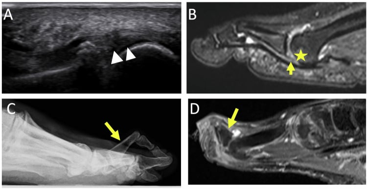
Deformity progression. Clinically, the deformity is staged:
- Stage 1: synovitis, mild dorsal subluxation
- Stage 2: plantar plate attenuation, reducible deformity
- Stage 3: cartilage loss, fixed deformity
- Stage 4: bone-on-bone, crossover toe, transfer lesions
Classification Systems
Two systems describe the joint itself. The clinical severity grade is the one that directs treatment, and it matters more for treatment decisions than the radiographic stage alone. Grades I-II typically respond to conservative measures for 6-12 months; failure of conservative treatment in Grade II, or presentation with Grade III-IV disease, prompts surgery, with the procedure matched to severity and to the patient's demands.
- Clinical Features
- Mild pain, minimal deformity, full ROM
- Radiographic Findings
- Normal joint space, no osteophytes
- Treatment
- Conservative: orthotics, NSAIDs
- Clinical Features
- Moderate pain, reducible deformity, limited ROM
- Radiographic Findings
- Mild joint space narrowing, early osteophytes
- Treatment
- Conservative or cheilectomy + osteotomy
- Clinical Features
- Severe pain, fixed deformity, stiff joint
- Radiographic Findings
- Significant joint space loss, sclerosis, large osteophytes
- Treatment
- Arthrodesis or arthroplasty
- Clinical Features
- Disabling pain, crossover toe, transfer lesions
- Radiographic Findings
- Complete joint destruction, subluxation, bone-on-bone
- Treatment
- Arthrodesis with metatarsal shortening and balancing
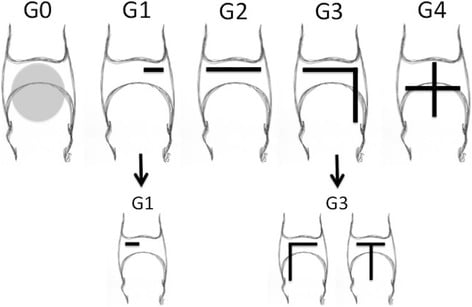
Plantar plate grading. The plantar plate has its own anatomical grading (Nery), which the plantar plate literature uses to match the procedure to the tear.
Clinical Assessment
History. The pain lies over the dorsal MTP joint and the plantar metatarsal head, and its onset is insidious or, after trauma, acute. Push-off, barefoot walking and stairs aggravate it; rest and supportive shoes relieve it. Establish the functional impact, particularly on athletic activity, what has already been tried (orthotics, injections, footwear changes), any associated hallux valgus or toe deformity, and screen for the systemic symptoms of inflammatory arthritis.
Examination.
- Inspection: swelling, erythema, hammertoe or crossover deformity
- Palpation: joint-line tenderness, osteophytes, a prominent metatarsal head
- Active and passive dorsiflexion and plantarflexion, compared with the other side
- Stability: the drawer test for plantar plate integrity
- Alignment: coronal deviation and rotation
- Transfer lesions: plantar calluses under the adjacent metatarsals
- Shoe wear, which shows how the load is distributed
- Technique
- Stabilise metatarsal, translate proximal phalanx dorsally
- Positive Finding
- Excessive dorsal translation (over 50% compared to normal)
- Interpretation
- Plantar plate tear or insufficiency
- Technique
- Hyperextend MTP while palpating plantar plate
- Positive Finding
- Pain, palpable defect, lack of firm endpoint
- Interpretation
- Plantar plate tear
- Technique
- Axial compression with circumduction of toe
- Positive Finding
- Crepitus, pain
- Interpretation
- Arthritis, cartilage damage
- Technique
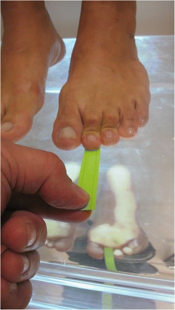
- Patient grips paper between affected toe and ground
- Positive Finding
- Inability to grip or pull paper from examiner
- Interpretation
- FDL weakness or plantar plate insufficiency
Reading the drawer. Judge the translation against a normal toe. Early tears are reducible; late ones become fixed.
Arthritis or plantar plate tear? An isolated plantar plate tear presents acutely, with a positive drawer test and instability, and its radiographs may initially be normal. Lesser MTP arthritis typically has an insidious onset, joint-line tenderness, reduced movement and radiographic change. Chronic plantar plate tears lead to arthritis, however, so the two sit on a spectrum, and MRI separates the acute tear that is amenable to repair from the chronic tear with arthritis that needs arthrodesis or arthroplasty.
- Typical Features
- Acute or attritional, instability, often normal early radiographs
- Distinguishing Test / Finding
- Positive drawer test, MRI tear without joint-space loss
- Typical Features
- Interdigital burning pain radiating to toes, no joint tenderness
- Distinguishing Test / Finding
- Positive Mulder click, tenderness in web space (not joint line)
- Typical Features
- Adolescent/young adult, second metatarsal head, dorsal stiffness
- Distinguishing Test / Finding
- Flattening/collapse of metatarsal head on radiograph
- Typical Features
- Activity-related shaft/neck pain, swelling, runners
- Distinguishing Test / Finding
- Focal shaft tenderness, callus or fracture line on imaging/MRI
- Typical Features
- Multiple joints, morning stiffness, systemic features
- Distinguishing Test / Finding
- Erosive radiographs, raised inflammatory markers / serology
- Typical Features
- Acute hot swollen joint, systemic upset
- Distinguishing Test / Finding
- Raised CRP/WCC, joint aspiration with organisms
- Typical Features
- Diffuse swelling, effusion, preserved joint space
- Distinguishing Test / Finding
- MRI synovitis without cartilage loss
Investigations
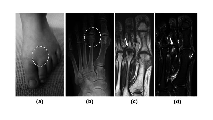
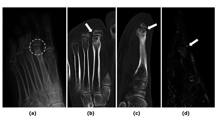
Radiographs. Weight-bearing AP, lateral and oblique views of the foot come first. Non-weight-bearing films underestimate joint-space narrowing and subluxation, so standing views are mandatory: they show true alignment and joint space under physiological load, and they are the standard of care for forefoot pathology. Look for:
- Joint-space narrowing
- Subchondral sclerosis
- Osteophytes
- Dorsal subluxation of the proximal phalanx
- The metatarsal length relationships
- Transfer lesions, seen as narrowing of the adjacent joints
Radiographs underestimate cartilage loss and cannot show the plantar plate. A joint may look Stage 1 on the film yet have extensive cartilage damage visible only on MRI or arthroscopy, so if the pain is disproportionate to the radiographs, consider MRI to assess the cartilage and plantar plate.
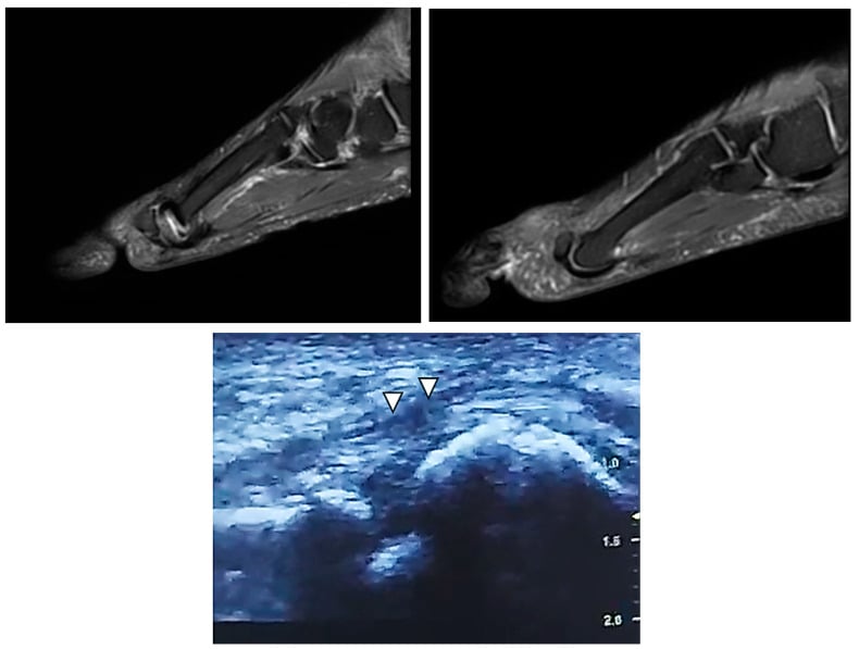
MRI. The gold standard for soft tissue and cartilage. Use it when a plantar plate tear is suspected, for early arthritis with normal radiographs, or for pre-operative planning. It shows:
- Plantar plate tear, high- or low-grade
- The extent of cartilage defects
- Bone marrow oedema, which suggests acute overload
- Synovitis, as T2 hyperintensity in the joint
- Collateral ligament integrity
Its accuracy for plantar plate tear rests on one cited series (Yamada, 45 joints against a surgical reference standard): pericapsular fibrosis was 91% sensitive, with a specificity resting on only 11 tear-free joints.
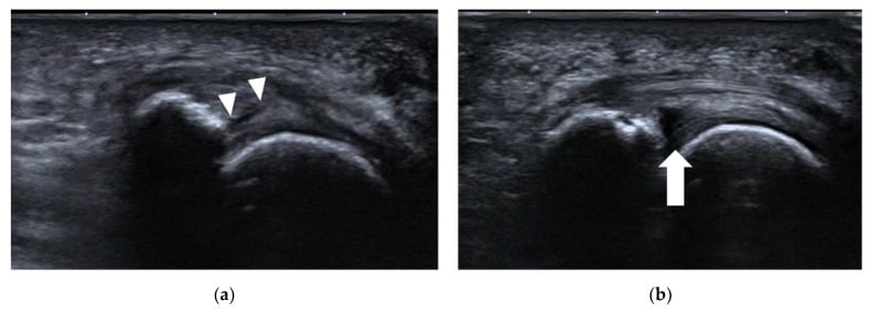
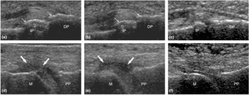
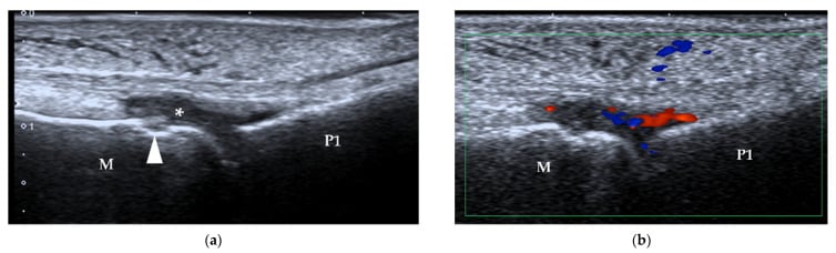
Ultrasound. A dynamic assessment of the plantar plate, and less expensive than MRI. It shows plate thickness (normal 3-4mm), tears and hyperaemia, but it is operator-dependent and less accurate than MRI for grading tear severity.
Blood tests. Isolated degenerative arthritis does not need blood work. If an inflammatory arthropathy is suspected:
- Rheumatoid factor and anti-CCP for rheumatoid arthritis
- Uric acid for gout
- ESR and CRP as inflammatory markers
- HLA-B27 for the spondyloarthropathies
Management Algorithm
Choosing the operation. The choice depends on disease severity, the patient's age and activity level, bone quality and the associated deformities.
- Patient and Grade
- Grade II with reducible deformity after failed conservative care; young or middle-aged, active, wanting to preserve motion
- Advantage
- Preserves motion, offloads joint
- Disadvantage
- Unpredictable pain relief, may progress to arthrodesis
- Patient and Grade
- Grade III-IV, fixed deformity, active patients, good bone stock
- Advantage
- Gold standard for pain relief, stable, predictable
- Disadvantage
- Loss of motion, risk transfer metatarsalgia, nonunion
- Patient and Grade
- Grade III-IV, elderly, low demand, poor or osteopenic bone
- Advantage
- Relieves pain, simple, preserves length
- Disadvantage
- Instability, floppy toe, recurrence
- Patient and Grade
- Grade III-IV, desire motion, moderate demand
- Advantage
- Preserves length and motion
- Disadvantage
- Higher complication rate, loosening, recurrence
Arthrodesis or arthroplasty. Arthrodesis provides pain relief and stability but risks transfer metatarsalgia; arthroplasty preserves motion but has a higher recurrence. Combine either with a metatarsal osteotomy for load balancing. For the elderly, low-demand patient with poor bone, arthroplasty, resection or implant, preserves length at the price of accepting instability.
Who. Mild to moderate symptoms, Grade I-II disease and no fixed deformity: the young, active patient with minimal deformity. The majority of early disease settles with 6-12 months of treatment, though no cited series gives a percentage. Trial it for at least 6 months before considering surgery.
Footwear, from the start.
- Rigid soles, to reduce MTP motion and dorsiflexion stress
- A rocker bottom, to offload the forefoot in gait
- A wide toe box, to accommodate deformity and reduce pressure
- A low heel, to minimise forefoot load transfer
Orthoses, weeks 0-4.
- Metatarsal pad, to offload the joint
- Custom orthotics, whose arch support redistributes load to the midfoot
- Toe spacers for crossover toe or deviation
- Accommodative padding for plantar calluses
The metatarsal pad goes proximal to the metatarsal heads, not under them. Placed correctly it unloads the MTP joint by transferring weight to the metatarsal shafts; placed under the heads it worsens symptoms.
Medication, weeks 0-12.
- NSAIDs first-line for pain and inflammation (ibuprofen 400mg TDS, naproxen 500mg BD)
- Topical NSAIDs as an alternative for patients with GI contraindications
- Intra-articular corticosteroid: a maximum of 2-3 injections, 3 months apart; the risks are plantar plate weakening and fat pad atrophy
Activity, ongoing. Avoid prolonged standing, running and high-impact activity, cross-train with cycling or swimming, and return gradually as symptoms improve.
Reassessment at 3-6 months. If symptoms persist despite 6 months of conservative measures, proceed to surgical consultation.
Surgical Technique
Indications. Grade III-IV arthritis in active patients, failed prior surgery, and instability. Contraindications are active infection, severe peripheral vascular disease and neuropathy.
Technique for the second MTP.
- Set-up. Supine with a bump under the ipsilateral hip; thigh or ankle tourniquet (250 mmHg); prepare to the knee with the foot draped free; C-arm positioned for AP and lateral views.
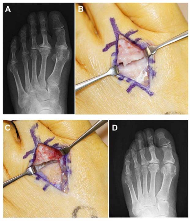
- Approach. A dorsal longitudinal incision of 3-4 cm centred over the joint. Split the extensor digitorum longus (EDL) longitudinally, incise the dorsal capsule longitudinally, preserving the collateral ligaments if possible, and retract the EDL to see the metatarsal head and phalangeal base.
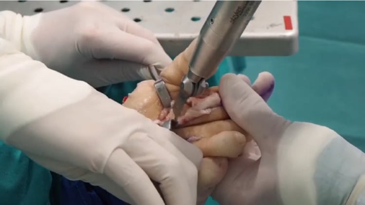
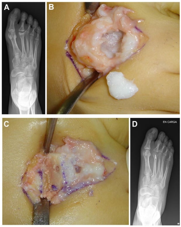
- Joint preparation. Remove dorsal and plantar osteophytes with a rongeur, then take the cartilage off both surfaces with an oscillating saw or burr. Make raw, bleeding subchondral surfaces, with perpendicular fish-scale cuts to increase the surface area, and resect more bone from the convex side if the toe is deviated.
- Balance the ray. If the second metatarsal is excessively long or the first ray short, resect 2-3mm from the metatarsal head as a Weil-type shortening. Failure to do so risks transferring the overload to the third MTP and creating new pathology.
- Provisional fixation. A 1.6mm K-wire from the tip of the phalanx across the fusion site into the metatarsal shaft, with AP and lateral fluoroscopy to confirm alignment and position.
- Definitive fixation, with AP and lateral fluoroscopy to confirm stable fixation and appropriate alignment.
- Closure. Repair the EDL split with absorbable suture and close the capsule if the tissue allows; 3-0 absorbable subcutaneous and 4-0 nylon interrupted or subcuticular skin sutures; a bulky dressing with the toe in slight plantarflexion.
Alignment.
- Neutral in the coronal plane, with no varus or valgus
- Sagittal position set by the goal, not a fixed angle: the toe must purchase the ground. Because the lesser metatarsal declines about 20 degrees, a few degrees of DORSIflexion relative to the metatarsal shaft is what leaves the toe plantigrade — check it against the adjacent toes with the foot held flat rather than measuring off the shaft
- Slight external rotation to match the adjacent toes
Fixation options.
- Plate, preferred for strength: a 1.3mm or 1.5mm mini plate, dorsal or dorsomedial, with two screws proximal and two distal and compression by lag screw technique
- Screw: a single countersunk 2.0-2.4mm lag screw from dorsal, or crossed 1.6mm K-wires (two) for temporary fixation
- Combination: a plate with a supplemental K-wire if the bone is soft
Dorsal plating provides greater biomechanical stability than K-wire fixation alone, allowing earlier weight-bearing, while K-wires are cheaper and simpler but need 6 weeks of pin-site care and delayed weight-bearing; plates may need removal if prominent. Be careful with fusion-rate figures: no series cited on this page reports a fusion rate for either construct in the lesser MTP joint, and the percentages that circulate are carried over from first MTP and other small-joint fusions. For exam purposes, know both techniques, when to choose each, and that the comparison is a biomechanical and practical one rather than a proven outcome difference.
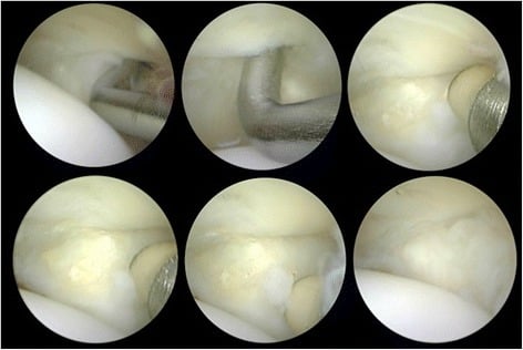
Complications
- Incidence
- 10-30%
- Risk Factors
- Isolated surgery without balancing, over-shortening
- Management
- Metatarsal offloading, consider revision with balancing osteotomy
- Incidence
- 5-10%
- Risk Factors
- Smoking, poor bone prep, inadequate fixation
- Management
- Revision arthrodesis with bone graft and plate fixation
- Incidence
- 5-15%
- Risk Factors
- Technical error, inadequate fixation
- Management
- Observation if asymptomatic; revision osteotomy if symptomatic
- Incidence
- 10-20% (arthroplasty)
- Risk Factors
- Failure to address underlying biomechanics
- Management
- Conversion to arthrodesis
- Incidence
- Variable
- Risk Factors
- Prolonged immobilisation, capsular scarring
- Management
- Physiotherapy, ROM exercises
- Incidence
- 1-2%
- Risk Factors
- Diabetes, peripheral vascular disease, smoking
- Management
- Antibiotics; debridement and hardware removal if deep
- Incidence
- 5-10% (plate)
- Risk Factors
- Prominent dorsal hardware
- Management
- Plate removal after fusion (minimum 6 months)
Transfer metatarsalgia, the most common complication. It occurs when an arthrodesis or shortening osteotomy alters the metatarsal parabola and shifts load to the adjacent metatarsals. Prevention is the key: assess the metatarsal length relationships on the pre-operative radiographs, perform balancing osteotomies when needed, and avoid over-shortening. Once established it is treated with offloading orthotics first, and persistent symptoms may need revision with a metatarsal osteotomy.
Postoperative Care and Rehabilitation
Arthrodesis is protected longer. The weight-bearing protocol after lesser MTP arthrodesis is more conservative than after forefoot osteotomies, and premature weight-bearing risks nonunion. Heel weight-bearing only for 2-4 weeks, flat-foot in the post-op shoe for 4-6 weeks, then supportive shoes at 6-8 weeks. The Weil osteotomy allows earlier weight-bearing, but stiffness is more common after it, so emphasise early range-of-motion exercises.
Days 0-2, hospital or day surgery.
- Elevate the foot above heart level; ice 20 minutes every 2 hours for the first 48 hours
- Multimodal analgesia: paracetamol, NSAIDs, opioids if needed
- DVT prophylaxis: aspirin 100mg daily, or LMWH if high risk
- Heel weight-bearing only in a post-op shoe; bulky dressing kept clean and dry
Weeks 0-2, early protection.
- Wound check and suture removal at day 10-14
- Heel weight-bearing in a rigid post-op shoe
- No active toe exercises; passive motion allowed
- Radiograph at 2 weeks for alignment and hardware position
Weeks 2-6, progressive loading.
- Flat-foot weight-bearing in the post-op shoe at 4 weeks if the radiographs show early healing
- Gentle passive ROM of the adjacent joints
- Compression stockings and continued elevation for swelling
- Radiograph at 6 weeks for fusion progress
Weeks 6-12, return to footwear.
- Rigid-soled supportive shoes at 6-8 weeks if fusion is progressing
- Full weight-bearing in normal shoes at 8-10 weeks
- Low-impact activity (walking, cycling) at 8 weeks
- Radiograph at 12 weeks to confirm fusion
Months 3-6, full activity.
- Radiographic fusion expected by 3-4 months
- Return to sport at 4-6 months, progressed gradually
- A symptomatic prominent plate is removed after confirmed fusion, at a minimum of 6 months
Outcomes and Prognosis
Conservative care. The textbook reference says conservative care "resolves the majority of early lesser MTP pain", which is the strongest supportable claim: no source cited on this page quantifies the success rate or the proportion who progress to surgery. Early presentation, compliance with orthotics and avoiding aggravating footwear predict success.
- What the evidence cited on this page shows
- Good/excellent in 86% of 37 feet at 30 months (Vandeputte) and 88% of 25 feet at 7 years (Hofstaetter); AOFAS 59→81 and 48→83
- Recognised trade-offs
- Range of motion significantly reduced; redislocation 8% at 1 year rising to 12% at 7; floating toe recognised but unquantified
- What the evidence cited on this page shows
- AOFAS 52→92 in 40 joints (Nery 2012); all tear grades improved significantly (Nery 2014)
- Recognised trade-offs
- Grade IV tears reached only AOFAS 72 with the least stable joints; the repair is never isolated from the osteotomy performed with it
- What the evidence cited on this page shows
- No series cited on this page reports a fusion rate, satisfaction rate or complication rate for lesser MTP arthrodesis
- Recognised trade-offs
- Conventionally reserved for end-stage degeneration or salvage; the plate-versus-K-wire comparison is not supported by any source cited here
- What the evidence cited on this page shows
- No series cited on this page quantifies it
- Recognised trade-offs
- Instability and recurrence are the recognised concerns; salvage procedure
Every comparative claim on this page rests on Level IV single-centre series without a control arm, and no cited study compares arthrodesis against a motion-sparing procedure, so "highest fusion rates and satisfaction" is a claim without a source here. Arthrodesis is the conventional choice for end-stage disease in an active patient: it trades MTP motion, often already minimal, for a stable, pain-free joint, at the cost of transfer metatarsalgia if the parabola is not balanced. Justify it in a viva on deformity, joint surface and patient demand rather than with a fusion-rate league table.
Predictors of a poor outcome.
- Failure to address the biomechanics: hallux valgus, metatarsal length discrepancy
- Smoking, which increases the nonunion risk
- Diabetes or peripheral vascular disease, with healing complications
- Inflammatory arthropathy, with higher recurrence
- The wrong procedure for the severity: cheilectomy fails in Grade IV disease
- Malalignment: malunion or uncorrected deformity
Defining the Metatarsal Parabola
Why define it. Restoring or balancing the metatarsal parabola, and shortening a "long" metatarsal, is the most-repeated surgical principle in this condition, so the normal parabola and how it is measured need defining.
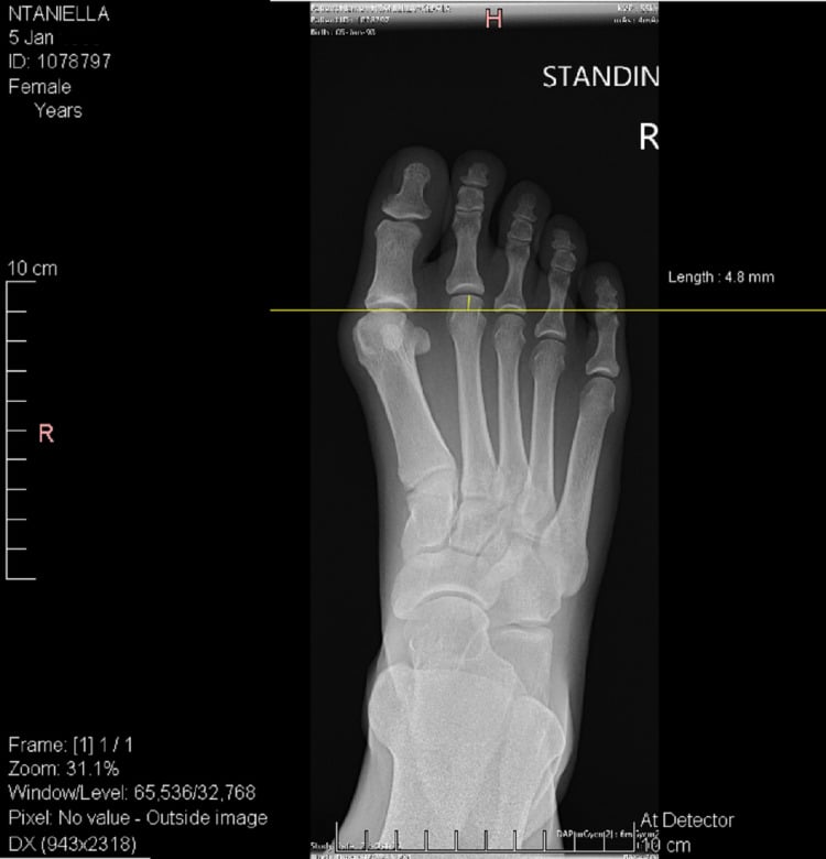
The parabola. The smooth, harmonious curve joining the tips of the metatarsal heads on a weight-bearing AP radiograph. Lengths decrease in an even progression: the second is usually the longest, or the first and second are roughly equal, and each of the third, fourth and fifth is a few millimetres shorter than the one medial to it. A break in the curve, most often a relatively long second metatarsal or a short first ray, concentrates load on the prominent head, and is the mechanical driver of overload referred to throughout this page.
The metatarsal formula. The index describes the first-to-second relationship:
- Index plus: first metatarsal longer than the second
- Index plus-minus: first and second equal
- Index minus: first shorter than the second
An index-minus foot, with a short first ray from hallux valgus or an over-shortened first metatarsal, transfers load to the second MTP: this is the first-ray insufficiency mechanism.
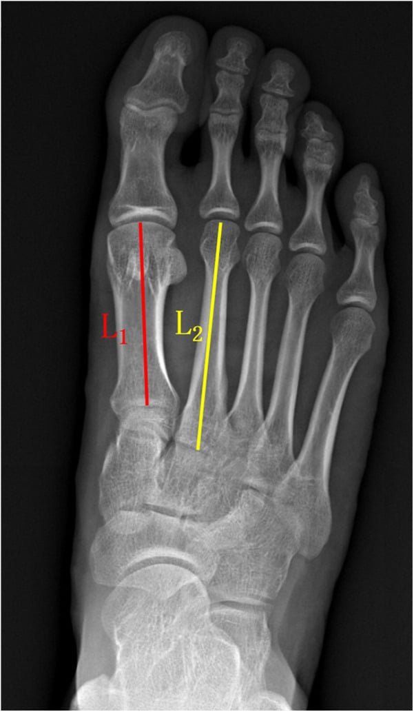
Maestro's criteria. A reproducible planning target. The lesser metatarsal heads should lie on a harmonious curve with the second at the apex, the fourth metatarsal head is referenced to the line joining the centres of the lateral sesamoid and the fourth metatarsal head, and there is a regular geometric decrement from the second through the fifth. Restoring this geometry, rather than simply shortening the sore ray, is what prevents transfer metatarsalgia.
In practice. Before any lesser MTP arthrodesis or Weil osteotomy, template the parabola on the weight-bearing AP. Shorten only enough to sit the operated head on the curve, and if the first ray is short, consider lengthening or plantarflexing it rather than shortening the lesser rays alone.
The Weil Osteotomy Floating Toe
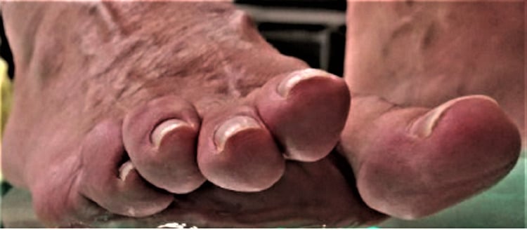
Floating toe and reduced MTP motion are the signature complications of the Weil osteotomy. Both follow directly from what the osteotomy does to the joint, so they can largely be designed out.
Why the toe floats. The Weil cut is made almost parallel to the sole, so as the metatarsal head is translated proximally it also drops (plantar-translates). That lowers the MTP joint axis, and the intrinsic muscles (interossei and lumbricals), whose tendons now pass dorsal to the new axis, lose their plantarflexion moment and can even act as extensors of the proximal phalanx. The toe then sits dorsiflexed and no longer purchases the ground, and MTP stiffness from the intra-articular osteotomy and capsular scarring compounds it.
Reducing the risk.
- Do not over-shorten or over-plantar-translate the head; take only what the parabola needs
- Elevate the head by removing a thin dorsal wedge (the "double" or triple Weil, a dorsal closing-wedge modification), so the head is shortened without being dropped and the joint axis stays up
- Balance the intrinsics: release a tight dorsal capsule and EDL, and add a flexor-to-extensor (Girdlestone-Taylor) transfer or a plantar plate repair where there is instability
- Start early active and passive MTP range of motion to limit stiffness
Why it matters. The Weil osteotomy is used both to offload an arthritic joint and to balance the parabola, so understanding the floating-toe mechanism is what lets you shorten safely and counsel the patient on the characteristic trade-off.
Guidelines, Registries & Global Practice
Lesser MTP arthritis and the closely related plantar plate instability spectrum are common globally, with female predominance, and the second ray most frequently involved. There is no single dedicated national guideline; practice is governed by foot-and-ankle society consensus, surgical textbooks (Mann's, Coughlin), and procedure-level registry and cohort data.
- Forefoot pain is among the commonest reasons for foot-and-ankle referral worldwide
- Second MTP most affected (about 63% of instability series), reflecting first-ray load transfer
- Female predominance and middle age typical across cohorts (Brazilian, European, North American series)
- Risk drivers: hallux valgus, long second metatarsal, inflammatory arthropathy, high-heeled/narrow footwear
- No implant registry captures lesser-MTP arthrodesis as a discrete entry (unlike hip/knee), so evidence rests on cohort series and systematic reviews
- Fusion rates for lesser MTP arthrodesis are not reported by any source cited on this page; the figures in circulation come from first MTP and other small-joint fusions
- Weil osteotomy durability shown to 7 years (Hofstaetter/Trnka), with floating toe as the signature complication
- MRI is the agreed reference imaging where plantar plate integrity is uncertain
- Region
- US
- Position on Lesser MTP Disease
- Conservative care first; restore metatarsal parabola surgically; arthrodesis for end-stage, motion-sparing for earlier disease
- Region
- UK / Europe
- Position on Lesser MTP Disease
- Stepwise non-operative management, address coexisting hallux valgus, individualised procedure choice
- Region
- Europe
- Position on Lesser MTP Disease
- Grade plantar plate tears (Nery classification) and match procedure to grade; Weil osteotomy widely used
- Region
- Global reference
- Position on Lesser MTP Disease
- Metatarsal pad proximal to head, biomechanical correction central, balance parabola at every step
- Routine weight-bearing radiographs plus MRI for plantar plate assessment
- Plate fixation and grade-specific plantar plate repair readily available
- Custom orthoses and formal physiotherapy pathways
- Reliance on clinical drawer test and plain radiographs; MRI often unavailable
- K-wire fixation and resection arthroplasty favoured where plates/implants are costly
- Off-the-shelf metatarsal pads and footwear modification as mainstay of conservative care
Whatever the setting, two principles are constant and examinable worldwide: exhaust biomechanically appropriate conservative care first, and protect or restore the metatarsal parabola at every surgical step to avoid transfer metatarsalgia. Consent should cover transfer metatarsalgia (10-30%), nonunion (5-10%), infection (1-2%) and possible hardware removal, with documented smoking-cessation advice given its effect on union.
Controversies and Areas of Uncertainty
Arthrodesis or motion-sparing surgery. No randomised trial compares lesser MTP arthrodesis with Weil osteotomy or implant arthroplasty. Arthrodesis is conventionally held to give the most reliable pain relief, at the cost of motion; the threshold at which to abandon joint preservation in degenerative, rather than purely unstable, disease remains opinion-based.
Durability of plantar plate repair. Grade-matched repair improves alignment and pain, but advanced (grade IV) tears retain residual instability and poorer scores. Whether direct repair alters the natural progression to arthritis, or simply delays it, is unproven.
The fixation construct. Plating is biomechanically more stable than K-wire fixation, but no series cited here reports a lesser MTP fusion rate for either construct, there is no high-level head-to-head trial, and hardware prominence drives a meaningful reoperation rate. The ideal construct (plate, screw, intramedullary device) is unsettled.
Defining and grading the disease. There is no universally accepted radiographic classification for lesser MTP arthritis itself; grading borrows from plantar plate (Nery) and clinical severity systems. That limits comparison between studies and complicates evidence synthesis.
State that high-level evidence is limited to cohort series and a small number of systematic reviews, that procedure choice is individualised to disease grade, bone quality, activity and the metatarsal parabola, and that the consistent, defensible principle is biomechanical correction with parabola preservation. Avoid claiming any single procedure is universally superior.
MCQ Practice Points
Q: Which lesser MTP joint is most commonly affected by degenerative arthritis? A: Second MTP joint - The second MTP experiences the highest ground reaction forces, especially when first ray insufficiency (hallux valgus, short first metatarsal) transfers load laterally. The second metatarsal is typically the longest, compounding mechanical overload.
Q: What does a positive drawer test of the lesser MTP joint indicate? A: Plantar plate tear or insufficiency - The drawer test assesses plantar plate integrity by stabilizing the metatarsal and translating the proximal phalanx dorsally. Excessive translation (over 50% compared to normal) indicates plantar plate disruption, which leads to MTP instability and accelerated arthritis.
Q: What is the success rate of conservative management for Grade I-II lesser MTP arthritis? A: Most early cases - conservative management with orthotics, metatarsal pads, rigid soled shoes and NSAIDs is the first-line approach for 6-12 months. Note that no series cited on this page puts a number on it: the textbook source says only that conservative care "resolves the majority of early lesser MTP pain", so quote it as the majority rather than as a percentage. Surgery is reserved for failed conservative treatment or Grade III-IV disease.
Q: What is the preferred fixation method for lesser MTP arthrodesis and why? A: Dorsal mini-plate fixation - Plate fixation provides greater biomechanical stability than K-wires and allows earlier weight-bearing; the trade-off is higher cost and potential hardware prominence requiring removal. Avoid quoting comparative fusion percentages: no series cited on this page reports a fusion rate for either construct in the lesser MTP joint.
Q: What is the most common complication after lesser MTP arthrodesis? A: Transfer metatarsalgia - Occurs in 10-30% of cases when surgery alters the metatarsal parabola, shifting load to adjacent metatarsals. Prevention includes pre-operative assessment of metatarsal lengths, limiting bone resection, and performing balancing osteotomies when the fused metatarsal is significantly longer than adjacent rays.
Q: What is the essential imaging requirement for diagnosing lesser MTP arthritis? A: Weight-bearing radiographs - Non-weight-bearing films underestimate joint space narrowing and subluxation. Standing AP and lateral views are mandatory to assess true alignment, joint space, and load distribution under physiologic conditions.
Exam Viva Scenarios
Practise clinical reasoning and management decisions out loud
“A 52-year-old female presents with 12 months of worsening pain under the second metatarsal head, worse with walking and wearing high heels. Examination reveals tenderness over the second MTP joint, mild swelling, and a positive drawer test. She has mild hallux valgus. Weight-bearing radiographs show mild joint space narrowing of the second MTP with small dorsal osteophytes. How would you assess and manage this patient?”
“A 58-year-old active male presents with disabling second MTP pain that has failed 9 months of conservative management including orthotics and injections. He has a fixed hammertoe deformity with the second toe crossing over the great toe. Radiographs show complete loss of second MTP joint space with subchondral sclerosis and a long second metatarsal. He wants to continue playing golf. What are your surgical options and preferred approach?”
“A 60-year-old patient underwent second MTP arthrodesis 4 months ago for end-stage arthritis. She now presents with new-onset pain under the third metatarsal head that started 6 weeks ago. Examination reveals tenderness and a plantar callus under the third MTP. Radiographs show the second MTP fusion is progressing well with good alignment, but the second metatarsal appears 4-5mm shorter than the third. How would you assess and manage this complication?”
Key Anatomy and Biomechanics
- Second MTP most affected - longest metatarsal, highest load (30% normally, 60%+ with hallux valgus)
- Plantar plate: fibrocartilaginous stabilizer, resists hyperextension, tears lead to instability
- First ray insufficiency (hallux valgus, hypermobility) transfers load to second MTP
- Metatarsal parabola: balanced load distribution requires graduated metatarsal lengths
Classification and Assessment
- Grade I: Mild pain, minimal deformity, normal joint space - conservative
- Grade II: Moderate pain, reducible deformity, mild narrowing - conservative or cheilectomy/Weil
- Grade III: Severe pain, fixed deformity, significant narrowing - arthrodesis or arthroplasty
- Grade IV: Disabling pain, crossover toe, bone-on-bone - arthrodesis with balancing
- Drawer test: assess plantar plate (over 50% dorsal translation = tear)
Conservative Management Algorithm
- First-line for Grade I-II: settles the majority of early cases
- Rigid soled shoes, rocker bottom, wide toe box, low heel
- Metatarsal pad PROXIMAL to metatarsal head (offloads joint)
- NSAIDs, corticosteroid injection (max 2-3, risk plantar plate weakening)
- 6-12 month trial before considering surgery
Surgical Decision-Making
- Cheilectomy + Weil: Grade II, young, desire motion, unpredictable pain relief
- MTP arthrodesis: Grade III-IV, active, conventional choice - no cited series here reports its fusion or satisfaction rate
- Arthroplasty (resection/implant): Elderly, low demand, poor bone - higher recurrence
- Must address metatarsal parabola: shorten long metatarsal or lengthen short first ray
Surgical Technique Pearls - Arthrodesis
- Dorsal approach, split EDL tendon longitudinally
- Alignment: neutral coronal, toe plantigrade and purchasing the ground, slight external rotation
- Plate fixation superior to K-wire (94% vs 83% fusion)
- Weil shortening 2-3mm if second metatarsal excessively long
- Post-op: heel WB 4 weeks, flat-foot 6-8 weeks, fusion 3-4 months
Complications and Management
- Transfer metatarsalgia 10-30% - most common, prevent with metatarsal balancing
- Nonunion 5-10% - plate fixation, smoking cessation, revision with bone graft
- Stiffness common with Weil osteotomy - early ROM exercises
- Hardware prominence 5-10% - remove plate after fusion (minimum 6 months)
Key Evidence and Exam Points
- Coughlin review: 91% fusion rate, plate better than K-wire
- Conservative management: 87% avoid surgery with 6-month trial
- Weight-bearing radiographs MANDATORY - non-WB underestimate severity
- Second MTP affected due to mechanical overload from first ray insufficiency
- Arthrodesis gold standard for end-stage disease - highest satisfaction
Evidence Base and Key Studies
Prospective Surgical Protocol for Lesser MTP Plantar Plate Tears
- Prospective series of 68 patients (100 lesser MTP joints) graded by anatomical plantar plate tear system
- Grade-matched surgery: grade 0-I radiofrequency shrinkage, grade II-III direct reinsertion, grade IV flexor-to-extensor transfer; all combined with Weil osteotomy
- Significant improvement in AOFAS and VAS across all grades (p less than .0001) at mean 2-year follow-up
- Grade IV tears had the poorest results (mean AOFAS 72) and least stable joints postoperatively
- Grade I, III and IV had lower rates of normal toe purchase and ground touch than grade 0 and II
Plantar Plate and Capsular Repair for Lesser MTP Instability
- Prospective study of 22 patients (40 MTP joints) with direct dorsal plantar plate repair plus Weil osteotomy
- Second MTP joint most commonly affected (63% of joints)
- Grade III (transverse and/or longitudinal extension) tear was the most frequent pattern
- Mean AOFAS improved from 52 preoperatively to 92 postoperatively
- Direct repair corrected medial, dorsal and dorsomedial toe deviation and restored alignment
Weil Osteotomy of the Lesser Metatarsals: Clinical and Pedobarographic Outcomes
- 32 patients (59 metatarsals) treated with distal shortening (Weil) osteotomy for plantar keratoses or dislocated lesser MTP joints
- Excellent or good result in 86% of feet; mean AOFAS improved from 59 to 81 (p less than .001)
- Pedobarography confirmed significantly reduced load under the operated metatarsal heads
- Mean shortening 5.9 mm with no nonunion, delayed union or malunion
- Only 2 symptomatic transfer lesions; recurrent dislocation in 15% and reduced MTP motion noted
Weil Osteotomy: Seven-Year Prospective Follow-Up
- Prospective evaluation of 25 feet (24 patients) with subluxed or dislocated MTP joints, followed to 7 years
- Good to excellent results in 84% at 1 year and 88% at 7 years
- Mean AOFAS improved from 48 preoperatively to 75 at 1 year and 83 at 7 years
- Redislocation in 8% at 1 year, rising to 12% at 7 years
- Floating toe and restricted MTP movement recognised but durable overall outcome
MRI Diagnostic Performance for Plantar Plate Tears
- 45 lesser MTP joints in 23 symptomatic patients, 1.5-T MRI assessed against surgical reference standard
- Pericapsular fibrosis was 91% sensitive, 91% specific and 91% accurate for plantar plate tear
- Increased plantar plate-to-proximal phalanx distance (cutoff 0.275 cm) was 91% specific but only 65% sensitive
- Several direct and indirect MRI features showed good to excellent diagnostic performance
- Supports MRI when clinical examination is equivocal for plantar plate integrity
Forefoot Pain, Plantar Keratoses and Lesser Toe Deformity (Expert Consensus / Textbook Standard)
- Conservative care (metatarsal pads placed proximal to the metatarsal head, stiff-soled/rocker shoes, activity modification, NSAIDs) is first-line and resolves the majority of early lesser MTP pain
- Restoration of the metatarsal parabola is the central principle preventing transfer metatarsalgia after any lesser-ray procedure
- Arthrodesis is reserved for end-stage degeneration or salvage; motion-sparing procedures suit earlier disease
- Address coexisting hallux valgus and first-ray insufficiency to avoid recurrence