High local levels, low systemic toxicity | PMMA vs calcium sulfate | biphasic elution | heat-stable antibiotics only
- PMMA is the non-absorbable gold standard but 90-95% of antibiotic stays permanently trapped in the matrix - only ~5-10% ever elutes
- Only heat-stable antibiotics (gentamicin, tobramycin, vancomycin) survive PMMA polymerization (exotherm 80-110°C; most denature above 60°C)
- Calcium sulfate is absorbable and osteoconductive, resorbing in 6-12 weeks to allow bone replacement - but can cause sterile drainage/hypercalcaemia
- Hand mixing raises cement porosity and increases elution versus vacuum mixing - but note the counterintuitive companion finding: antibiotic you add and mix yourself elutes LESS than industrially pre-loaded cement, so for a reliable prophylactic dose the commercial premixed cement is the better choice (Neut 2003)
- Prophylactic antibiotic cement in primary arthroplasty reduces infection rates (Norwegian Registry evidence)
- “Elution is biphasic: a high initial burst then a low sustained tail; local levels can reach 200-1000× MIC near the carrier
- “Two-stage revision arthroplasty uses a gentamicin/vancomycin PMMA spacer to deliver antibiotic and maintain the joint space
- “Antibiotic beads manage osteomyelitis dead space; absorbable carriers avoid a second removal operation
- “Gentamicin-PMMA was introduced by Buchholz in the 1970s - a classic viva fact
Local Antibiotic Delivery Systems
Overview
Local antibiotic delivery puts the drug into the infected or contaminated site itself, released from a carrier over days to weeks. The reward is concentration: local levels of 100-1000× MIC, from 10-100× in the surrounding tissue to 200-1000× immediately around the carrier, against the 2-10× MIC that systemic dosing achieves before its toxicity becomes dose-limiting, and with serum levels low enough that systemic toxicity is minimal. That is why the technique is fundamental to managing prosthetic joint infection, osteomyelitis and open-fracture contamination.
How it works. The carrier is either permanent (PMMA) or absorbable (calcium sulfate, bioabsorbable polymers), and the antibiotic elutes from it into the tissue. Around a loaded carrier the zone of inhibition extends 2-5mm, and the drug reaches the relatively avascular environment of infected bone and soft tissue that systemic antibiotics struggle to penetrate.
History. The carriers arrived in sequence:
- 1970: Buchholz and Engelbrecht describe gentamicin-loaded PMMA bone cement, the foundation of all modern local antibiotic delivery
- 1980s: antibiotic-impregnated beads for osteomyelitis
- 1990s: calcium sulfate pellets as absorbable carriers
- 2000s: bioabsorbable polymer systems and commercial products
- Current era: combination carriers and growth-factor-loaded systems
The Target: Bone, Dead Space and Biofilm
Where the drug has to reach. Cortical bone is avascular and poorly penetrated; cancellous bone distributes drug better. The dead space left by debridement is avascular, and it is the primary target for local delivery: the carrier is placed directly into it.
The infection microenvironment. Biofilm on an implant shields its bacteria, sequestra create avascular zones that no systemic drug reaches, and local pH changes reduce antibiotic efficacy. A sequestrum has no blood supply and is debrided first, with the carrier filling the space the debridement leaves.
Biofilm. Eradicating biofilm requires 100-1000× MIC. Only local delivery reaches that level, so systemic antibiotics alone are insufficient against an established biofilm.
Contraindications
Absolute. For PMMA beads or spacers:
- Known allergy to the antibiotic being loaded
- Inadequate soft-tissue coverage, leaving cement exposed
- Severe renal impairment with high-dose aminoglycoside loading
For calcium sulfate:
- Known hypersensitivity to calcium sulfate
- Areas requiring immediate structural support
- Severe renal impairment, because of the hypercalcaemia risk with large volumes
Relative. For PMMA:
- An organism resistant to the available heat-stable antibiotics
- A need for MRI, because of the metal beads on wire
- A patient unable to tolerate the second surgery for removal
For calcium sulfate:
- High-risk wounds with poor soft-tissue coverage or questionable healing
- Large cavitary defects requiring structural support
- Sites where prolonged wound drainage would be a problem, such as cosmetic areas
For bioabsorbable polymers:
- Cost constraints and limited insurance coverage
- An uncertain local environment, such as ischaemic tissue with poor vascularity
Classification of Carriers
Carriers are classified by two properties: whether they resorb, and how they release the drug.
By resorption. Non-absorbable carriers, meaning PMMA, are permanent and need a second procedure for removal. Absorbable carriers, calcium sulfate and the bioabsorbable polymers, resorb and need no removal.
By release mechanism. PMMA and calcium sulfate release by passive diffusion. The bioabsorbable polymers offer controlled release that follows the polymer's degradation.
The distinction that decides loading. PMMA releases only 5-10% of the antibiotic it carries; calcium sulfate releases 90-100%. That difference affects how much drug goes into each.
- PMMA Cement
- Non-absorbable (permanent)
- Calcium Sulfate
- 6-12 weeks complete
- Bioabsorbable Polymer
- Variable (weeks to months)
- PMMA Cement
- Yes (beads/spacers)
- Calcium Sulfate
- No
- Bioabsorbable Polymer
- No
- PMMA Cement
- 5-10% total content
- Calcium Sulfate
- 90-100% total content
- Bioabsorbable Polymer
- 70-95% controlled release
- PMMA Cement
- Days to weeks (low level)
- Calcium Sulfate
- 2-6 weeks
- Bioabsorbable Polymer
- Weeks to months (programmable)
- PMMA Cement
- High (load-bearing)
- Calcium Sulfate
- Low (dead space only)
- Bioabsorbable Polymer
- Variable
- PMMA Cement
- None
- Calcium Sulfate
- Yes (resorbs to bone)
- Bioabsorbable Polymer
- Variable
- PMMA Cement
- Yes (80-110°C)
- Calcium Sulfate
- No (cold-mixed)
- Bioabsorbable Polymer
- No (cold-mixed)
- PMMA Cement
- Low to moderate
- Calcium Sulfate
- Moderate to high
- Bioabsorbable Polymer
- High
- PMMA Cement
- Yes (multiple products)
- Calcium Sulfate
- Yes (Osteoset)
- Bioabsorbable Polymer
- Limited (investigational)
PMMA: Polymerisation and Elution
The polymerisation exotherm. PMMA polymerises exothermically, reaching 80-110°C as liquid monomer and powder react. Many antibiotics denature above 60°C, so only heat-stable agents survive incorporation. The cured cement is a dense polymer matrix with antibiotic distributed throughout it.
Heat-stable, suitable for PMMA:
- Gentamicin: most common, stable to 110°C
- Tobramycin: stable, similar profile to gentamicin
- Vancomycin: stable to 100°C
- Clindamycin: moderately stable
- Erythromycin: moderately stable
Heat-labile, not suitable for PMMA:
- Cephalosporins: denature above 60°C
- Penicillins: unstable
- Fluoroquinolones: variable stability
Heat-labile agents can be used only with cold-setting carriers such as calcium sulfate or the bioabsorbable polymers.
Biphasic elution. Release comes in two phases:
- Initial burst (0-24 hours): rapid release of surface antibiotic, giving peak local concentrations
- Sustained low-level release (days to weeks): slow diffusion from deeper layers through microporosity
About 80% is released in the first 24 hours, a figure that refers to the fraction that ever leaves the cement, and that fraction is small: 90-95% of the incorporated antibiotic stays permanently bound in the matrix. The sub-inhibitory tail and the retained drug have implications for resistance and for long-term biofilm formation on cement surfaces.
What changes elution. Release rises with:
- Higher antibiotic loading
- Hand-mixed cement, which is more porous than vacuum-mixed
- Glycine, glucose or dextran added to the mix, which increase porosity
- Smaller beads, which have a higher surface-area-to-volume ratio
- Lower cement viscosity
Release falls with:
- Vacuum mixing, which reduces porosity
- High-viscosity formulations
- Thick cement mantles
- Smooth cement surfaces
The hand-mixing caveat. Porosity is one of several determinants of elution, not the sole driver. Neut (2003) compared six commercial antibiotic-loaded cements and found that vacuum mixing reduced porosity but only minimally reduced total gentamicin release, that antibiotic added by hand to plain cement released less than industrially pre-loaded cement, and that release varied substantially between brands. The hand-mix advantage is therefore smaller than often quoted, and for a reliable prophylactic dose the commercial premixed cement is the better choice.
Why two antibiotics. The standard infected-arthroplasty spacer carries both vancomycin and an aminoglycoside (tobramycin or gentamicin), for two reasons. Vancomycin covers the gram-positive organisms that dominate prosthetic joint infection (staphylococci, including MRSA and coagulase-negative species) while the aminoglycoside adds gram-negative cover, so together they empirically span the likely pathogens while cultures are awaited or optimised. And the pair elute more of each other than either does alone: as each drug leaves, it raises the porosity of the cement ("passive opportunistic release"), so dual loading raises the local concentration and prolongs release beyond simple addition.
High-dose dual loading weakens the cement and raises the risk of systemic absorption and acute kidney injury. That is why high-dose loading belongs in non-load-bearing spacers and beads for established infection, and not in prophylactic primary cement.
Choosing the Antibiotic
The ideal agent for local delivery has these properties:
- Broad-spectrum activity against common orthopaedic pathogens
- Bactericidal, not merely bacteriostatic
- Heat-stable, if the carrier is PMMA
- Available as a powder
- Minimal local tissue toxicity
- Low systemic absorption
Gentamicin is the most common choice: broad gram-negative cover with good staphylococcal activity, heat stability, a well-studied elution profile, a readily available powder and minimal local tissue toxicity.
Vancomycin gives excellent MRSA cover and is heat-stable. It costs more than gentamicin, and it can reduce the mechanical properties of cement more than gentamicin or tobramycin do.
Tobramycin has a similar spectrum to gentamicin with slightly better Pseudomonas cover, is heat-stable, and is the aminoglycoside in some commercial antibiotic cements. Gentamicin and tobramycin are the first-line choices for PMMA on heat stability, spectrum and extensive clinical experience.
Other agents. Clindamycin adds anaerobic cover. Daptomycin covers MRSA and VRE but is heat-labile, so it is confined to calcium sulfate. Rifampicin penetrates biofilm and is never used alone, because of resistance.
Loading by indication. The dose follows the job the cement has to do:
- Prophylaxis in primary arthroplasty: low-dose gentamicin, 0.5-1g per 40g cement packet
- Treatment of infected arthroplasty: vancomycin 1-2g plus tobramycin 2.4-4.8g per 40g cement, up to 10% total antibiotic by weight, adjusted to organism sensitivity
- Osteomyelitis, calcium sulfate: 1-2g antibiotic per 10cc calcium sulfate, culture-specific where possible, with no mechanical concern because the use is non-load-bearing and with heat-labile agents allowed because the carrier is cold-mixed
Investigations Before Local Delivery
Microbiology. Obtain cultures before any antibiotic is started; tissue specimens are more accurate than swabs, extended cultures are needed for slow-growing organisms, and removed implants can be sonicated. Sensitivities guide the choice of antibiotic and, through heat stability, the choice of carrier.
Bloods. CRP and ESR are taken at baseline and serially to monitor response. Renal function is checked pre-operatively and during treatment because of aminoglycoside toxicity. The white cell count is often normal in chronic infection.
Management: Choosing the Carrier
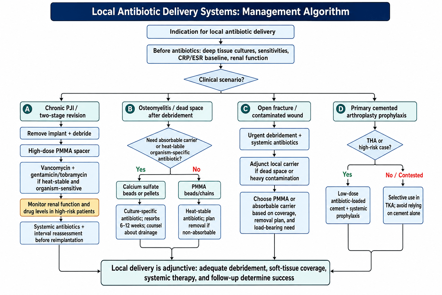
Three questions choose the carrier. Does it need to come out, or should it resorb? Does the site need load-bearing strength? And is there a heat-stable antibiotic to which the organism is sensitive?
By scenario. The answers map onto three standard pairings:
- Prosthetic joint infection: high-dose PMMA spacer, vancomycin plus gentamicin, in a two-stage revision
- Osteomyelitis: calcium sulfate or PMMA beads, loaded to culture
- Prophylaxis in primary arthroplasty: low-dose gentamicin in the cement
By organism. MRSA must include vancomycin. For Pseudomonas, tobramycin is preferred. When the organism-specific drug is heat-labile, use calcium sulfate.
Surgical Technique
Preparing the cement. Add the antibiotic powder to the polymer powder and mix thoroughly before the monomer goes in, so the drug is evenly distributed. Hand-mix for porosity, bearing in mind the caveat above about how much that gains.
Making beads. Roll the cement during the doughy phase into beads of 6-8mm diameter, the size that gives the best surface area, and thread 4-6 beads on each surgical wire.
Building a spacer. Mould the cement around the femoral component and ensure the antibiotic is adequately distributed through the construct.
Complications
PMMA: mechanical failure. Spacer fracture occurs in 5-15% with high antibiotic loading, following the loss of compressive strength above 10% loading described earlier, and is more common with vancomycin than gentamicin.
PMMA: resistance. The concern with prophylactic cement is theoretical: sub-inhibitory levels follow the initial burst, and biofilm can form on retained cement surfaces. Registry data have not shown increased resistance to date.
PMMA: systemic toxicity. Rare with standard loading. Acute kidney injury has been reported with high-dose spacers, and is more common with renal impairment and with multiple spacers.
Renal Impairment: High-dose antibiotic spacers (particularly aminoglycosides) can cause systemic absorption and nephrotoxicity in patients with pre-existing renal dysfunction. A systematic review of two-stage revision reported an average acute kidney injury incidence of 4.8% (Luu 2013, PMID 23578491). Monitor renal function and antibiotic levels in high-risk patients with spacers in situ.
Polymers. A foreign-body reaction to degradation products can produce a sterile seroma or abscess, and is more common with the rapid-degrading PGA. Release is less predictable than from the older carriers: kinetics vary with local pH and vascularity, and acidic degradation products may themselves affect the release rate.
The draining wound. A draining wound after local antibiotic delivery is a common viva scenario. The decision is distinguishing expected, self-limiting calcium-sulfate seepage from true ongoing infection that mandates a return to theatre.
- Typical Features
- Clear/serous, peaks early, settles as pellets resorb (weeks)
- Cultures
- Negative (sterile)
- Management
- Conservative wound care; reassure; avoid overpacking next time
- Typical Features
- Serous collection, no systemic upset, normal/falling CRP
- Cultures
- Negative
- Management
- Observe; aspirate only if symptomatic
- Typical Features
- Purulent, rising CRP/ESR, pain, systemic signs, sinus tract
- Cultures
- Positive (organism)
- Management
- Debridement, repeat sampling, revise antibiotic strategy
- Typical Features
- Bloody, early, may be tense
- Cultures
- Negative unless secondarily infected
- Management
- Evacuate if large/tense; correct coagulopathy
- Typical Features
- Sterile drainage with rapid-degrading PGA
- Cultures
- Negative
- Management
- Usually self-limiting; debride if abscess forms
Key discriminator: Calcium-sulfate drainage is clear/serous, sterile, and time-limited to the resorption phase. Purulent drainage, rising inflammatory markers, or a positive culture point to true infection and warrant return to theatre, not reassurance.
Postoperative Care
PMMA spacer or beads. Follow serial inflammatory markers, with a weekly CRP that should fall, inspect the wound daily for drainage and healing, and check creatinine weekly for nephrotoxicity if an aminoglycoside was used. Plan the bead removal from the outset.
Calcium sulfate. Expect the sterile, self-limiting drainage described above and manage it conservatively; it rarely needs reoperation. Watch for the rare hypercalcaemia.
Outcomes
Two-stage revision. The gold standard for prosthetic joint infection: infection is eradicated in 85-95%, functional success is achieved in 70-80%, and reinfection occurs in 5-15%.
Osteomyelitis. Calcium sulfate eradicates infection in 75-90% and PMMA beads in 80-90%: comparable outcomes, with calcium sulfate sparing the removal surgery.
Prophylaxis in primary arthroplasty. The absolute risk reduction from antibiotic cement is 0.5-1% on registry data.
What decides success. Adequate debridement is the most important factor, followed by appropriate antibiotic selection, a sufficient local concentration, and concurrent systemic antibiotics. Local delivery is adjunctive to all four and cannot replace them.
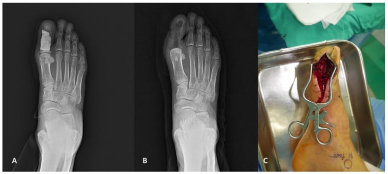
Clinical Applications of PMMA
Two-stage revision arthroplasty. The most established use of antibiotic-loaded PMMA is the articulating spacer for infected total joint arthroplasty. The standard protocol:
- Stage 1: implant removal, debridement, placement of the antibiotic spacer
- Interval: 6-12 weeks with the spacer in situ and systemic antibiotics
- Stage 2: spacer removal and reimplantation of a new prosthesis
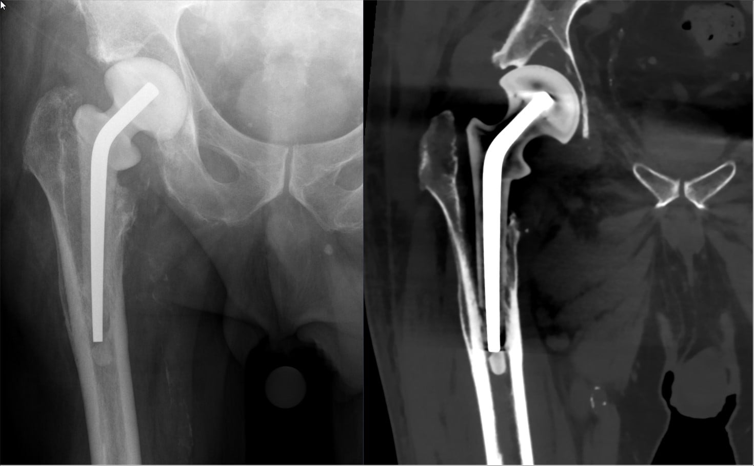
Loading and strength. Antibiotic loading over 10% by weight significantly reduces compressive strength, and a static spacer requires less structural integrity than an articulating one.
Articulating or static. An articulating spacer allows joint motion between stages. It preserves range of motion, soft-tissue and capsular length and quadriceps function, eases the eventual reimplantation, and gives better interim function and final outcomes in most patients. The trade-off is a more complex construct that can fracture, dislocate or wear, and it needs reasonable bone stock and a competent extensor mechanism and soft-tissue envelope.
A static block spacer immobilises the joint. It is chosen for severe bone loss, an incompetent extensor mechanism or collateral ligaments, a compromised soft-tissue envelope, or where stability is paramount. It sacrifices motion and causes more arthrofibrosis and quadriceps shortening, which makes reimplantation harder, but it is mechanically safer in a deficient knee. Articulating by default for function; static when the bone or soft tissue cannot support a mobile spacer or maximal stability is needed.
Prophylactic antibiotic cement. Low-dose gentamicin cement in primary arthroplasty remains controversial. In favour: reduced revision for infection in cemented THA on meta-analysis (RR 0.66, Farhan-Alanie 2021), the lowest infection-revision rate in the Norwegian Register when cement and systemic antibiotics were combined (Espehaug/Engesaeter 1997), cost-effectiveness in high-risk populations, and minimal mechanical compromise at low doses. Against: no significant benefit for primary TKA in the same meta-analysis (RR 0.92, not significant), concern for antibiotic resistance, a low absolute risk reduction in low-risk patients, and added cost, with some systematic reviews arguing that plain cement saves health-system expense.
The strongest evidence is for cemented THA, while the TKA question is genuinely contested. Reserve antibiotic cement for higher-risk patients (diabetes, immunosuppression, prior infection, revision) where the evidence is least ambiguous, and combine it with systemic prophylaxis rather than relying on either alone.
Beads and chains. Beads threaded on surgical wire manage dead space in osteomyelitis, deliver antibiotic at fracture sites, and give temporary wound coverage with antibiotic elution. They can be used with negative pressure wound therapy. Being PMMA, they need a second procedure for removal at 2-4 weeks.
Masquelet (induced-membrane) technique. This is the major trauma application of an antibiotic spacer, and a two-stage reconstruction in its own right. At the first stage, after radical debridement of an infected segmental bone defect, an antibiotic-loaded PMMA spacer is placed in the defect. It does two jobs at once: it delivers high local antibiotic to the dead space, and it provokes a foreign-body reaction that, over 6-8 weeks, forms a vascular induced membrane around itself.
At the second stage the spacer is carefully removed, preserving the membrane, and the defect is filled with bone graft (autograft, often with an osteoconductive expander). The membrane is the point: it secretes osteoinductive and angiogenic factors and, crucially, prevents the graft from being resorbed, which is why a large defect grafted without a membrane fails. The membrane is delicate and biologically active, so at the second stage open it longitudinally, keep it intact, and close it over the graft rather than debriding it away.
Antibiotic-coated intramedullary nails. For intramedullary infection or an infected long-bone nonunion, a nail can serve as both implant and carrier. A standard or custom nail is coated with antibiotic-loaded PMMA, usually by injecting the cement into a chest-tube or silicone mould around the nail and letting it set, then inserted after reaming and debridement. It provides stable fixation and sustained local antibiotic simultaneously, which is exactly what an infected nonunion needs: stability to heal, and drug at the infected interface. The coating can fracture or debond on insertion, and the nail must still be exchanged once infection is controlled, so it is a staged implant rather than a definitive one.
Calcium Sulfate
The material. Calcium sulfate is medical-grade plaster of Paris, used in orthopaedics since the 1890s for bone-defect filling; modern formulations are designed for antibiotic delivery with controlled resorption. Setting converts the hemihydrate to the dihydrate (CaSO₄·½H₂O + H₂O → CaSO₄·2H₂O) in 5-15 minutes depending on formulation. The reaction is exothermic but reaches only 37-42°C, which is why any antibiotic, heat-labile ones included, can be mixed into it.
Biology. Calcium sulfate is an osteoconductive scaffold for bone ingrowth that resorbs completely over 6-12 weeks and is replaced by host bone, so there is no second surgery for removal. The rapid resorption gives near-complete antibiotic elution, and it releases calcium and sulfate ions, with the potential for a local acidosis that may inhibit osteogenesis.
The same resorption is the weakness. It can create a transient dead space before new bone forms, which matters most in load-bearing areas needing mechanical support during healing, and may require bone grafting later if healing is inadequate. Mechanical strength is limited, so the carrier is for non-load-bearing use only, and wound drainage is common.
Osteomyelitis. Beads or pellets are placed into the debrided cavity in a single-stage procedure that needs no removal, gives high local antibiotic concentrations, leaves an osteoconductive scaffold, and accepts any antibiotic. The technique:
- Thorough surgical debridement first
- Mix the antibiotic powder directly into the calcium sulfate, at the loading above
- Pack into the dead space without overpacking
- Consider closed suction drainage
Open fractures. Use in contaminated open fractures remains controversial. The potential benefits are local antibiotic at the contaminated fracture site, reduced infection rates in some studies, and dead-space management. The concerns are wound-drainage complications, cost, limited high-quality RCT evidence, and possible interference with fracture healing in some cases.
Complications. Wound drainage is the most common, in 15-30% of cases. It is usually sterile and culture-negative, can persist for 2-4 weeks, is managed with local wound care and rarely requires reoperation; avoiding overpacking and placing a drain reduce it. A local inflammatory reaction follows the acidic pH of resorption and can cause a seroma, which is usually self-limiting. Hypercalcaemia is rare, reported with very large volumes (over 100cc), usually transient and asymptomatic, and worth monitoring in patients with renal impairment.
Bioabsorbable Polymers
Polylactic and polyglycolic acid. These synthetic polymers degrade by hydrolysis to lactic and glycolic acid, which are metabolised through the Krebs cycle. Degradation rate depends on composition:
- PLLA (poly-L-lactic acid): slow, 12-24 months
- PLGA (poly-lactic-co-glycolic acid): faster, 6-12 months
- PGA (polyglycolic acid): rapid, 1-3 months
Antibiotic release follows the degradation profile, so the release rate is programmable through polymer composition, and controlled degradation can sustain therapeutic levels for weeks to months.
Collagen carriers. Resorbable collagen sponges or fleece impregnated with gentamicin (Collatamp G sponge, Septocoll fleece) resorb in 4-12 weeks, are haemostatic and conformable to a wound bed, and have limited mechanical strength.
The caution this product carries: the largest trial of the gentamicin-collagen sponge (SWIPE, Bennett-Guerrero NEJM 2010, colorectal surgery) found it did not reduce surgical-site infection and paradoxically caused more (30.0% vs 20.9%, p=0.01), with more wound-related returns. That population was abdominal, so direct extrapolation to orthopaedics is limited, but it is a landmark warning that a resorbable antibiotic carrier cannot be assumed beneficial; its value must be demonstrated for each indication rather than inferred from the drug it carries.
Chitosan and natural polymers. Emerging carriers from natural sources:
- Chitosan: derived from crustacean shells, with antimicrobial properties of its own
- Hyaluronic acid: a viscosupplement carrier for antibiotics
- Fibrin glue: a carrier for antibiotics in wound beds
Where they stand. Current uses, with mixed evidence:
- Gentamicin-collagen sponges: widely available, but orthopaedic and spine data are lower quality and inconsistent after the colorectal result above
- Sternal wound infection prophylaxis in cardiac surgery, also studied with conflicting results
- High-risk orthopaedic wounds, where carrier benefit must be demonstrated per indication
Investigational uses are fracture fixation with antibiotic-coated implants, antibiotic-loaded bone graft substitutes, and combined growth-factor and antibiotic delivery. The barriers are manufacturing complexity and cost, regulatory hurdles with few FDA-approved products, release kinetics that vary with the local environment, a potential inflammatory response to degradation products, a high cost compared with PMMA or calcium sulfate, limited long-term outcome data, and an unclear advantage over established systems in most applications.
Guidelines, Registries & Global Practice
Global Epidemiology and Practice Variation
Periprosthetic joint infection complicates roughly 1-2% of primary and up to 4-15% of revision arthroplasties worldwide, and is a leading cause of revision in every major registry. Local antibiotic delivery is near-universal in two-stage revision, but prophylactic antibiotic-loaded cement (ALBC) in primary arthroplasty varies enormously by region: it is routine across much of Europe (driven by Scandinavian register data) yet historically optional in parts of North America, where many surgeons reserved it for high-risk cases. The strongest registry-derived signal is in cemented THA; the primary TKA evidence is genuinely contested.
- Position
- Two-stage revision with antibiotic spacer is a standard option for chronic PJI; surgeon-directed local antibiotics
- Evidence Level
- Consensus / moderate
- Position
- Systemic prophylaxis mandatory; ALBC not mandated routinely - clinician judgement
- Evidence Level
- Low-moderate
- Position
- IV prophylactic antibiotics as soon as possible, ideally within 1 hour of injury, per a published network guideline; makes no recommendation on local antibiotics
- Evidence Level
- Consensus
- Position
- Debridement plus local antibiotic carrier (PMMA or bioabsorbable) for dead space
- Evidence Level
- Consensus
- Position
- Routine ALBC in cemented arthroplasty widely endorsed
- Evidence Level
- Register-based
Registry Evidence
- Norwegian Arthroplasty Register: combined systemic + antibiotic cement gave the lowest revision-for-infection rate; cement without antibiotic increased infection risk that ALBC neutralised (Espehaug/Engesaeter 1997; Engesaeter 2006).
- Pooled register/meta-analysis (Farhan-Alanie 2021): ALBC protective for revision-for-PJI in THA (RR 0.66) but not significant in TKA.
- AOANJRR (Australia): tracks cement type and revision for infection and is used to inform local ALBC practice, particularly in cemented fixation and higher-risk patients.
Commonly Used Commercial Products
These antibiotic-loaded cements and resorbable carriers are available internationally (brand availability varies by region):
- PMMA (factory-loaded): Palacos R+G (gentamicin) and Simplex with Tobramycin
- Calcium sulfate (surgeon-loaded): Osteoset and Stimulan, which accept any antibiotic mixed intraoperatively
- Type
- PMMA
- Antibiotic
- Gentamicin 0.5g
- Type
- PMMA
- Antibiotic
- Tobramycin 1g
- Type
- Calcium sulfate
- Antibiotic
- Surgeon-loaded (any antibiotic)
Examiner Favorite: "What factors increase antibiotic elution from PMMA cement?" Answer systematically: Higher antibiotic loading, hand-mixing (vs vacuum), additives like glycine, smaller bead size (higher surface area), and lower cement viscosity. Then contrast with calcium sulfate which releases 90-100% regardless of these factors due to complete resorption.
MCQ Practice Points
Q: What is the most commonly used antibiotic in PMMA bone cement for arthroplasty infection prophylaxis?
A: Gentamicin (tobramycin in some regions). Commercial antibiotic-loaded cements contain 0.5-1g gentamicin per 40g cement. This provides local concentrations 100-1000x higher than MIC for staphylococci while maintaining low systemic levels. For treatment of established infection, higher doses (3-4g per 40g cement) are hand-mixed. Vancomycin 1-2g is added for MRSA coverage.
Q: What are the advantages of local antibiotic delivery compared to systemic administration?
A: (1) Local concentrations 100-1000x higher than achievable systemically, (2) Minimal systemic absorption and toxicity, (3) Effective in avascular areas where systemic antibiotics cannot penetrate, (4) Sustained release over weeks. Limitations: only heat-stable antibiotics survive cement polymerization (gentamicin, vancomycin, tobramycin - NOT beta-lactams), requires surgical placement.
Q: What is the recommended antibiotic-loaded cement spacer regimen for a two-stage revision of an infected total knee arthroplasty?
A: High-dose antibiotic cement: vancomycin 3-4g + gentamicin 3-4g per 40g PMMA cement. Articulating spacer preferred over static spacer (maintains soft tissue tension, easier revision). Cement spacer remains in situ for 6-12 weeks while systemic antibiotics administered. Consider antibiotic holiday (2-6 weeks) before reimplantation to confirm infection clearance.
Q: What are the properties of an ideal local antibiotic delivery system?
A: (1) Heat-stable (survives cement polymerization at 70-100°C), (2) Water-soluble (for elution from cement), (3) Broad-spectrum coverage, (4) Bactericidal, (5) Low allergenicity, (6) Minimal systemic absorption, (7) Prolonged elution kinetics. Gentamicin, tobramycin, and vancomycin meet these criteria. Beta-lactams are heat-labile and not suitable.
Q: What is the role of antibiotic-impregnated calcium sulfate beads in osteomyelitis management?
A: Biodegradable alternative to PMMA beads that does not require removal surgery. Calcium sulfate resorbs over 4-12 weeks, releasing antibiotics and being replaced by bone. Can deliver vancomycin, gentamicin, or tobramycin. Useful for: dead space management, osteomyelitis debridement, open fracture void filling. Complication: transient hypercalcemia, prolonged wound drainage during resorption.
Clinical Decision Scenarios
Practise clinical reasoning and management decisions out loud
“A 68-year-old woman with diabetes presents 14 months after primary total knee arthroplasty with chronic sinus drainage and pain. Aspiration grows MRSA. You plan a two-stage revision. Discuss your antibiotic spacer strategy.”
“You are managing chronic osteomyelitis of the tibia following an open fracture. After debridement, you have a 50cc bone defect. The organism is pan-sensitive Staphylococcus aureus. Discuss local antibiotic delivery options.”
“A colleague asks whether they should routinely use antibiotic-loaded bone cement (ALBC) in all primary cemented total knee and hip arthroplasties. Discuss the evidence.”
PMMA Antibiotic Cement
Calcium Sulfate Carriers
Bioabsorbable Polymers
Evidence-Based Practice
Complications to Mention
Evidence Base
- Finding
- Systemic + ALBC lowest infection revision
- Level
- Level 2
- Finding
- ALBC protective in THA, not TKA
- Level
- Level 1
- Finding
- CaSO4 = PMMA, fewer reops
- Level
- Level 2
- Finding
- Gent-collagen sponge increased SSI
- Level
- Level 1
The evidence base has its limits: it is mostly observational and registry data, there are few RCTs for antibiotic cement, the patient populations are heterogeneous, and follow-up periods vary.
Norwegian Register: Systemic Plus Antibiotic Cement Lowers Revision for Infection
- Combined systemic + antibiotic cement gave the lowest revision rate for infection
- Systemic antibiotics alone: 4.3x higher revision rate for infection (95% CI 1.7-11.0)
- Antibiotic in cement only: 6.3x higher (95% CI 1.6-25.0)
- No antibiotics: 11.5x higher (95% CI 2.1-63.0)
Antibiotic-Loaded Cement and Revision for Infection: Hip vs Knee Differ
- THA: ALBC protective against revision for PJI (RR 0.66, 95% CI 0.56-0.77)
- THA: no significant difference in all-cause revision (RR 0.62, 95% CI 0.35-1.09)
- TKA: no significant difference for PJI (RR 0.92, 95% CI 0.59-1.45)
- TKA: no significant difference for all-cause revision (RR 0.73, 95% CI 0.53-1.02)
Calcium Sulfate vs PMMA Beads in Chronic Osteomyelitis (RCT)
- Infection eradicated in 86% (12/14) in both calcium sulfate and PMMA groups
- Fewer reoperations with calcium sulfate (7 vs 15, p=0.04)
- Comparable union rates (7/8 vs 6/8 nonunions)
- Calcium sulfate avoids a second procedure for bead removal
Calcium Sulfate Bone Substitute in Infected Long-Bone Defects
- Infection eradicated in 92% (23/25)
- Pellets radiographically resorbed at a mean of 2.7 months
- Isolated bony defects healed in all 9 patients without further treatment
- 8 patients developed sterile draining sinuses that healed on resorption
Gentamicin-Collagen Sponge Does NOT Prevent SSI (and may worsen it)
- SSI higher in the sponge group (30.0%) than control (20.9%), p=0.01
- Superficial SSI 20.3% vs 13.6% (p=0.03)
- More wound-related ER/surgeon visits in the sponge group (19.7% vs 11.0%)
- No reduction in deep SSI
Acute Kidney Injury Risk From Antibiotic Spacers
- Pooled infection control rate 91%
- Average acute kidney injury incidence 4.8%
- Average infection persistence/recurrence 11%
- Marked heterogeneity in spacer antibiotic dosing across studies
Vacuum vs Hand Mixing and Gentamicin Elution
- Vacuum mixing reduced porosity but only minimally reduced total gentamicin release
- Hand-added antibiotic released less than industrially pre-loaded cement
- Release varied substantially between commercial brands
- Porosity is one of several determinants of elution, not the sole driver
Cross-References
Related Basic Science Topics:
- PMMA Bone Cement (polymerization, mechanical properties, thermal effects)
- Calcium Phosphate Cements (alternative osteoconductive carriers)
- Bioabsorbable Materials (polymer degradation kinetics, tissue response)
- Common Pathogens in Orthopaedics (organism-specific antibiotic selection)
- Osteomyelitis Pathophysiology (infection biology, antibiotic penetration)
Related Clinical Topics:
- Periprosthetic Joint Infection (two-stage revision, spacer technique)
- Pediatric Acute Osteomyelitis (dead space management, antibiotic delivery)
- Open Fracture Management (local antibiotic prophylaxis)
- Surgical Site Infection Prevention (prophylactic antibiotic strategies)
Related Surgical Topics:
- Two-Stage Revision Arthroplasty (spacer fabrication and implantation)
- Debridement Techniques for Osteomyelitis (preparation for antibiotic bead placement)
This comprehensive understanding of local antibiotic delivery systems is essential for managing orthopaedic infections and is frequently tested in orthopaedic fellowship examinations across multiple stations.