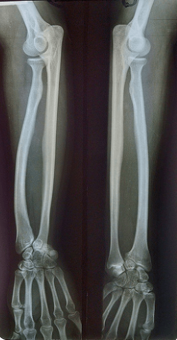
Growth disturbance of the volar-ulnar distal radial physis | Tethered by Vickers ligament | Linked to Leri-Weill dyschondrosteosis and SHOX defects | Bilateral, female, adolescent
- Core lesion: a partial growth arrest of the volar and ulnar corner of the distal radial physis, so the radius keeps growing on its dorsal-radial side but not its volar-ulnar side - this twists the joint surface and lets the carpus slide volar and ulnar
- Vickers ligament: an abnormal thick band running from the volar distal radius to the lunate. It tethers the volar physis and is the structure released at surgery
- Who gets it: typically an adolescent girl with bilateral wrists, presenting with deformity, a prominent ulnar head, pain, and loss of forearm rotation and wrist extension
- The big association: Leri-Weill dyschondrosteosis (a mesomelic short-stature dwarfism) caused by SHOX gene defects - but many cases are idiopathic, post-traumatic, post-infective, or part of other dysplasias
- Treatment depends on skeletal maturity: in the growing child, release Vickers ligament and free the radial physis (physiolysis) to slow progression; in the mature, symptomatic wrist, a dome osteotomy of the radius corrects the deformity, and severe joint damage may need a salvage procedure
- “Madelung deformity is the classic exam wrist that is bilateral, female, adolescent and associated with short stature - say all four
- “The deforming tether is Vickers ligament running from the volar radius to the lunate; releasing it is the key growth-modulating step in children
- “Think SHOX and Leri-Weill dyschondrosteosis, but remember Madelung-like deformity can also follow trauma, infection, or other skeletal dysplasias (the reverse Madelung points the other way)
- “Match the operation to the skeleton: physiolysis and ligament release while growing; osteotomy once mature
Overview
Madelung deformity is a developmental abnormality of the wrist in which the distal radius grows in the wrong shape because part of its growth plate stops working. It is named after Otto Madelung, who described it in the 19th century. The typical patient is an adolescent girl who develops a bent-looking wrist with a prominent bump on the little-finger side (the ulnar head), often in both wrists, with pain and reduced movement.
The lesion in one idea. Think of the distal radial growth plate as having a corner that fails. The volar (palmar) and ulnar corner of the physis stops growing while the rest of the radius carries on, so the joint surface tilts towards the ulna and towards the palm, the carpus slides into the gap that forms, and the ulna ends up looking too long because the radius has shortened on that side. Running across the front of the failing physis is an abnormal thick band, the Vickers ligament, which tethers the lunate to the volar radius and is central to both the pathology and the surgery.
Why examiners like it. The topic links genetics (the SHOX gene and Leri-Weill dyschondrosteosis), growth-plate biology, three-dimensional deformity and a maturity-dependent treatment decision. Get the classic picture, the Vickers ligament and the growing-versus-mature treatment split, and you have answered most of what an examiner will ask.
Madelung deformity is the wrist condition. Madelung disease (benign symmetric lipomatosis) is a completely different fatty-neck condition: same surname, unrelated.
Anatomy and Pathophysiology
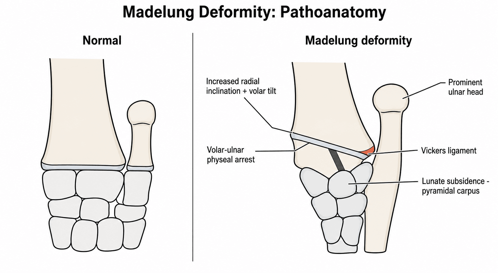
The physis. The distal radius normally grows from a physis that contributes most of the length of the radius. In Madelung deformity the volar-ulnar part of this physis is abnormal and arrests early while the dorsal-radial part keeps growing, and this asymmetric growth twists the distal radius:
- Coronal plane: the joint surface tilts so its ulnar side is lower, an increased radial inclination that points the carpus towards the ulna
- Sagittal plane: the joint surface tilts towards the palm, an increased volar (palmar) tilt, so the carpus and lunate are carried forwards (palmar carpal displacement)
- The ulna: the radius shortens on the affected side while the ulna grows normally, so the ulna becomes relatively long and its head sits prominent and often dorsal at the wrist
The carpus adapts. The lunate sinks into the notch that forms between the tilted radius and the ulna (lunate subsidence), and the proximal carpal row takes on a wedge or pyramid/triangle shape pointing proximally, a classic descriptive sign on the radiograph.
The Vickers ligament. This abnormal structure is at the centre of it all. Histopathological study of patients with Leri-Weill dyschondrosteosis and a confirmed SHOX abnormality showed widespread disorganisation of the radial growth plate, the normally neat parallel columns of cartilage cells disrupted, and confirmed that the ligament blends with the triangular fibrocartilage complex (TFCC) (Munns et al., Hand Surg 2001, DOI). The volar tether is thought to bowstring across the volar physis, both reflecting and reinforcing the local growth disturbance.
The core sentence to memorise: "Madelung deformity is a partial arrest of the volar-ulnar distal radial physis, tethered by an abnormal Vickers ligament running from the volar radius to the lunate, producing increased radial inclination, increased volar tilt, lunate subsidence, and a relatively prominent ulna."
Aetiology and Genetics
Not all Madelung deformity has the same cause. Split it into a genetic/dysplastic group, where the physis is intrinsically abnormal, and an acquired group, where something damages a previously normal physis. Naming the group guides whether you look for short stature, a family history or a previous injury.
- Examples
- SHOX gene defect (deletion or mutation), mesomelic short stature
- Key point
- The classic association; bilateral, female predominant, true Vickers ligament
- Examples
- Turner syndrome, multiple hereditary exostoses, other skeletal dysplasias
- Key point
- Madelung-like deformity as part of a wider skeletal picture
- Examples
- Physeal injury to the volar-ulnar distal radius
- Key point
- Post-traumatic arrest mimics the genetic shape
- Examples
- Osteomyelitis affecting the physis, prior radiation
- Key point
- Damaged physis behaves like an arrest
- Examples
- No identifiable cause or syndrome
- Key point
- Common - do not assume every case is genetic
Where the gene sits. SHOX (short-stature homeobox) lies in the pseudoautosomal region (PAR1) at the tips of the X and Y chromosomes, so it escapes X-inactivation and is normally present as two active copies in everyone. Because both copies are normally active it behaves in a pseudoautosomal dominant fashion: losing one working copy (haploinsufficiency) is enough to cause disease, with variable penetrance and a tendency for Madelung deformity to be more frequent and more marked in females.
What one lost copy does. SHOX haploinsufficiency causes Leri-Weill dyschondrosteosis, a mesomelic (middle-segment) short-stature dwarfism in which Madelung deformity is the most characteristic limb feature, and accounts for around 80% of the genetic causes of Leri-Weill dyschondrosteosis. SHOX-deficient bone shows a disrupted columnar growth plate with an expanded hypertrophic layer and altered programmed cell death of the cartilage cells, the cellular basis of the deformity (Seki et al., Clin Pediatr Endocrinol 2014, DOI).
The gene-dose spectrum. Examiners like the spectrum because the phenotype follows the number of working copies:
- Condition
- Leri-Weill dyschondrosteosis
- Phenotype
- Mesomelic short stature plus Madelung deformity - the classic association
- Condition
- Langer mesomelic dysplasia
- Phenotype
- Severe mesomelic AND rhizomelic dwarfism - the most severe end of the spectrum
- Condition
- Turner syndrome
- Phenotype
- Short stature and an increased incidence of Madelung deformity, among the other features
- Condition
- Isolated / idiopathic short stature
- Phenotype
- Short stature with little or no Madelung change
Why it matters clinically. Bilateral Madelung deformity in a short child should prompt genetic testing (SHOX deletion/duplication analysis such as MLPA, plus sequencing) and endocrine referral. Growth hormone is an approved treatment for the short stature of SHOX deficiency and Leri-Weill dyschondrosteosis, so an early diagnosis has a non-orthopaedic payoff. Add the caveat, when you present the case, that Madelung-like deformity can also be acquired (trauma, infection) or part of other dysplasias; it shows you are not pattern-matching blindly.
Frame SHOX as a dose-response gene: one copy lost gives Leri-Weill dyschondrosteosis (mesomelic short stature plus Madelung), both copies lost gives Langer mesomelic dysplasia (severe dwarfism), and 45,X (Turner) loses a copy too, which is why those girls are short and prone to Madelung. It is pseudoautosomal dominant with variable, female-predominant penetrance.
Classification
There is no single universally used classification, but examiners expect you to categorise by cause and to describe the deformity in a structured way. A practical scheme uses four axes:
- By cause: dysplastic/genetic (Leri-Weill dyschondrosteosis and SHOX, other dysplasias), post-traumatic, post-infective and idiopathic, as set out above
- By direction: the typical (volar) Madelung has the radius tilting volar and ulnar with the carpus drawn towards the palm. A reverse (dorsal) Madelung is uncommon and tilts the opposite way, with a dorsally subluxed distal ulna, worth a mention to show you know it exists
- By severity and symptoms: from a mild, painless cosmetic deformity found incidentally, through progressive deformity in a growing child, to an established painful wrist in the mature patient with restricted rotation and grip. Severity and symptoms, not the radiograph alone, drive treatment
- By radiographic measurement: radial (ulnar) tilt, lunate subsidence, lunate fossa angle and palmar carpal displacement, the same numbers used to plan and to judge surgical correction; how each is measured is set out under Investigations
Clinical Presentation
The story. An adolescent girl, commonly in her early teens, notices that her wrists look bent and that there is a bump on the ulnar side, the prominent ulnar head. It is frequently bilateral, and there may be a family history and short stature if there is underlying Leri-Weill dyschondrosteosis. In acquired cases, ask about trauma or infection of the distal radius.
Symptoms. Deformity and cosmetic concern, especially the prominent dorsal ulnar head, are often the main reason for presenting. Pain is typically ulnar-sided, worse with loading and activity, and often emerges in adolescence as the deformity matures. Stiffness takes wrist extension and especially forearm rotation (supination), with loss of radial deviation, and symptomatic wrists are weak in grip.
Madelung deformity is a leading congenital cause of ulnar-sided wrist pain and loss of supination in an adolescent. If you are given that triad with a prominent ulnar head and bilateral involvement, Madelung deformity should be at the top of your list.
Examination. The sequence is the usual one, with the findings that belong to this condition at each step:
- Look: inspect both wrists from the front, side and above. Note the volar-tilted "bayonet" or step contour with the hand looking displaced towards the palm, the dorsally prominent ulnar head, any forearm shortening (mesomelia) and surgical scars. Always expose and compare both sides
- Feel: palpate the prominent ulnar head and the distal radioulnar joint (DRUJ) for tenderness; assess DRUJ stability (ballottement) and ulnar-sided tenderness over the TFCC
- Move: measure active and passive wrist flexion/extension and especially forearm pronation/supination and radial deviation, which are characteristically reduced; compare with the other side. Check grip strength
- Special tests and completing the examination: examine the DRUJ (piano-key sign), do a focused neurovascular check, then examine the rest of the child
A bilateral Madelung deformity in a short adolescent is a clue to an underlying skeletal dysplasia. Examine the whole child (height, limb proportions, the other limbs and the contralateral wrist) and have a low threshold for genetic/endocrine referral: the wrist may be the presenting sign of Leri-Weill dyschondrosteosis.
Investigations
Radiographs. Posteroanterior and lateral wrist films are the cornerstone and usually diagnostic. Look for:
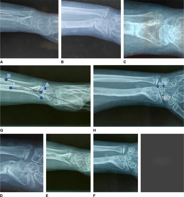
- Increased radial inclination, the joint surface tilting towards the ulna
- Increased volar tilt and palmar carpal displacement on the lateral view
- Lunate subsidence into a V-shaped notch between radius and ulna, with the proximal carpal row taking a pyramid/triangle shape
- A relatively long, dorsally prominent ulna and changes in the sigmoid notch
- Bowing of the radial shaft, and shortening
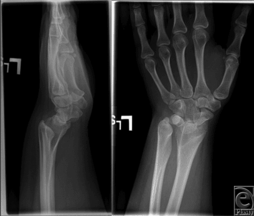
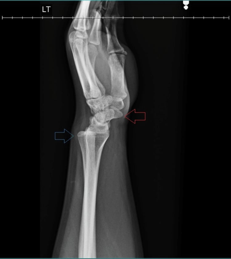
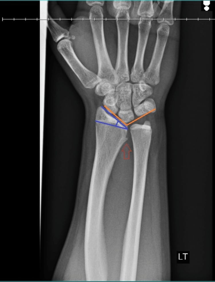
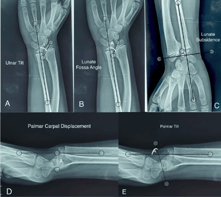
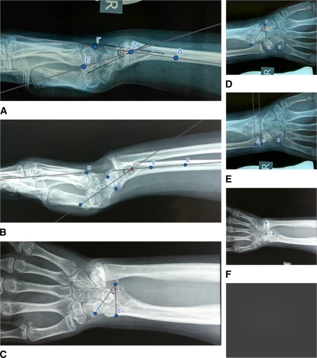
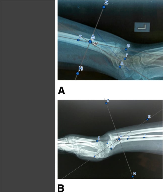
Measuring the deformity. Ulnar (radial) tilt, lunate subsidence, lunate fossa angle and palmar carpal displacement quantify the deformity, let you follow progression and judge surgical correction, and are what an examiner may ask you to demonstrate on the film:
- Ulnar tilt (radial inclination): on the PA view, the angle between the distal radial articular surface and a line perpendicular to the long axis of the radius. It is increased in Madelung, the joint surface sloping towards the ulna
- Lunate subsidence: on the PA view, how far the proximal pole of the lunate has sunk proximally between the radius and ulna, where it sits in the V-shaped notch, measured against the distal ulna/radius as a reference
- Lunate fossa angle: the angulation of the lunate fossa of the distal radius, reflecting the abnormal slope of the articular surface that the carpus sits on
- Palmar carpal displacement: on the lateral view, how far the carpus (lunate) lies volar to the long axis of the radius, the radiographic counterpart of the clinical palmar carpal shift
There is no single agreed abnormal cut-off. Each value is interpreted against the normal or contralateral wrist and, more usefully, tracked over time and before and after surgery to judge progression and the adequacy of correction. These same four measurements are the ones reported in the surgical literature, for example the dome-osteotomy and growth-modulation series cited below.
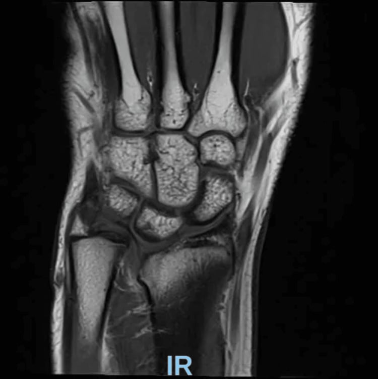
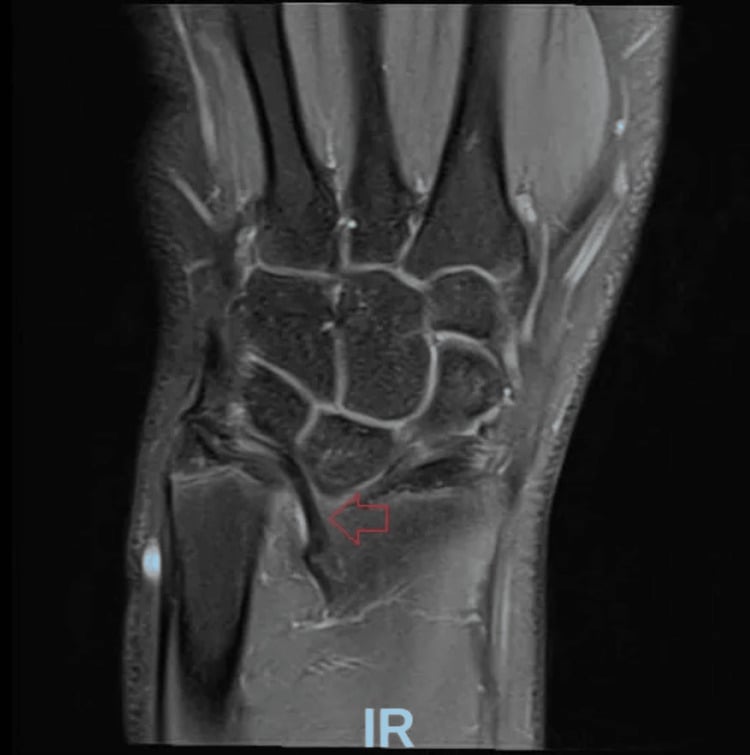
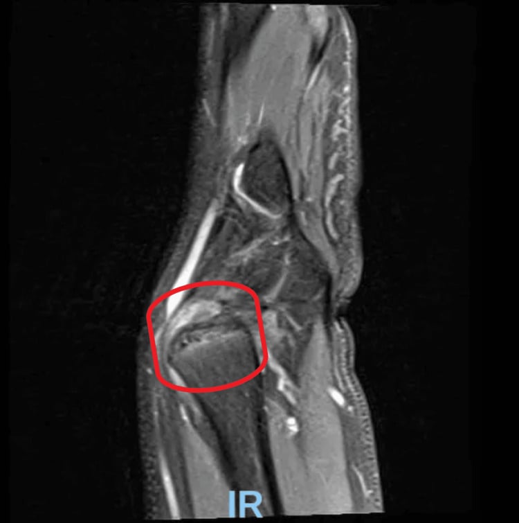
MRI demonstrates the Vickers ligament and the abnormal volar physis directly, assesses the cartilage and TFCC, and helps preoperative planning, especially in younger patients where you want to confirm the tether before release.
CT, often with 3D reconstruction, characterises the bony deformity in three planes, the sigmoid notch and any joint incongruity, and is increasingly used for patient-specific osteotomy planning in the mature wrist.
Differential Diagnosis
Madelung deformity is the classic congenital cause of a bent, ulnar-prominent adolescent wrist, but several conditions mimic it. The discriminators are laterality, sex, the presence of bony lesions, systemic features and a trauma history, and the single most useful of them is the whole-forearm radiograph: osteochondromas point to multiple hereditary exostoses (a Masada forearm deformity), a unilateral deformity with a physeal bar points to old trauma or infection, and a symmetric bilateral volar-ulnar physeal arrest in a short girl points to true (often SHOX-related) Madelung deformity.
- Discriminating feature
- Bilateral, female, short stature; volar-ulnar physeal arrest with a Vickers ligament
- Best test
- PA and lateral radiographs; MRI for the tether; SHOX testing
- Discriminating feature
- Visible osteochondromas tethering the distal ulna/radius (Masada forearm deformity); often male; lesions elsewhere; family history
- Best test
- Radiographs of the whole forearm / skeletal survey showing exostoses
- Discriminating feature
- History of distal radius trauma; usually unilateral; no systemic features
- Best test
- Radiographs; CT or MRI for a physeal bar
- Discriminating feature
- Wider skeletal or syndromic stigmata; short stature with other features
- Best test
- Skeletal survey; karyotype / genetics
- Discriminating feature
- History of distal radius osteomyelitis or sepsis; often unilateral
- Best test
- Radiographs; history and inflammatory markers
- Discriminating feature
- Radius tilts dorsally with a dorsally subluxed ulna - the opposite tilt
- Best test
- Lateral radiograph showing dorsal rather than volar carpal displacement
Management
The decision. The single most important determinant is skeletal maturity: a growing physis can be modulated, whereas an established deformity in a mature wrist can only be corrected by cutting and realigning bone. Severity and symptoms then refine the plan.
Non-operative. Many patients, especially those with mild, painless deformity, never need surgery. The natural history is unpredictable, so mild asymptomatic deformity warrants a period of non-surgical management: observation with serial radiographs, activity modification, analgesia and splinting for symptom control (Kozin and Zlotolow, J Hand Surg Am 2015, DOI).
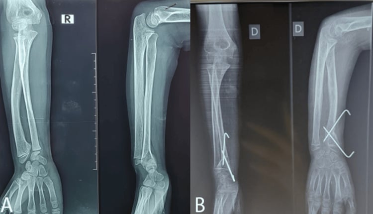
The growing child. In a child with growth remaining and progressive deformity or symptoms, the aim is to stop the tether and free the physis before more deformity develops:
- Vickers ligament release: dividing the abnormal volar tether
- Radial physiolysis: resecting the abnormal bony bar/diseased physis to allow the remaining physis to grow more normally, sometimes with interposition (for example a fat graft)
- Ulnar epiphysiodesis may be added to stop the ulna outgrowing the shortened radius
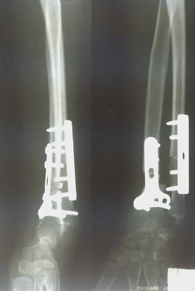
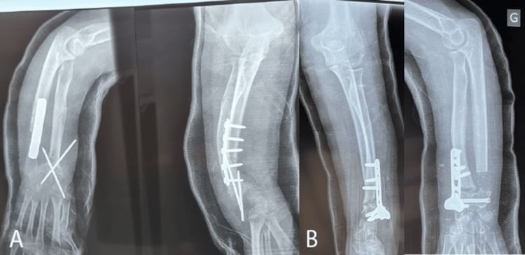
The mature wrist. Once the patient is skeletally mature, growth modulation is no longer possible and the deformity is corrected directly. The dome (or wedge) osteotomy of the distal radius is the workhorse corrective operation: it allows three-dimensional realignment of the tilted joint surface and is usually fixed with a plate. Ulnar-sided procedures, an ulnar shortening osteotomy or a distal-ulna procedure (for example Sauve-Kapandji or Darrach-type), address the prominent, long ulna and the distal radioulnar joint.
Salvage. For a severely damaged joint or sigmoid notch, partial wrist arthrodesis (such as radioscapholunate fusion, sometimes with a reconstructed distal radioulnar joint) is an option.
- Typical approach
- Observation, serial X-rays, splint/analgesia
- Aim
- Avoid unnecessary surgery; many never progress
- Typical approach
- Vickers ligament release + radial physiolysis (+/- ulnar epiphysiodesis)
- Aim
- Slow progression and let remaining physis grow
- Typical approach
- Dome osteotomy of distal radius (+/- ulnar shortening)
- Aim
- Correct deformity, offload the ulnar wrist, restore rotation
- Typical approach
- Salvage: ulnar procedure, partial arthrodesis, DRUJ reconstruction
- Aim
- Reliable pain relief when reconstruction is not possible
The examiner wants to hear the maturity split out loud: "If she is still growing I would release the Vickers ligament and free the radial physis to modulate growth; if she is skeletally mature with a symptomatic deformity I would correct it with a dome osteotomy of the distal radius, addressing the ulna as needed." That single sentence carries most of the marks.
Complications
- Why it happens
- Ongoing asymmetric physeal growth and tethering
- Key point
- Natural history is unpredictable; serial X-rays in mild cases
- Why it happens
- Abnormal radioulnar mechanics, TFCC and DRUJ loading
- Key point
- Common indication for surgery; main complaint in adolescents
- Why it happens
- Distorted distal radioulnar joint and sigmoid notch
- Key point
- Supination and wrist extension are typically most affected
- Why it happens
- Joint incongruity and chronic abnormal loading
- Key point
- May ultimately need salvage (arthrodesis, DRUJ reconstruction)
- Why it happens
- Growth modulation does not fully normalise the physis
- Key point
- Long-term studies show further surgery may be needed after growth
- Why it happens
- Nonunion, malunion, hardware irritation, neurovascular risk
- Key point
- Standard risks of distal radius corrective osteotomy
Clinical Relevance
Madelung deformity sits at the crossroads of paediatric hand surgery, genetics and growth-plate biology, which is why it recurs in examinations. The everyday skills it tests, reading the deformity on plain films, deciding who needs surgery, and choosing between ligament release, physiolysis, osteotomy and salvage, are the same judgements that come up across deformity correction in the upper limb.
Guidelines, Registries and Global Practice
- No high-level guideline or registry governs Madelung deformity - it is rare, so the evidence base is case series (level IV) and a systematic review of them, and practice is guided by paediatric hand surgery experience rather than randomised trials.
- Consistent global principle: treat symptoms and progression, not the X-ray alone. Mild, painless deformity is observed with serial radiographs everywhere; surgery is reserved for pain, functional loss, or progressive deformity.
- Maturity-based strategy is universal: growth modulation (Vickers ligament release with radial physiolysis, sometimes ulnar epiphysiodesis) in the skeletally immature wrist, and corrective osteotomy (commonly a distal radial dome osteotomy, with ulnar-sided procedures as needed) once mature. Salvage arthrodesis is reserved for severely damaged joints.
- Multidisciplinary input: because of the strong link with SHOX-related Leri-Weill dyschondrosteosis, bilateral disease in a short child prompts genetic and endocrine assessment in addition to orthopaedic care - a point valued internationally.
Exam Viva Scenarios
Practise clinical reasoning and management decisions out loud
“A 14-year-old girl is referred with deformity of both wrists and a prominent bump on the little-finger side. She is short for her age. The examiner asks for your diagnosis and assessment.”
“Two patients have symptomatic Madelung deformity: a 10-year-old with open physes and a 19-year-old who is skeletally mature. The examiner asks how your surgical plan differs and why.”
The Core Lesion
- Partial arrest of the volar-ulnar distal radial physis
- Abnormal Vickers ligament tethers volar radius to lunate
- Increased radial inclination + volar tilt + palmar carpal displacement
- Lunate subsidence, pyramid carpus, relatively long prominent ulna
Classic Patient and Association
- Adolescent girl, bilateral wrists, short stature
- Leri-Weill dyschondrosteosis from SHOX gene defects
- But also post-traumatic, post-infective, idiopathic, other dysplasias
- Presents with ulnar-sided pain and loss of supination/extension
Investigations
- PA + lateral wrist X-rays - usually diagnostic
- Measure radial tilt, lunate subsidence, palmar carpal displacement
- MRI to show Vickers ligament and plan; CT/3D for osteotomy planning
- Genetic/endocrine referral if syndromic (SHOX testing)
Treatment by Maturity
- Mild/painless: observe with serial X-rays, splint, analgesia
- Growing child: Vickers release + physiolysis (+/- ulnar epiphysiodesis)
- Mature wrist: dome osteotomy of distal radius (+/- ulnar shortening)
- Destroyed joint: salvage (partial arthrodesis, DRUJ reconstruction)
Evidence
The following studies support the modern, maturity-dependent approach to Madelung deformity.
Surgical Management of Madelung Deformity: A Systematic Review
- Systematic review of 25 studies (all case series, level IV evidence) describing surgery for Madelung deformity
- The main indication for surgery was wrist pain, not deformity alone
- Procedures fall into radial lengthening/correction, ulnar shortening, or a combination of both
- All studies reported postoperative pain reduction and most reported improved range of motion, but inconsistent outcome reporting prevented pooling of results
Long-Term Outcomes After Vickers Ligament Release and Growth Modulation
- Six skeletally immature patients (8 wrists) with symptomatic Madelung deformity treated by Vickers ligament release and distal radial physiolysis, median follow-up 10.6 years
- Pain relief was durable - 6 of 8 wrists were completely pain-free at final follow-up
- Range of motion was preserved compared with before surgery
- Lunate subsidence still progressed over time and 2 of 6 patients needed a later procedure (one radial dome osteotomy, one ulnar shortening osteotomy)
Dome Osteotomy with Vickers Ligament Section in Children: Esthetic and Functional Outcomes
- Retrospective series of 6 wrists treated with distal radial dome osteotomy plus Vickers ligament section
- Radiographic correction achieved across ulnar tilt, lunate subsidence, lunate fossa angle, and palmar carpal displacement
- Good postoperative motion preserved in flexion, extension, pronation, and supination
- Low residual pain and disability (median DASH 0), with high patient satisfaction
Radioscapholunate Arthrodesis with a Neo-DRUJ as a Salvage Option
- Six wrists with a severely abnormal sigmoid notch treated by radioscapholunate arthrodesis with construction of a neo-distal radioulnar joint, compared with 7 reverse wedge osteotomies
- No difference in postoperative pain, grip strength, or most range of motion between the two salvage approaches
- Quality of life and Michigan Hand Outcomes Questionnaire scores were similar between groups
- Extension was reduced after the arthrodesis-with-neo-DRUJ procedure